Downloaded 176 times

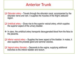
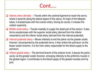
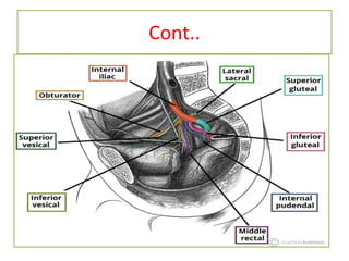
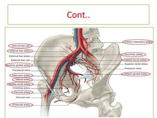
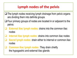
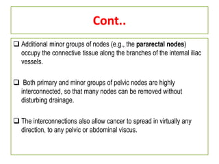
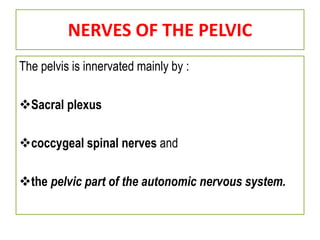
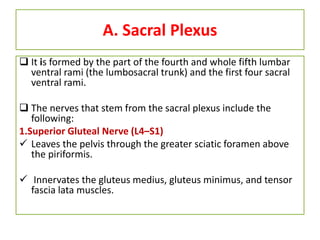
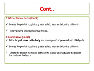
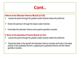
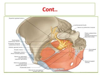

The major arteries, veins and nerves of the pelvis are described. The internal iliac artery and its branches supply the pelvic organs and structures. It has anterior and posterior trunks that give rise to arteries like the obturator, internal pudendal and inferior gluteal. The internal iliac vein drains deoxygenated blood from the pelvis. Lymph nodes in the pelvis include external iliac, internal iliac, sacral and common iliac nodes. The sacral plexus and coccygeal plexus provide motor and sensory innervation through nerves such as the pudendal and obturator nerves.


![ANATOMY OF THE LOWER URINARY TRACT AND MALE [Autosaved] [Autosaved].pptx](https://cdn.slidesharecdn.com/ss_thumbnails/anatomyofthelowerurinarytractandmaleautosavedautosaved-240526080531-9d6371e3-thumbnail.jpg?width=640&height=640&fit=bounds)


