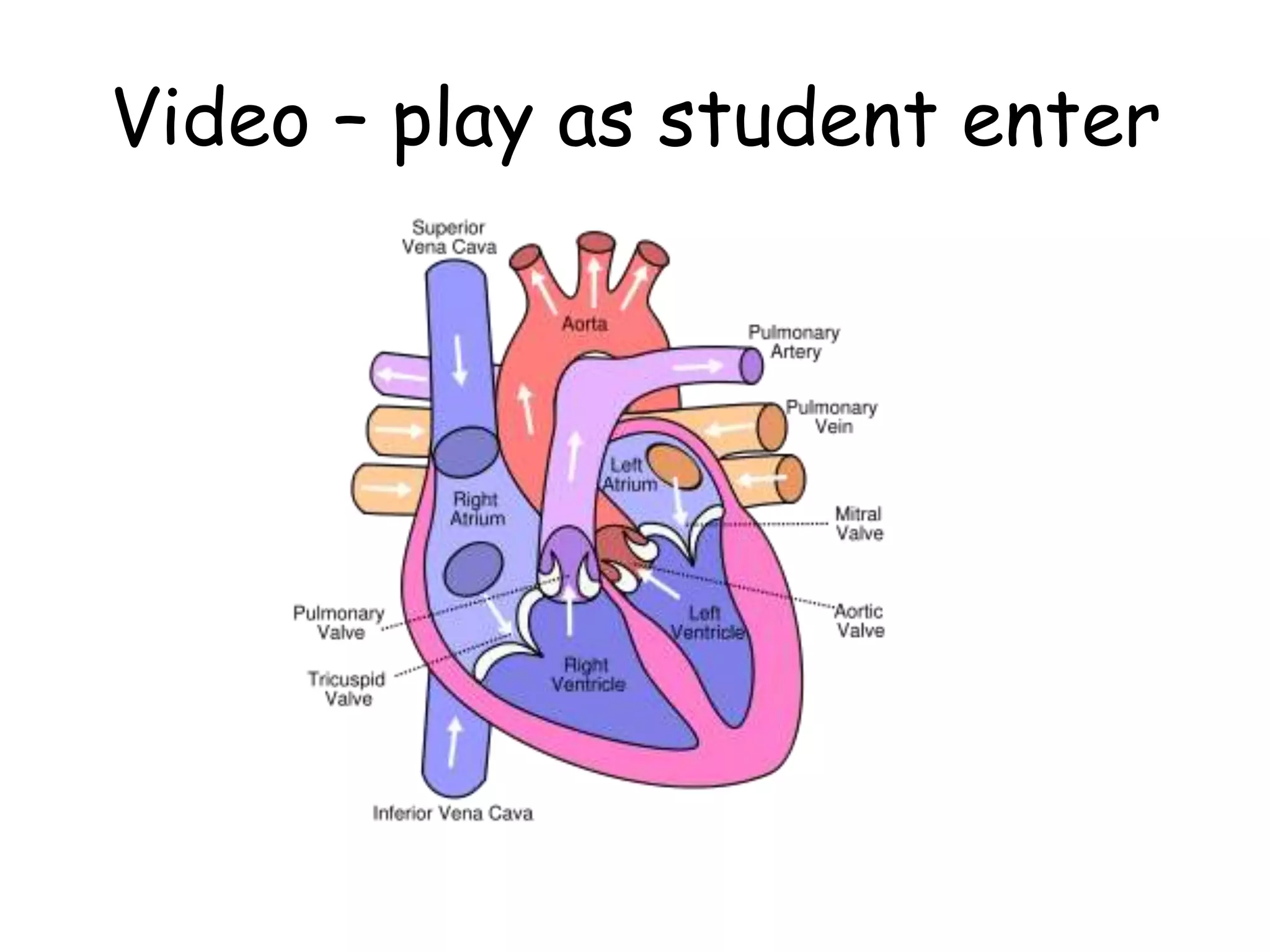
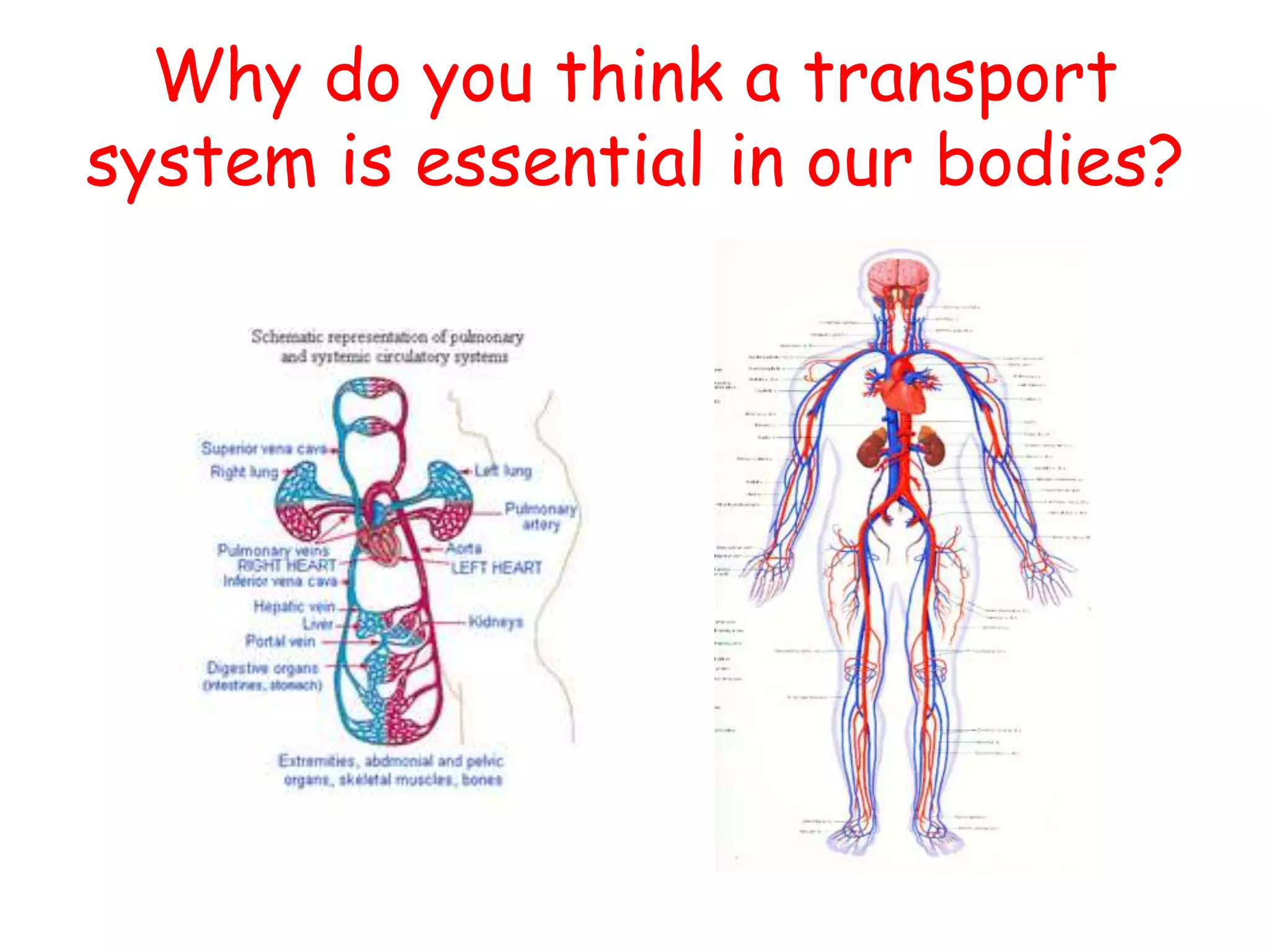
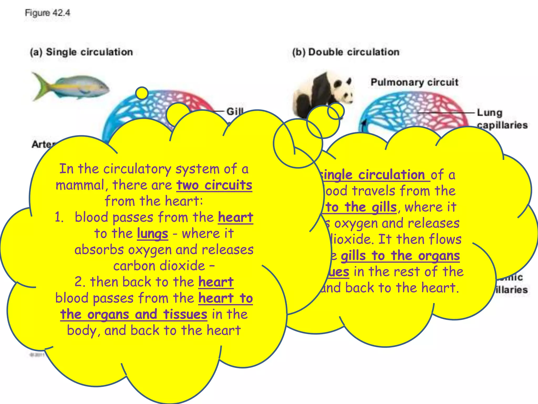
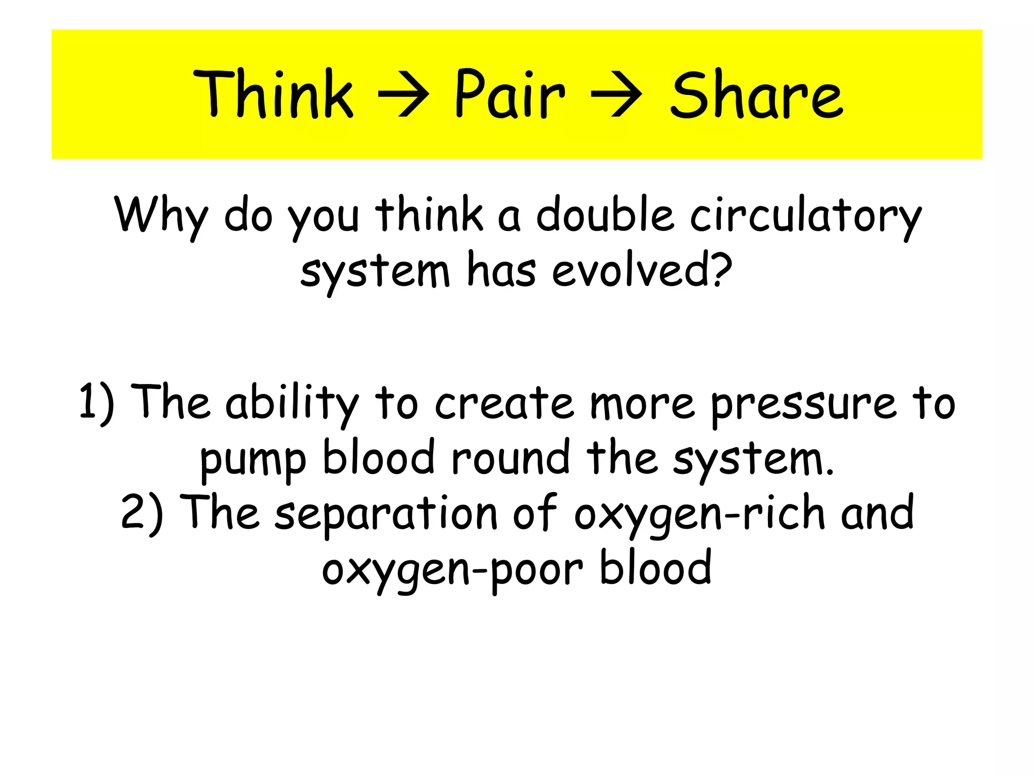
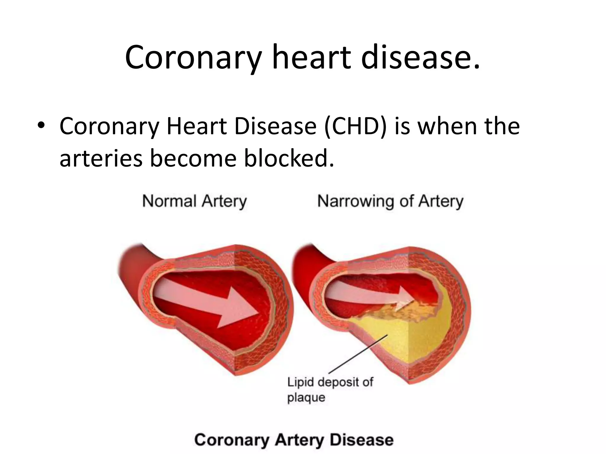
Unit 9 – Transport in animals
Learning Outcomes
• Describe the circulatory system
• Describe single and double circulatory systems
• Explain the advantages of a double circulation
How are things transported around
the body?
• The main transport system of all mammals is
the blood system, which is also known as the
circulatory system.
Video – play as student enter
Why do you think a transport
system is essential in our bodies?
The circulatory system is a system
of tubes with a pump and valves to
ensure one way flow of blood.
In a single circulation of a
fish blood travels from the
heart to the gills, where it
absorbs oxygen and releases
carbon dioxide. It then flows
from the gills to the organs
and tissues in the rest of the
body, and back to the heart.
In the circulatory system of a
mammal, there are two circuits
from the heart:
1. blood passes from the heart
to the lungs - where it
absorbs oxygen and releases
carbon dioxide –
2. then back to the heart
blood passes from the heart to
the organs and tissues in the
body, and back to the heart
What is meant by a ‘double
circulation’?
This means we have two transport systems.
• One carries blood from your heart to your
lungs and back again. What is the function of
this part?
Exchange oxygen and carbon dioxide with the
air. Low pressure
• The other carries oxygen around the body
and back again. What is the function of this
part?
Carry oxygen to tissues and cells that need it
and remove carbon dioxide. High pressure
Think  Pair  Share
Why do you think a double circulatory
system has evolved?
Think  Pair  Share
Why do you think a double circulatory
system has evolved?
1) The ability to create more pressure to
pump blood round the system.
2) The separation of oxygen-rich and
oxygen-poor blood
The Heart
Learning Outcomes
• Name and identify the structures of the heart.
• State the flow of blood to and from the heart.
Worksheet
• Label the heart
Heartbeat animation
Heartbeat animation
Stage 1:
A heartbeat begins
with the heart muscle relaxed
and valves
closed. Diastole
Blood flows into the two atria
and both sides fill up with
blood.
Stage 2:
The atria contract and the
blood is squeezed which
causes the valves leading to
the ventricles to open.
Systole.
Blood then flows from the atria
into the ventricles.
Stage 2 (continued):
The valves between the atria
and the ventricles close.
This prevents any backflow.
Stage 3:
Almost immediately, the
ventricles contract and the
blood is squeezed again.
The pressure of the blood
forces open the valves
leading out of the heart.
Blood is pumped out
of the heart.
Stage 3 (continued):
When the ventricles are
empty, the valves leading out
of the
heart close and the heart
muscle relaxes.
This completes the sequence
of contraction and relaxation in
one heartbeat.
Stage 1 (again):
The atria fill up with blood as
the heartbeat sequence
begins again.
Cut and Stick
Cut and stick in order to explain how
blood moves through the circulatory
system.
The correct order (Part 1)
• Start: Deoxygenated blood enters vena cava
• Blood enters right atrium
• Blood passes through atrioventricular valve
• Blood enters right ventricle
• Blood passes through the semilunar valve
• Blood leaves the heart via the pulmonary
artery and travels to the lungs
The correct order (Part 2)
• Oxygenated blood returns to the heart via the
pulmonary vein
• Blood enters the left atrium
• Blood passes through atrioventricular valve
• Blood enters the left ventricle
• Blood passes through the semilunar valve
• Blood leaves the heart via the aorta under
high pressure to the rest of the body
Coursebook questions
• Page 109
Monitoring the heart
Learning Outcomes
• State that the activity of the heart may be
monitored by ECG
• Investigate the effect of physical activity on
the pulse rate.
Measuring your heart activity.
• monitored by ECG, electrocardiogram
• pulse rate
• listening to sounds of valves closing
Investigating the effect of physical
activity on pulse rate.
• Practical worksheet.
• You need a watch.
Explain why pulse rate increases
during exercise.
• Exercise increases the rate at which energy is
needed from food.
• This increases the need for both food and oxygen
in the body.
• Your pulse rate increases so blood can be
pumped around the body faster.
• Your heart speeds up to pump extra food and
oxygen to the muscles for respirateion.
• Breathing speeds up to get more oxygen and to
get rid of more carbon dioxide.
Worksheet 2
• Effect of activity on heart
Heart problems
Coronary Arteries
• The blood vessels on
the outside of the
heart are called
coronary arteries
• They supply blood to
the heart muscles.
Heart attack.
• The heart muscles need a constant supply of
nutrients and oxygen, so it can keep
contracting and relaxing.
• If the arteries get blocked, the cardiac muscles
run short of oxygen.
• They can’t respire and will eventually stop
beating.
• This is called a heart attack or cardiac arrest.
Coronary heart disease.
• Coronary Heart Disease (CHD) is when the
arteries become blocked.
Factors that increase the risk of CHD
What can increase the chance of CHD?
• Smoking Cigarettes
• Diet
• Obesity
• Stress
• Genes
How can you prevent CHD?
• Regular exercise
– Keeps you fits
– Prevents excessive weight gain
– Decrease blood pressure
– Destress
• Stop smoking
• Diet
– Avoid diets high in animal fats
Preventing CHD
• Statins
– Statins are a type of drug that helps reduce the
cholesterol levels in the blood.
– However, they sometimes have unpleasant side
effects.
Treating CHD
• Doctors can prescribe statins to help reduce
cholesterol.
• Also, other drugs to lower blood pressure.
– For example: aspirin
• Surgery –
– bypass (see page 111 figure 9.8)
– Stent: a small tube is put inside the artery to keep it
open.
– Angioplasty – a small balloon is put in the artery and
inflated using water to open the artery and then
removed
Blood and lymphatic vessels
Learning Outcomes
• Describe the structure and function of
arteries, veins and capillaries.
• Name the blood vessels to and from the heart,
lungs and kidneys
• State the function of arterioles, venules and
shunt vessels.
• Outline the lymphatic system.
Types of blood vessel
There are 3 main types:
• Arteries – take blood away from the heart.
• Veins – take blood back to the heart
• Capillaries – involved in the exchange of
materials at the tissues.
Arteries
Veins
Veins
• They have valves to keep the blood moving in
one direction.
Capillaries
Naming blood vessels
Learn these
3 words
Joining blood vessels together
What are arterioles?
• Arterioles are vessels that small arteries
branch out into.
Function?
• To regulate the flow of blood into different
tissues.
What are venules?
• Small vessels that are formed when capillaries
come together. Many venules unite to form a
vein.
Function?
• Allow deoxygenated blood to return from the
capillary beds to the veins, which then
transports the blood back to the heart.
What are shunt vessels?
• A blood vessel that links an artery directly to a
vein, allowing blood to bypass the capillaries
in certain areas.
Function of shunt vessels
• Provide channels that bypass capillary beds.
Lymphatic Vessels
What is the lymphatic system?
• The lymphatic system is a network of tubes
throughout the body that drains fluid (called
lymph) from tissues and empties it back into
the bloodstream. Lymph is filtered through
the spleen, thymus and lymph nodes before
being emptied into the blood.
The role of the lymphatic system.
1) Fluid balance/ circulation of body fluids: to
return tissue fluid to the blood
2) Protection from infection: Produce white
blood cells (lymphocytes)
3) Absorption of fats: transport digested fats
from villi to blood stream
4) Filtering out bacteria
Coursebook
• Page 123 - Questions
Learning Outcomes
• List the components of blood.
• State the functions of each component of
blood.
• State the functions of lymphocytes and
phagocytes
• Describe the process of clotting.
• State the roles of blood clotting.
Blood >
• Red blood cells
• White blood cells
• Plasma
• Platelets
• Page 119 – table 9.3
White Blood Cells
• This is called PHAGOCYTOSIS
a) Blood vessel gets damaged
b) Platelets begin adhering to damaged area to begin to
form a plug.
c) To reinforce the plug platelets and damaged cells
release a group of proteins called clotting factors into
the plasma a wound site.
d) Clotting factors activate the enzyme thrombin from
its inactive form prothrombin
e) Thrombin is an enzyme which catalyses the conversion
of the soluble plasma protein fibrinogen into its insoluble
fibrous form fibrin.
f) Fibrin binds together platelets and blood cells to form
a solid 'plug' for the wound. This plug is called a clot.
Blood clotting >
Moving from blood to tissues >
Unit 9   transport in animals

Unit 9 transport in animals

  • 1.
    Unit 9 –Transport in animals
  • 2.
    Learning Outcomes • Describethe circulatory system • Describe single and double circulatory systems • Explain the advantages of a double circulation
  • 3.
    How are thingstransported around the body? • The main transport system of all mammals is the blood system, which is also known as the circulatory system.
  • 4.
    Video – playas student enter
  • 5.
    Why do youthink a transport system is essential in our bodies?
  • 6.
    The circulatory systemis a system of tubes with a pump and valves to ensure one way flow of blood.
  • 8.
    In a singlecirculation of a fish blood travels from the heart to the gills, where it absorbs oxygen and releases carbon dioxide. It then flows from the gills to the organs and tissues in the rest of the body, and back to the heart. In the circulatory system of a mammal, there are two circuits from the heart: 1. blood passes from the heart to the lungs - where it absorbs oxygen and releases carbon dioxide – 2. then back to the heart blood passes from the heart to the organs and tissues in the body, and back to the heart
  • 9.
    What is meantby a ‘double circulation’? This means we have two transport systems. • One carries blood from your heart to your lungs and back again. What is the function of this part? Exchange oxygen and carbon dioxide with the air. Low pressure • The other carries oxygen around the body and back again. What is the function of this part? Carry oxygen to tissues and cells that need it and remove carbon dioxide. High pressure
  • 10.
    Think  Pair Share Why do you think a double circulatory system has evolved?
  • 11.
    Think  Pair Share Why do you think a double circulatory system has evolved? 1) The ability to create more pressure to pump blood round the system. 2) The separation of oxygen-rich and oxygen-poor blood
  • 14.
  • 15.
    Learning Outcomes • Nameand identify the structures of the heart. • State the flow of blood to and from the heart.
  • 16.
  • 18.
  • 19.
    Stage 1: A heartbeatbegins with the heart muscle relaxed and valves closed. Diastole Blood flows into the two atria and both sides fill up with blood.
  • 20.
    Stage 2: The atriacontract and the blood is squeezed which causes the valves leading to the ventricles to open. Systole. Blood then flows from the atria into the ventricles.
  • 21.
    Stage 2 (continued): Thevalves between the atria and the ventricles close. This prevents any backflow.
  • 22.
    Stage 3: Almost immediately,the ventricles contract and the blood is squeezed again. The pressure of the blood forces open the valves leading out of the heart. Blood is pumped out of the heart.
  • 23.
    Stage 3 (continued): Whenthe ventricles are empty, the valves leading out of the heart close and the heart muscle relaxes. This completes the sequence of contraction and relaxation in one heartbeat.
  • 24.
    Stage 1 (again): Theatria fill up with blood as the heartbeat sequence begins again.
  • 25.
    Cut and Stick Cutand stick in order to explain how blood moves through the circulatory system.
  • 26.
    The correct order(Part 1) • Start: Deoxygenated blood enters vena cava • Blood enters right atrium • Blood passes through atrioventricular valve • Blood enters right ventricle • Blood passes through the semilunar valve • Blood leaves the heart via the pulmonary artery and travels to the lungs
  • 27.
    The correct order(Part 2) • Oxygenated blood returns to the heart via the pulmonary vein • Blood enters the left atrium • Blood passes through atrioventricular valve • Blood enters the left ventricle • Blood passes through the semilunar valve • Blood leaves the heart via the aorta under high pressure to the rest of the body
  • 28.
  • 29.
  • 30.
    Learning Outcomes • Statethat the activity of the heart may be monitored by ECG • Investigate the effect of physical activity on the pulse rate.
  • 31.
    Measuring your heartactivity. • monitored by ECG, electrocardiogram • pulse rate • listening to sounds of valves closing
  • 32.
    Investigating the effectof physical activity on pulse rate. • Practical worksheet. • You need a watch.
  • 33.
    Explain why pulserate increases during exercise. • Exercise increases the rate at which energy is needed from food. • This increases the need for both food and oxygen in the body. • Your pulse rate increases so blood can be pumped around the body faster. • Your heart speeds up to pump extra food and oxygen to the muscles for respirateion. • Breathing speeds up to get more oxygen and to get rid of more carbon dioxide.
  • 34.
    Worksheet 2 • Effectof activity on heart
  • 35.
  • 36.
    Coronary Arteries • Theblood vessels on the outside of the heart are called coronary arteries • They supply blood to the heart muscles.
  • 37.
    Heart attack. • Theheart muscles need a constant supply of nutrients and oxygen, so it can keep contracting and relaxing. • If the arteries get blocked, the cardiac muscles run short of oxygen. • They can’t respire and will eventually stop beating. • This is called a heart attack or cardiac arrest.
  • 38.
    Coronary heart disease. •Coronary Heart Disease (CHD) is when the arteries become blocked.
  • 39.
    Factors that increasethe risk of CHD What can increase the chance of CHD? • Smoking Cigarettes • Diet • Obesity • Stress • Genes
  • 40.
    How can youprevent CHD? • Regular exercise – Keeps you fits – Prevents excessive weight gain – Decrease blood pressure – Destress • Stop smoking • Diet – Avoid diets high in animal fats
  • 41.
    Preventing CHD • Statins –Statins are a type of drug that helps reduce the cholesterol levels in the blood. – However, they sometimes have unpleasant side effects.
  • 42.
    Treating CHD • Doctorscan prescribe statins to help reduce cholesterol. • Also, other drugs to lower blood pressure. – For example: aspirin • Surgery – – bypass (see page 111 figure 9.8) – Stent: a small tube is put inside the artery to keep it open. – Angioplasty – a small balloon is put in the artery and inflated using water to open the artery and then removed
  • 43.
  • 44.
    Learning Outcomes • Describethe structure and function of arteries, veins and capillaries. • Name the blood vessels to and from the heart, lungs and kidneys • State the function of arterioles, venules and shunt vessels. • Outline the lymphatic system.
  • 45.
    Types of bloodvessel There are 3 main types: • Arteries – take blood away from the heart. • Veins – take blood back to the heart • Capillaries – involved in the exchange of materials at the tissues.
  • 46.
  • 47.
  • 48.
    Veins • They havevalves to keep the blood moving in one direction.
  • 49.
  • 50.
  • 52.
    Joining blood vesselstogether What are arterioles? • Arterioles are vessels that small arteries branch out into. Function? • To regulate the flow of blood into different tissues.
  • 53.
    What are venules? •Small vessels that are formed when capillaries come together. Many venules unite to form a vein. Function? • Allow deoxygenated blood to return from the capillary beds to the veins, which then transports the blood back to the heart.
  • 54.
    What are shuntvessels? • A blood vessel that links an artery directly to a vein, allowing blood to bypass the capillaries in certain areas. Function of shunt vessels • Provide channels that bypass capillary beds.
  • 55.
    Lymphatic Vessels What isthe lymphatic system? • The lymphatic system is a network of tubes throughout the body that drains fluid (called lymph) from tissues and empties it back into the bloodstream. Lymph is filtered through the spleen, thymus and lymph nodes before being emptied into the blood.
  • 56.
    The role ofthe lymphatic system. 1) Fluid balance/ circulation of body fluids: to return tissue fluid to the blood 2) Protection from infection: Produce white blood cells (lymphocytes) 3) Absorption of fats: transport digested fats from villi to blood stream 4) Filtering out bacteria
  • 57.
  • 59.
    Learning Outcomes • Listthe components of blood. • State the functions of each component of blood. • State the functions of lymphocytes and phagocytes • Describe the process of clotting. • State the roles of blood clotting.
  • 60.
    Blood > • Redblood cells • White blood cells • Plasma • Platelets
  • 62.
    • Page 119– table 9.3
  • 65.
  • 66.
    • This iscalled PHAGOCYTOSIS
  • 69.
    a) Blood vesselgets damaged b) Platelets begin adhering to damaged area to begin to form a plug. c) To reinforce the plug platelets and damaged cells release a group of proteins called clotting factors into the plasma a wound site. d) Clotting factors activate the enzyme thrombin from its inactive form prothrombin e) Thrombin is an enzyme which catalyses the conversion of the soluble plasma protein fibrinogen into its insoluble fibrous form fibrin. f) Fibrin binds together platelets and blood cells to form a solid 'plug' for the wound. This plug is called a clot. Blood clotting >
  • 70.
    Moving from bloodto tissues >