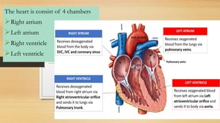
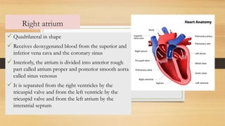
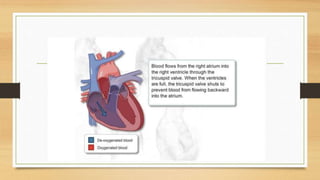
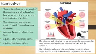
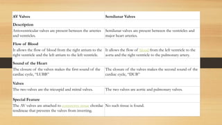
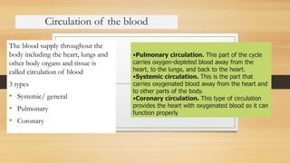
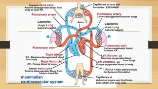
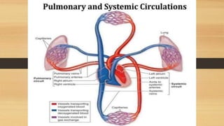
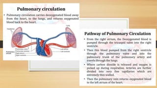
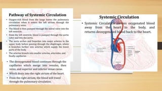
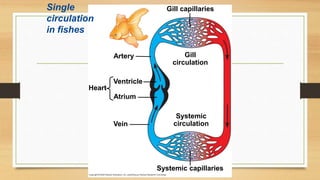
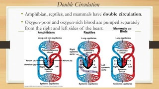
Cardiovascular system
cardiovascular system delivers
oxygen, nutrients, hormones,
and other important
substances to cells and organs
in the body. It plays an
important role in helping the
body meet the demands of
activity, exercise, and stress.
functions- transportation, protection,
fluid balance and thermoregulation.
• blood-vascular, or simply the
circulatory, system
• Heart-a muscular pumping device, a
• closed system of vessels called
arteries, veins, and capillaries.
• The heart is a hollow, muscular organ
situated in the space between lungs in he
thoracic cavity
• It lies obliquely, little more to the left
that the right
• Apex is about 9cm to the left
• About the size of a clenched fist
• Composed pf endocardium,
myocardium and pericardium
• Endocardium – the inner layer
and it consists of endothelial
tissue that lines the inner
surface of the heart and the
cardiac valves
• Myocardium – the middle layer
and composed of muscle fibers
that enable the heart to pump
• Epicardium-outer layer, tightly
adherent to the heart ad the
base of the great vessels
• Pericardium-thin, fibrous,
double layered sac that
surrounds the heart
The heart is consist of 4 chambers
➢Right atrium
➢Left atrium
➢Right ventricle
➢Left ventricle
Right atrium
✓ Quadrilateral in shape
✓ Receives deoxygenated blood from the superior and
inferior vena cava and the coronary sinus
✓ Interiorly, the atrium is divided into anterior rough
part called atrium proper and posterior smooth aorta
called sinus venosus
✓ It is separated from the right ventricles by the
tricuspid valve and from the left ventricle by the
tricuspid valve and from the left atrium by the
interatrial septum
Right ventricle
• It is a triangular chamber situated to left and right
atrium which receives blood from right atrium
and pumps blood into the pulmonary trunk
• The interior is rough due to muscular rings called
as trabeculae carnae
• It contains cords connecting cusps of valves and
the papillary muscles known as cordae tendanae
• It is separated from the right atrium and left
ventricle by tricuspid calve ad interventricular
septum respectively
Heart valves
• The cardiac valves are composed of
fibrous tissue and allow blood to
flow in one direction they prevent
regurgitation of the blood
• The valves open and close as a
result of blood flow and pressure
differences
• there are 2 pairs of valves in the
heart
• A pair of atrioventricular valves
• A pair of semilunar valves
• The tricuspid and mitral valves are known as the atrioventricular
valve because they are located between the atria and the
ventricles
• The pulmonic and aortic valves are known as the semilunar
valves because each has three leaflets shaped like half-moons
AV Valves Semilunar Valves
Description
Atrioventricular valves are present between the arteries
and ventricles.
Semilunar valves are present between the ventricles and
major heart arteries.
Flow of Blood
It allows the flow of blood from the right atrium to the
right ventricle and the left atrium to the left ventricle.
It allows the flow of blood from the left ventricle to the
aorta and the right ventricle to the pulmonary artery.
Sound of the Heart
The closure of the valves makes the first sound of the
cardiac cycle, “LUBB”
The closure of the valves makes the second sound of the
cardiac cycle, “DUB”
Valves
The two valves are the tricuspid and mitral valves. The two valves are aortic and pulmonary valves.
Special Feature
The AV valves are attached to connective tissue chordae
tendineae that prevents the valves from inverting.
No such tissue is found.
Circulation of the blood
The blood supply throughout the
body including the heart, lungs and
other body organs and tissue is
called circulation of blood
3 types
• Systemic/ general
• Pulmonary
• Coronary
•Pulmonary circulation. This part of the cycle
carries oxygen-depleted blood away from the
heart, to the lungs, and back to the heart.
•Systemic circulation. This is the part that
carries oxygenated blood away from the heart and
to other parts of the body.
•Coronary circulation. This type of circulation
provides the heart with oxygenated blood so it can
function properly
mammalian
cardiovascular system
Superior vena cava
Returns deoxygenated blood from
body to heart RA
Pulmonary artery
Capillaries
of right Lung
GAS EXCHANGE
3
7
3
8
9
2
4
11
5
1
10
Aorta
Pulmonary vein
Right Atrium
RA - Receives deoxygenated blood
from body
Right Ventricle
RV - Pumps blood to lungs
Inferior vena cava
Returns deoxygenated blood from
body to heart RA
Capillaries of
abdominal organs and hind limbs
EXCHANGE with body cells
Pulmonary vein
Carries oxygenated blood
to heart: LA
Left Atrium - LA
Receives oxygenated blood
from lungs
Left Ventricle - LV
Pumps oxygenated blood to body
Aorta = main artery to body
for Systemic Circulation
Capillaries
of left Lung
GAS EXCHANGE
Pulmonary artery
Carries deoxygenated blood to lungs
Capillaries of head and
Forelimbs - EXCHANGE
Coronary circulation is the circulation
of blood in the arteries and veins that
supply the heart muscle (myocardium).
Coronary
arteries supply oxygenated blood to the
heart muscle. Cardiac veins then drain
away the blood after it has been
deoxygenated. Because the rest of the
body, and most especially the brain,
needs a steady supply of oxygenated
blood that is free of all but the slightest
interruptions, the heart is required to
function continuously.
5 types of blood vessels
Circulatory systems link exchange surfaces with cells throughout the body
• In small and/or thin animals, cells can exchange
materials directly with the surrounding medium.
• In most animals, transport systems connect
the organs of exchange with the body cells.
• Most complex animals have internal transport
systems that circulate fluid.
Gastrovascular Cavities
• Simple animals, such as cnidarians, have a body wall
that is only two cells thick and that encloses a
gastrovascular cavity.
• This cavity functions in both digestion and
distribution of substances throughout the body.
• Some cnidarians, such as jellies, have elaborate
gastrovascular cavities.
• Flatworms have a gastrovascular cavity and a large
surface area to volume ratio.
Internal transport in gastrovascular cavities
Circular
canal
Radial canal
Mouth
(a) The moon jelly Aurelia, a cnidarian The planarian Dugesia, a
flatworm
(b)
Mouth
Pharynx
2 mm
5 cm
Open and Closed Circulatory Systems
• More complex animals have
either open or closed
circulatory systems.
• Both systems have three basic
components:
• A circulatory fluid = blood or
hemolymph.
• A set of tubes = blood vessels.
• A muscular pump = the heart.
• In insects, other arthropods, and most
molluscs, blood bathes the organs directly
in an open circulatory system.
• In an open circulatory system, there is no
distinction between blood and interstitial
fluid, and this general body fluid is more
correctly called hemolymph.
• In a closed circulatory system, the
blood is confined to vessels and is distinct
from the interstitial fluid.
• Closed systems are more efficient at
transporting circulatory fluids to tissues
and cells.
Open and closed circulatory systems
Heart
Hemolymph in
sinuses
surrounding organs
Heart
Interstitial
fluid
Small branch vessels
In each organ
Blood
Dorsal vessel
(main heart)
Auxiliary hearts Ventral vessels
(b) A closed circulatory system
(a) An open circulatory system
Tubular heart
Pores
Organization of Vertebrate Closed
Circulatory Systems
• Humans and other vertebrates have a closed
circulatory system, often called the cardiovascular
system.
• The three main types of blood vessels are:
arteries - away from the heart.
veins - toward the heart.
capillaries - exchange with body cells.
• Arteries branch into arterioles and carry blood to
capillaries.
• Networks of capillaries called capillary beds are the
sites of chemical exchange between the blood and
interstitial fluid.
• Venules converge into veins and return blood from
capillaries to the heart.
• Vertebrate hearts contain two or more chambers.
• Blood enters through an atrium and is pumped out
through a ventricle.
Atria - receive blood
Ventricles - pump blood
Single Circulation
• Bony fishes, rays, and sharks have single circulation
with a two-chambered heart.
• In single circulation, blood leaving the heart passes
through two capillary beds before returning.
Single
circulation
in fishes
Artery
Ventricle
Atrium
Heart
Vein
Systemic capillaries
Systemic
circulation
Gill
circulation
Gill capillaries
Double Circulation
• Amphibian, reptiles, and mammals have double circulation.
• Oxygen-poor and oxygen-rich blood are pumped separately
from the right and left sides of the heart.
Amphibians
Lung and skin capillaries
Pulmocutaneous
circuit
Atrium (A)
Ventricle (V)
Atrium (A)
Systemic
circuit
Right Left
Systemic capillaries
Reptiles
Lung capillaries
Pulmonary
circuit
Right
systemic
aorta
Right Left
Left
systemic
aorta
Systemic capillaries
A A
V
V
Systemic capillaries
Pulmonary
circuit
Systemic
circuit
Right Left
A A
V
V
Lung capillaries
Mammals and
Birds
• In reptiles and mammals, oxygen-poor blood flows
through the pulmonary circuit to pick up oxygen
through the lungs.
• In amphibians, oxygen-poor blood flows through a
pulmocutaneous circuit to pick up oxygen through
the lungs and skin.
• Oxygen-rich blood delivers oxygen through the
systemic circuit.
• Double circulation maintains higher blood pressure in
the organs than does single circulation.
Adaptations of Double Circulatory Systems
Amphibians:
• Frogs / amphibians have a three-
chambered heart: 2 atria and 1
ventricle.
• The ventricle pumps blood into a
forked artery that splits the
ventricle’s output into the
pulmocutaneous circuit and the
systemic circuit.
• Underwater, blood flow to the
lungs is nearly shut off.
• Turtles, snakes, and lizards have a
three-chambered heart: two atria
and one ventricle.
• In alligators, caimans, and other
crocodilians a septum - partially
or fully divides the ventricle.
• Reptiles have double circulation,
with a pulmonary circuit - lungs
and a systemic circuit.
• Mammals and birds have a four-chambered heart with two atria and two
ventricles.
• The left side of the heart pumps and receives only oxygen-rich blood, while
the right side receives and pumps only oxygen-poor blood.
• Mammals and birds are endotherms and require more O2 than ectotherms.
Cardiovascular System and Excretory system

Cardiovascular System and Excretory system

  • 1.
  • 2.
    cardiovascular system delivers oxygen,nutrients, hormones, and other important substances to cells and organs in the body. It plays an important role in helping the body meet the demands of activity, exercise, and stress. functions- transportation, protection, fluid balance and thermoregulation. • blood-vascular, or simply the circulatory, system • Heart-a muscular pumping device, a • closed system of vessels called arteries, veins, and capillaries.
  • 3.
    • The heartis a hollow, muscular organ situated in the space between lungs in he thoracic cavity • It lies obliquely, little more to the left that the right • Apex is about 9cm to the left • About the size of a clenched fist • Composed pf endocardium, myocardium and pericardium
  • 4.
    • Endocardium –the inner layer and it consists of endothelial tissue that lines the inner surface of the heart and the cardiac valves • Myocardium – the middle layer and composed of muscle fibers that enable the heart to pump • Epicardium-outer layer, tightly adherent to the heart ad the base of the great vessels • Pericardium-thin, fibrous, double layered sac that surrounds the heart
  • 5.
    The heart isconsist of 4 chambers ➢Right atrium ➢Left atrium ➢Right ventricle ➢Left ventricle
  • 6.
    Right atrium ✓ Quadrilateralin shape ✓ Receives deoxygenated blood from the superior and inferior vena cava and the coronary sinus ✓ Interiorly, the atrium is divided into anterior rough part called atrium proper and posterior smooth aorta called sinus venosus ✓ It is separated from the right ventricles by the tricuspid valve and from the left ventricle by the tricuspid valve and from the left atrium by the interatrial septum
  • 7.
    Right ventricle • Itis a triangular chamber situated to left and right atrium which receives blood from right atrium and pumps blood into the pulmonary trunk • The interior is rough due to muscular rings called as trabeculae carnae • It contains cords connecting cusps of valves and the papillary muscles known as cordae tendanae • It is separated from the right atrium and left ventricle by tricuspid calve ad interventricular septum respectively
  • 9.
    Heart valves • Thecardiac valves are composed of fibrous tissue and allow blood to flow in one direction they prevent regurgitation of the blood • The valves open and close as a result of blood flow and pressure differences • there are 2 pairs of valves in the heart • A pair of atrioventricular valves • A pair of semilunar valves • The tricuspid and mitral valves are known as the atrioventricular valve because they are located between the atria and the ventricles • The pulmonic and aortic valves are known as the semilunar valves because each has three leaflets shaped like half-moons
  • 10.
    AV Valves SemilunarValves Description Atrioventricular valves are present between the arteries and ventricles. Semilunar valves are present between the ventricles and major heart arteries. Flow of Blood It allows the flow of blood from the right atrium to the right ventricle and the left atrium to the left ventricle. It allows the flow of blood from the left ventricle to the aorta and the right ventricle to the pulmonary artery. Sound of the Heart The closure of the valves makes the first sound of the cardiac cycle, “LUBB” The closure of the valves makes the second sound of the cardiac cycle, “DUB” Valves The two valves are the tricuspid and mitral valves. The two valves are aortic and pulmonary valves. Special Feature The AV valves are attached to connective tissue chordae tendineae that prevents the valves from inverting. No such tissue is found.
  • 11.
    Circulation of theblood The blood supply throughout the body including the heart, lungs and other body organs and tissue is called circulation of blood 3 types • Systemic/ general • Pulmonary • Coronary •Pulmonary circulation. This part of the cycle carries oxygen-depleted blood away from the heart, to the lungs, and back to the heart. •Systemic circulation. This is the part that carries oxygenated blood away from the heart and to other parts of the body. •Coronary circulation. This type of circulation provides the heart with oxygenated blood so it can function properly
  • 12.
    mammalian cardiovascular system Superior venacava Returns deoxygenated blood from body to heart RA Pulmonary artery Capillaries of right Lung GAS EXCHANGE 3 7 3 8 9 2 4 11 5 1 10 Aorta Pulmonary vein Right Atrium RA - Receives deoxygenated blood from body Right Ventricle RV - Pumps blood to lungs Inferior vena cava Returns deoxygenated blood from body to heart RA Capillaries of abdominal organs and hind limbs EXCHANGE with body cells Pulmonary vein Carries oxygenated blood to heart: LA Left Atrium - LA Receives oxygenated blood from lungs Left Ventricle - LV Pumps oxygenated blood to body Aorta = main artery to body for Systemic Circulation Capillaries of left Lung GAS EXCHANGE Pulmonary artery Carries deoxygenated blood to lungs Capillaries of head and Forelimbs - EXCHANGE
  • 16.
    Coronary circulation isthe circulation of blood in the arteries and veins that supply the heart muscle (myocardium). Coronary arteries supply oxygenated blood to the heart muscle. Cardiac veins then drain away the blood after it has been deoxygenated. Because the rest of the body, and most especially the brain, needs a steady supply of oxygenated blood that is free of all but the slightest interruptions, the heart is required to function continuously.
  • 17.
    5 types ofblood vessels
  • 52.
    Circulatory systems linkexchange surfaces with cells throughout the body • In small and/or thin animals, cells can exchange materials directly with the surrounding medium. • In most animals, transport systems connect the organs of exchange with the body cells. • Most complex animals have internal transport systems that circulate fluid.
  • 53.
    Gastrovascular Cavities • Simpleanimals, such as cnidarians, have a body wall that is only two cells thick and that encloses a gastrovascular cavity. • This cavity functions in both digestion and distribution of substances throughout the body. • Some cnidarians, such as jellies, have elaborate gastrovascular cavities. • Flatworms have a gastrovascular cavity and a large surface area to volume ratio.
  • 54.
    Internal transport ingastrovascular cavities Circular canal Radial canal Mouth (a) The moon jelly Aurelia, a cnidarian The planarian Dugesia, a flatworm (b) Mouth Pharynx 2 mm 5 cm
  • 55.
    Open and ClosedCirculatory Systems • More complex animals have either open or closed circulatory systems. • Both systems have three basic components: • A circulatory fluid = blood or hemolymph. • A set of tubes = blood vessels. • A muscular pump = the heart. • In insects, other arthropods, and most molluscs, blood bathes the organs directly in an open circulatory system. • In an open circulatory system, there is no distinction between blood and interstitial fluid, and this general body fluid is more correctly called hemolymph. • In a closed circulatory system, the blood is confined to vessels and is distinct from the interstitial fluid. • Closed systems are more efficient at transporting circulatory fluids to tissues and cells.
  • 56.
    Open and closedcirculatory systems Heart Hemolymph in sinuses surrounding organs Heart Interstitial fluid Small branch vessels In each organ Blood Dorsal vessel (main heart) Auxiliary hearts Ventral vessels (b) A closed circulatory system (a) An open circulatory system Tubular heart Pores
  • 57.
    Organization of VertebrateClosed Circulatory Systems • Humans and other vertebrates have a closed circulatory system, often called the cardiovascular system. • The three main types of blood vessels are: arteries - away from the heart. veins - toward the heart. capillaries - exchange with body cells.
  • 58.
    • Arteries branchinto arterioles and carry blood to capillaries. • Networks of capillaries called capillary beds are the sites of chemical exchange between the blood and interstitial fluid. • Venules converge into veins and return blood from capillaries to the heart.
  • 59.
    • Vertebrate heartscontain two or more chambers. • Blood enters through an atrium and is pumped out through a ventricle. Atria - receive blood Ventricles - pump blood
  • 60.
    Single Circulation • Bonyfishes, rays, and sharks have single circulation with a two-chambered heart. • In single circulation, blood leaving the heart passes through two capillary beds before returning.
  • 61.
  • 62.
    Double Circulation • Amphibian,reptiles, and mammals have double circulation. • Oxygen-poor and oxygen-rich blood are pumped separately from the right and left sides of the heart. Amphibians Lung and skin capillaries Pulmocutaneous circuit Atrium (A) Ventricle (V) Atrium (A) Systemic circuit Right Left Systemic capillaries Reptiles Lung capillaries Pulmonary circuit Right systemic aorta Right Left Left systemic aorta Systemic capillaries A A V V Systemic capillaries Pulmonary circuit Systemic circuit Right Left A A V V Lung capillaries Mammals and Birds
  • 63.
    • In reptilesand mammals, oxygen-poor blood flows through the pulmonary circuit to pick up oxygen through the lungs. • In amphibians, oxygen-poor blood flows through a pulmocutaneous circuit to pick up oxygen through the lungs and skin. • Oxygen-rich blood delivers oxygen through the systemic circuit. • Double circulation maintains higher blood pressure in the organs than does single circulation.
  • 64.
    Adaptations of DoubleCirculatory Systems Amphibians: • Frogs / amphibians have a three- chambered heart: 2 atria and 1 ventricle. • The ventricle pumps blood into a forked artery that splits the ventricle’s output into the pulmocutaneous circuit and the systemic circuit. • Underwater, blood flow to the lungs is nearly shut off. • Turtles, snakes, and lizards have a three-chambered heart: two atria and one ventricle. • In alligators, caimans, and other crocodilians a septum - partially or fully divides the ventricle. • Reptiles have double circulation, with a pulmonary circuit - lungs and a systemic circuit. • Mammals and birds have a four-chambered heart with two atria and two ventricles. • The left side of the heart pumps and receives only oxygen-rich blood, while the right side receives and pumps only oxygen-poor blood. • Mammals and birds are endotherms and require more O2 than ectotherms.