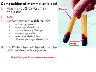
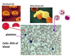
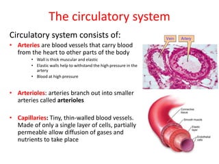
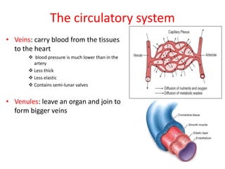
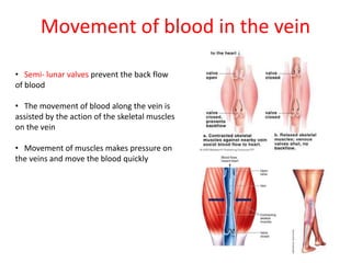
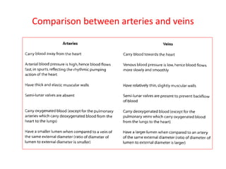
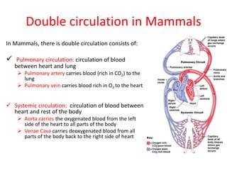
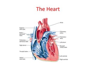
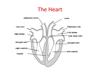
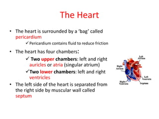
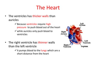
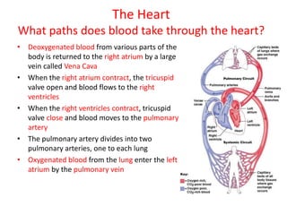
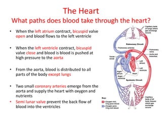
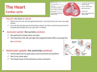
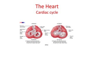
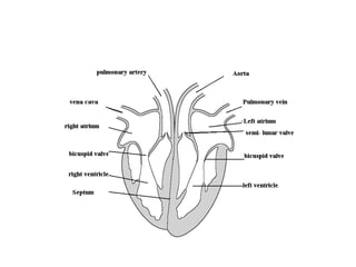
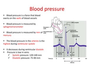
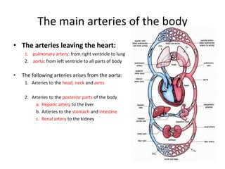
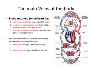
Transport in
Humans
The heart & its blood vessels
Composition of mammalian blood
I. Plasma (55% by volume)
contains:
1. water
2. soluble substances which include:
a. nutrients, e.g. glucose
b. wastes, e.g. carbon dioxide
c. plasma proteins, e.g. fibrinogen
d. hormones, e.g. insulin
e. antibodies for body defense,
f. dissolves gases, e.g. carbon dioxide
II. Cells (45% by volume) which include: red blood
cells, white blood cells and platelets
Blood cells produced in the bone marrow
The Transport Function of blood
• Blood transports the following:
 Digested food: glucose, amino acids, vitamins
etc..
Excretory products from tissues
 Nitrogenous wastes: urea uric acid (to the kidney)
Carbon dioxide ( to the lung)
 Hormones: insulin
Heat
oxygen
Red blood cells
White blood cells
platelets
Cells: 45% of
blood
What are the different types of blood cells?
• RBCs: contain red hemoglobin which enables RBCs to carry
oxygen and some carbon dioxide
• WBCs: lymphocytes & phagocytes, protect us from diseases
• Platelets: broken cell fragments, help in blood clotting
The Mechanism of Blood Clotting
1. Damaged tissue and platelets produce
thrombokinases (enzyme)
2. Thrombokinase converts the protein
prothrombin into thrombin
3. Thrombin is an enzyme which converts
fibrinogen into fibrin
4. Fibrin threads form a clot
Normally, undamaged blood vessels does
not clot. This is due to the absence of
anti-clotting substance called heparin
How does blood transport oxygen to the cells of the body?
1. Oxygen diffuses from the alveoli into the blood
2. Hemoglobin combines with oxygen to form oxyhemoglobin
3. Blood transports oxyhemoglobin to all tissues of the body
4. In the tissues oxyhemoglobin release oxygen
Lymphocytes & phagocytes, protect us from diseases
1. Lymphocytes: produce antibodies which acts by:
I. Destroying the bacteria
II. Causing bacteria agglutinate together, so it is easily to
ingested by phagocytes
III. Neutralizing the harmful substances (toxin)
IV. Attaching to virus, prevent them to bind to the body cell
2. Phagocytes: it engulfs the bacteria and then ingests it
by a process called phagocytosis
Phagocytosis: the process of engulfing and ingesting foreign
particles, such as bacteria, by the white blood cells
Immunization: dead bacteria injected to animal body to induce
antibodies production. Then these antibodies are injected to the
human body to provide immunity against pathogens
Organ transplant
• Organs can be transplanted from a donor to a recipient
• The recipient’s lymphocytes may respond by producing
antibodies to destroy the transplanted organ
How can tissue rejection be prevented?
• The tissues of both donor and recipient must be genetically close
(transplant between relatives)
• Reduce the risk of tissue rejection by using immunosuppressive drugs
Blood Groups
The circulatory system
Circulatory system consists of:
• Arteries are blood vessels that carry blood
from the heart to other parts of the body
• Wall is thick muscular and elastic
• Elastic walls help to withstand the high pressure in the
artery
• Blood at high pressure
• Arterioles: arteries branch out into smaller
arteries called arterioles
• Capillaries: Tiny, thin-walled blood vessels.
Made of only a single layer of cells, partially
permeable allow diffusion of gases and
nutrients to take place
The circulatory system
• Veins: carry blood from the tissues
to the heart
 blood pressure is much lower than in the
artery
 Less thick
 Less elastic
 Contains semi-lunar valves
• Venules: leave an organ and join to
form bigger veins
Movement of blood in the vein
• Semi- lunar valves prevent the back flow
of blood
• The movement of blood along the vein is
assisted by the action of the skeletal muscles
on the vein
• Movement of muscles makes pressure on
the veins and move the blood quickly
Comparison between arteries and veins
Double circulation in Mammals
In Mammals, there is double circulation consists of:
 Pulmonary circulation: circulation of blood
between heart and lung
 Pulmonary artery carries blood (rich in CO2) to the
lung
 Pulmonary vein carries blood rich in O2 to the heart
 Systemic circulation: circulation of blood between
heart and rest of the body
 Aorta carries the oxygenated blood from the left
side of the heart to all parts of the body
 Venae Cava carries deoxygenated blood from all
parts of the body back to the right side of heart
The Heart
The Heart
The Heart
• The heart is surrounded by a ‘bag’ called
pericardium
Pericardium contains fluid to reduce friction
• The heart has four chambers:
 Two upper chambers: left and right
auricles or atria (singular atrium)
Two lower chambers: left and right
ventricles
• The left side of the heart is separated from
the right side by muscular wall called
septum
The Heart
• The ventricles has thicker walls than
auricles
Because ventricles require high
pressure to push blood out of the heart
while auricles only push blood to
ventricles
• The right ventricle has thinner walls
than the left ventricle
It pumps blood to the lungs which are a
short distance from the heart
The Heart
What paths does blood take through the heart?
• Deoxygenated blood from various parts of the
body is returned to the right atrium by a large
vein called Vena Cava
• When the right atrium contract, the tricuspid
valve open and blood flows to the right
ventricles
• When the right ventricles contract, tricuspid
valve close and blood moves to the pulmonary
artery
• The pulmonary artery divides into two
pulmonary arteries, one to each lung
• Oxygenated blood from the lung enter the left
atrium by the pulmonary vein
The Heart
What paths does blood take through the heart?
• When the left atrium contract, bicuspid valve
open and blood flows to the left ventricle
• When the left ventricle contract, bicuspid
valve close and blood is blood is pushed at
high pressure to the aorta
• From the aorta, blood is distributed to all
parts of the body except lungs
• Two small coronary arteries emerge from the
aorta and supply the heart with oxygen and
nutrients
• Semi lunar valve prevent the back flow of
blood into the ventricles
The Heart
Cardiac cycle
• Diastole: the heart is relaxed
 Blood comes into the atria through the pulmonary veins (left atrium) and vena cava (right
atrium).
 From the left and right atria the blood flows directly into both ventricles because both the
bicuspid and tricuspid valves are open (80 % of blood).
• Auricular systole: the auricles contract.
 Bicuspid and tricuspid valves are open
 The blood from the left and right atria (approximately 20%) is pumped into
the ventricles.
• Ventricular systole: the ventricles contract
 the bicuspid and tricuspid valves close to prevent any backflow
 Semi-lunar valve open
 The blood moves to the pulmonary artery and aorta
The Heart
Cardiac cycle
Blood pressure
 Blood pressure is a force that blood
exerts on the walls of blood vessels
 Blood pressure is measured by
sphygmomanometer
 Blood pressure is measured by mm of
mercury
 The blood pressure in the arteries is the
highest during ventricular systole
 It decreases during ventricular diastole
 Pressure is low in veins
 Systolic pressure: 120-140 mm
 Diastolic pressure: 75-90 mm
The main arteries of the body
• The arteries leaving the heart:
1. pulmonary artery: from right ventricle to lung
2. aorta: from left ventricle to all parts of body
• The following arteries arises from the aorta:
1. Arteries to the head, neck and arms
2. Arteries to the posterior parts of the body
a. Hepatic artery to the liver
b. Arteries to the stomach and intestine
c. Renal artery to the kidney
The main Veins of the body
• Blood returned to the heart by:
1. pulmonary vein: from lung to the left atrium
2. anterior or superior vena cava: from head,
neck and arms to the right atrium
3. Posterior or inferior vena cava: from the lower
parts to the right atrium
• The inferior vena cava collects blood from
various veins. Among these are:
1. Renal vein: bringing blood from kidney
2. Hepatic vein bringing blood from the liver
Tissue and Lymph
• Tissue fluid is a colorless liquid found in the tiny space
between cells.
How are substances transferred between capillaries and
tissue cells?
1. Dissolved food substances (nutrients) and oxygen
diffuse from the blood capillaries to the tissue fluid and
then into the cells
2. Waste products diffuse from the cells into the tissue
fluid and then through the blood capillary walls into the
blood
How tissue fluid is formed?
Blood plasma is forced out through the capillary walls
into the spaces between the cells as tissue fluid
Tissue fluid contains white blood cells (phagocytes)
and plasma with no red blood cells
Coronary heart disease
• The most common heart disease is
coronary heart disease
• During heart attack, blood flow to a
particular part of the heart may be
completely blocked.
• Due to this block, part of the heart
does not receive sufficient oxygen and
nutrients
• This region of the heart dies
Coronary heart disease
Causes of heart attack:
• Fatty acid (cholesterol) deposits on the
inner surface of the coronary artery. This is
called atherosclerosis
• This narrows the artery and increase the
blood pressure
• This affects the artery and develops rough
inner surface
• This increase the risk of blood clot called
thrombosis
• The supply of blood and oxygen to the
heart muscles may be completely cut off
• This leads to death of heart muscles cells
and heart attack occurs
Factors that increase the risk of coronary heart
disease
1. Diet rich in cholesterol
2. stress
3. smoking
 Nicotine: increase blood pressure
 Carbon monoxide: increase fatty deposits
4. hereditary factors
5. Increasing age
6. Sedentary life (not doing exercise)
7. Overweight (obesity)
8. Diabetes
9. High blood pressure
The circulatory system

The circulatory system

  • 1.
    Transport in Humans The heart& its blood vessels
  • 2.
    Composition of mammalianblood I. Plasma (55% by volume) contains: 1. water 2. soluble substances which include: a. nutrients, e.g. glucose b. wastes, e.g. carbon dioxide c. plasma proteins, e.g. fibrinogen d. hormones, e.g. insulin e. antibodies for body defense, f. dissolves gases, e.g. carbon dioxide II. Cells (45% by volume) which include: red blood cells, white blood cells and platelets Blood cells produced in the bone marrow
  • 3.
    The Transport Functionof blood • Blood transports the following:  Digested food: glucose, amino acids, vitamins etc.. Excretory products from tissues  Nitrogenous wastes: urea uric acid (to the kidney) Carbon dioxide ( to the lung)  Hormones: insulin Heat oxygen
  • 4.
    Red blood cells Whiteblood cells platelets Cells: 45% of blood
  • 5.
    What are thedifferent types of blood cells? • RBCs: contain red hemoglobin which enables RBCs to carry oxygen and some carbon dioxide • WBCs: lymphocytes & phagocytes, protect us from diseases • Platelets: broken cell fragments, help in blood clotting
  • 6.
    The Mechanism ofBlood Clotting 1. Damaged tissue and platelets produce thrombokinases (enzyme) 2. Thrombokinase converts the protein prothrombin into thrombin 3. Thrombin is an enzyme which converts fibrinogen into fibrin 4. Fibrin threads form a clot Normally, undamaged blood vessels does not clot. This is due to the absence of anti-clotting substance called heparin
  • 7.
    How does bloodtransport oxygen to the cells of the body? 1. Oxygen diffuses from the alveoli into the blood 2. Hemoglobin combines with oxygen to form oxyhemoglobin 3. Blood transports oxyhemoglobin to all tissues of the body 4. In the tissues oxyhemoglobin release oxygen
  • 8.
    Lymphocytes & phagocytes,protect us from diseases 1. Lymphocytes: produce antibodies which acts by: I. Destroying the bacteria II. Causing bacteria agglutinate together, so it is easily to ingested by phagocytes III. Neutralizing the harmful substances (toxin) IV. Attaching to virus, prevent them to bind to the body cell 2. Phagocytes: it engulfs the bacteria and then ingests it by a process called phagocytosis Phagocytosis: the process of engulfing and ingesting foreign particles, such as bacteria, by the white blood cells Immunization: dead bacteria injected to animal body to induce antibodies production. Then these antibodies are injected to the human body to provide immunity against pathogens
  • 9.
    Organ transplant • Organscan be transplanted from a donor to a recipient • The recipient’s lymphocytes may respond by producing antibodies to destroy the transplanted organ How can tissue rejection be prevented? • The tissues of both donor and recipient must be genetically close (transplant between relatives) • Reduce the risk of tissue rejection by using immunosuppressive drugs
  • 10.
  • 11.
    The circulatory system Circulatorysystem consists of: • Arteries are blood vessels that carry blood from the heart to other parts of the body • Wall is thick muscular and elastic • Elastic walls help to withstand the high pressure in the artery • Blood at high pressure • Arterioles: arteries branch out into smaller arteries called arterioles • Capillaries: Tiny, thin-walled blood vessels. Made of only a single layer of cells, partially permeable allow diffusion of gases and nutrients to take place
  • 12.
    The circulatory system •Veins: carry blood from the tissues to the heart  blood pressure is much lower than in the artery  Less thick  Less elastic  Contains semi-lunar valves • Venules: leave an organ and join to form bigger veins
  • 13.
    Movement of bloodin the vein • Semi- lunar valves prevent the back flow of blood • The movement of blood along the vein is assisted by the action of the skeletal muscles on the vein • Movement of muscles makes pressure on the veins and move the blood quickly
  • 14.
  • 15.
    Double circulation inMammals In Mammals, there is double circulation consists of:  Pulmonary circulation: circulation of blood between heart and lung  Pulmonary artery carries blood (rich in CO2) to the lung  Pulmonary vein carries blood rich in O2 to the heart  Systemic circulation: circulation of blood between heart and rest of the body  Aorta carries the oxygenated blood from the left side of the heart to all parts of the body  Venae Cava carries deoxygenated blood from all parts of the body back to the right side of heart
  • 16.
  • 17.
  • 19.
    The Heart • Theheart is surrounded by a ‘bag’ called pericardium Pericardium contains fluid to reduce friction • The heart has four chambers:  Two upper chambers: left and right auricles or atria (singular atrium) Two lower chambers: left and right ventricles • The left side of the heart is separated from the right side by muscular wall called septum
  • 20.
    The Heart • Theventricles has thicker walls than auricles Because ventricles require high pressure to push blood out of the heart while auricles only push blood to ventricles • The right ventricle has thinner walls than the left ventricle It pumps blood to the lungs which are a short distance from the heart
  • 21.
    The Heart What pathsdoes blood take through the heart? • Deoxygenated blood from various parts of the body is returned to the right atrium by a large vein called Vena Cava • When the right atrium contract, the tricuspid valve open and blood flows to the right ventricles • When the right ventricles contract, tricuspid valve close and blood moves to the pulmonary artery • The pulmonary artery divides into two pulmonary arteries, one to each lung • Oxygenated blood from the lung enter the left atrium by the pulmonary vein
  • 22.
    The Heart What pathsdoes blood take through the heart? • When the left atrium contract, bicuspid valve open and blood flows to the left ventricle • When the left ventricle contract, bicuspid valve close and blood is blood is pushed at high pressure to the aorta • From the aorta, blood is distributed to all parts of the body except lungs • Two small coronary arteries emerge from the aorta and supply the heart with oxygen and nutrients • Semi lunar valve prevent the back flow of blood into the ventricles
  • 23.
    The Heart Cardiac cycle •Diastole: the heart is relaxed  Blood comes into the atria through the pulmonary veins (left atrium) and vena cava (right atrium).  From the left and right atria the blood flows directly into both ventricles because both the bicuspid and tricuspid valves are open (80 % of blood). • Auricular systole: the auricles contract.  Bicuspid and tricuspid valves are open  The blood from the left and right atria (approximately 20%) is pumped into the ventricles. • Ventricular systole: the ventricles contract  the bicuspid and tricuspid valves close to prevent any backflow  Semi-lunar valve open  The blood moves to the pulmonary artery and aorta
  • 24.
  • 26.
    Blood pressure  Bloodpressure is a force that blood exerts on the walls of blood vessels  Blood pressure is measured by sphygmomanometer  Blood pressure is measured by mm of mercury  The blood pressure in the arteries is the highest during ventricular systole  It decreases during ventricular diastole  Pressure is low in veins  Systolic pressure: 120-140 mm  Diastolic pressure: 75-90 mm
  • 27.
    The main arteriesof the body • The arteries leaving the heart: 1. pulmonary artery: from right ventricle to lung 2. aorta: from left ventricle to all parts of body • The following arteries arises from the aorta: 1. Arteries to the head, neck and arms 2. Arteries to the posterior parts of the body a. Hepatic artery to the liver b. Arteries to the stomach and intestine c. Renal artery to the kidney
  • 28.
    The main Veinsof the body • Blood returned to the heart by: 1. pulmonary vein: from lung to the left atrium 2. anterior or superior vena cava: from head, neck and arms to the right atrium 3. Posterior or inferior vena cava: from the lower parts to the right atrium • The inferior vena cava collects blood from various veins. Among these are: 1. Renal vein: bringing blood from kidney 2. Hepatic vein bringing blood from the liver
  • 29.
    Tissue and Lymph •Tissue fluid is a colorless liquid found in the tiny space between cells. How are substances transferred between capillaries and tissue cells? 1. Dissolved food substances (nutrients) and oxygen diffuse from the blood capillaries to the tissue fluid and then into the cells 2. Waste products diffuse from the cells into the tissue fluid and then through the blood capillary walls into the blood How tissue fluid is formed? Blood plasma is forced out through the capillary walls into the spaces between the cells as tissue fluid Tissue fluid contains white blood cells (phagocytes) and plasma with no red blood cells
  • 30.
    Coronary heart disease •The most common heart disease is coronary heart disease • During heart attack, blood flow to a particular part of the heart may be completely blocked. • Due to this block, part of the heart does not receive sufficient oxygen and nutrients • This region of the heart dies
  • 31.
    Coronary heart disease Causesof heart attack: • Fatty acid (cholesterol) deposits on the inner surface of the coronary artery. This is called atherosclerosis • This narrows the artery and increase the blood pressure • This affects the artery and develops rough inner surface • This increase the risk of blood clot called thrombosis • The supply of blood and oxygen to the heart muscles may be completely cut off • This leads to death of heart muscles cells and heart attack occurs
  • 32.
    Factors that increasethe risk of coronary heart disease 1. Diet rich in cholesterol 2. stress 3. smoking  Nicotine: increase blood pressure  Carbon monoxide: increase fatty deposits 4. hereditary factors 5. Increasing age 6. Sedentary life (not doing exercise) 7. Overweight (obesity) 8. Diabetes 9. High blood pressure