Human Circulatory System
LectureOutline:
Circulatory System
Parts of the Circulatory System
The Pathway of Blood Circulation
Systemic Circuit
Pulmonary Circuit
Disorders
3.
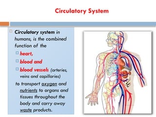
Circulatory System
Circulatorysystem in
humans, is the combined
function of the
heart,
blood and
blood vessels (arteries,
veins and capillaries)
to transport oxygen and
nutrients to organs and
tissues throughout the
body and carry away
waste products.
4.
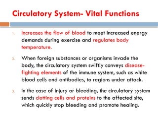
Circulatory System- VitalFunctions
1. Increases the flow of blood to meet increased energy
demands during exercise and regulates body
temperature.
2. When foreign substances or organisms invade the
body, the circulatory system swiftly conveys disease-
fighting elements of the immune system, such as white
blood cells and antibodies, to regions under attack.
3. In the case of injury or bleeding, the circulatory system
sends clotting cells and proteins to the affected site,
which quickly stop bleeding and promote healing.
5.
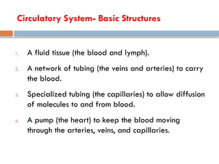
Circulatory System- BasicStructures
1. A fluid tissue (the blood and lymph).
2. A network of tubing (the veins and arteries) to carry
the blood.
3. Specialized tubing (the capillaries) to allow diffusion
of molecules to and from blood.
4. A pump (the heart) to keep the blood moving
through the arteries, veins, and capillaries.
6.
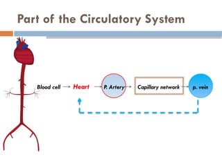
Part of theCirculatory System
Blood cell Heart P. Artery Capillary network p. vein
7.
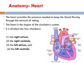
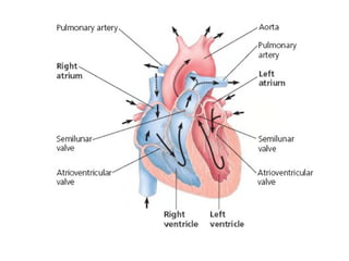
Anatomy- Heart
Theheart provides the pressure needed to keep the blood flowing
through the network of tubing.
The heart is the engine of the circulatory system.
It is divided into four chambers:
(1) the right atrium,
(2) the right ventricle,
(3) the left atrium, and
(4) the left ventricle.
8.
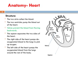
8
Structure:
The twoatria collect the blood.
The two ventricles pump the blood out
of the heart.
Valves prevent the blood from flowing
backwards.
The septum separates the two sides of
the heart.
The right side of the heart pumps de-
oxygenated blood to the lungs to pick
up oxygen.
The left side of the heart pumps the
oxygenated blood from the lungs
around the rest of the body.
Right
atrium
Left
atrium
Right
ventricle
Left
ventricle
Valves
Septum
Anatomy- Heart
9.
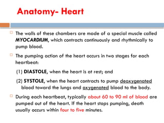
The wallsof these chambers are made of a special muscle called
MYOCARDIUM, which contracts continuously and rhythmically to
pump blood.
The pumping action of the heart occurs in two stages for each
heartbeat:
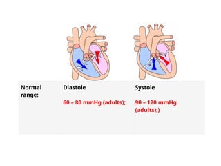
(1) DIASTOLE, when the heart is at rest; and
(2) SYSTOLE, when the heart contracts to pump deoxygenated
blood toward the lungs and oxygenated blood to the body.
During each heartbeat, typically about 60 to 90 ml of blood are
pumped out of the heart. If the heart stops pumping, death
usually occurs within four to five minutes.
Anatomy- Heart
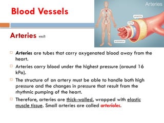
Arteries ধমনী
Arteriesare tubes that carry oxygenated blood away from the
heart.
Arteries carry blood under the highest pressure (around 16
kPa).
The structure of an artery must be able to handle both high
pressure and the changes in pressure that result from the
rhythmic pumping of the heart.
Therefore, arteries are thick-walled, wrapped with elastic
muscle tissue. Small arteries are called arterioles.
Blood Vessels
12.
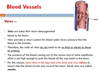
Veins শিরা
Veinsare tubes that return deoxygenated
blood to the heart.
Veins provide a return system for blood under lower pressure that the
blood in the arteries.
Therefore, the walls of veins do not need to be as thick or elastic as those
of arteries.
The pressure of the blood coming out of the venous end of some capillaries
often is not high enough to push the blood all the way back to the heart
For this reason, some veins in the legs and arms have one-way valves to
ensure that the blood travels only toward the heart. Small veins are called
venules.
Blood Vessels
13.
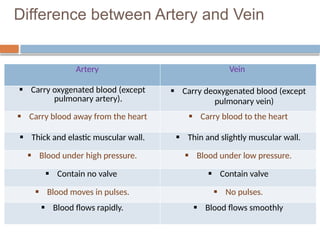
Difference between Arteryand Vein
Artery Vein
Carry oxygenated blood (except
pulmonary artery).
Carry deoxygenated blood (except
pulmonary vein)
Carry blood away from the heart Carry blood to the heart
Thick and elastic muscular wall. Thin and slightly muscular wall.
Blood under high pressure. Blood under low pressure.
Contain no valve Contain valve
Blood moves in pulses. No pulses.
Blood flows rapidly. Blood flows smoothly
14.
Blood Circulation inHeart:
Oxygenated blood from lungs to heart– Pulmonary
vein left Atrium [diastole] Left Ventricle [systole]
Aorta (to body)
Deoxygenated blood from body to heart – Superior and
Inferior Vena cava Right Atrium [diastole] Right
Ventricle [systole] Pulmonary Artery (to lungs)
15.
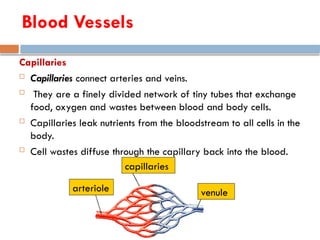
Capillaries
Capillaries connectarteries and veins.
They are a finely divided network of tiny tubes that exchange
food, oxygen and wastes between blood and body cells.
Capillaries leak nutrients from the bloodstream to all cells in the
body.
Cell wastes diffuse through the capillary back into the blood.
arteriole venule
capillaries
Blood Vessels
16.
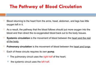
The Pathway ofBlood Circulation
Blood returning to the heart from the arms, head, abdomen, and legs has little
oxygen left in it.
As a result, the pathway that the blood follows should put more oxygen into the
blood and then direct the re-oxygenated blood back out to the body tissues.
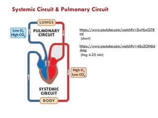
Systemic circulation is the movement of blood between the heart and the rest
of the body.
Pulmonary circulation is the movement of blood between the heart and lungs.
Each of these circuits requires its own pump.
The pulmonary circuit uses the right half of the heart;
the systemic circuit uses the left half.
19
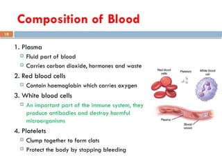
Composition of Blood
1.Plasma
Fluid part of blood
Carries carbon dioxide, hormones and waste
2. Red blood cells
Contain haemoglobin which carries oxygen
3. White blood cells
An important part of the immune system, they
produce antibodies and destroy harmful
microorganisms
4. Platelets
Clump together to form clots
Protect the body by stopping bleeding
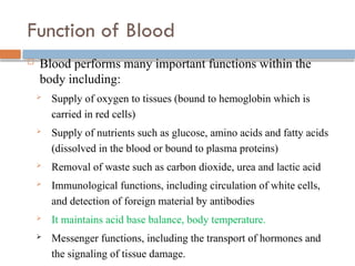
Function of Blood
Blood performs many important functions within the
body including:
Supply of oxygen to tissues (bound to hemoglobin which is
carried in red cells)
Supply of nutrients such as glucose, amino acids and fatty acids
(dissolved in the blood or bound to plasma proteins)
Removal of waste such as carbon dioxide, urea and lactic acid
Immunological functions, including circulation of white cells,
and detection of foreign material by antibodies
It maintains acid base balance, body temperature.
Messenger functions, including the transport of hormones and
the signaling of tissue damage.
22.
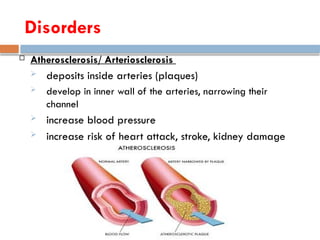
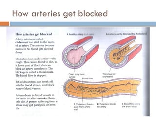
Atherosclerosis/ Arteriosclerosis
deposits inside arteries (plaques)
develop in inner wall of the arteries, narrowing their
channel
increase blood pressure
increase risk of heart attack, stroke, kidney damage
Disorders
23.
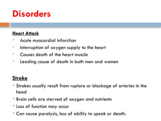
Heart Attack
Acutemyocardial infarction
Interruption of oxygen supply to the heart
Causes death of the heart muscle
Leading cause of death in both men and women
Stroke
Strokes usually result from rupture or blockage of arteries in the
head
Brain cells are starved of oxygen and nutrients
Loss of function may occur
Can cause paralysis, loss of ability to speak or death.
Disorders
How to avoidheart diseases
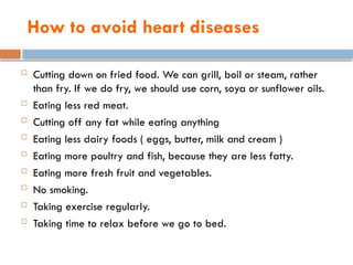
Cutting down on fried food. We can grill, boil or steam, rather
than fry. If we do fry, we should use corn, soya or sunflower oils.
Eating less red meat.
Cutting off any fat while eating anything
Eating less dairy foods ( eggs, butter, milk and cream )
Eating more poultry and fish, because they are less fatty.
Eating more fresh fruit and vegetables.
No smoking.
Taking exercise regularly.
Taking time to relax before we go to bed.
26.
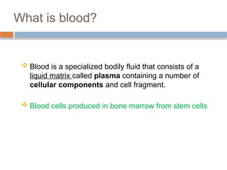
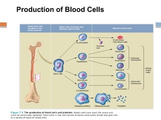
What is blood?
Blood is a specialized bodily fluid that consists of a
liquid matrix called plasma containing a number of
cellular components and cell fragment.
Blood cells produced in bone marrow from stem cells
27.
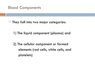
They fallinto two major categories:
1) The liquid component (plasma) and
2) The cellular component or formed
elements (red cells, white cells, and
platelets)
Blood Components
28.
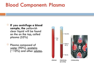
Blood Component: Plasma
If you centrifuge a blood
sample, the yellowish
clear liquid will be found
on the on the top, called
plasma (55%)
Plasma composed of
water (90%), proteins
(~10%) and other solutes.
29.
Plasma
The largestgroup of solutes in plasma consists of plasma proteins,
which serve a variety of functions.
Important plasma proteins include albumins, globulins, and clotting
proteins.
Albumins:
2/3 of total protein
Maintain the proper water balance between blood and the
interstitial fluid.
Manufactured in the liver.
Albumins also bind to certain molecules (such as bilirubin and fatty
acids) and drugs (such as penicillin) and assist in their transport in
30.
Plasma
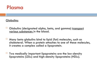
Globulins:
Globulins (designatedalpha, beta, and gamma) transport
various substances in the blood.
Many beta globulins bind to lipid (fat) molecules, such as
cholesterol. When a protein attaches to one of these molecules,
it creates a complex called a lipoprotein.
Two medically important lipoproteins are the low-density
lipoproteins (LDLs) and high-density lipoproteins (HDLs).
31.
Plasma
Globulins:
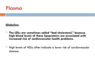
The LDLsare sometimes called “bad cholesterol,” because
high blood levels of these lipoproteins are associated with
increased risk of cardiovascular health problems.
High levels of HDLs often indicate a lower risk of cardiovascular
disease.
32.
Plasma
Clotting proteins:
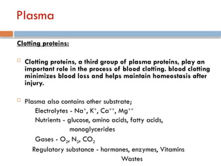
Clottingproteins, a third group of plasma proteins, play an
important role in the process of blood clotting. blood clotting
minimizes blood loss and helps maintain homeostasis after
injury.
Plasma also contains other substrate;
Electrolytes - Na+
, K+
, Ca++
, Mg++
Nutrients - glucose, amino acids, fatty acids,
monoglycerides
Gases - O2, N2, CO2
Regulatory substance - hormones, enzymes, Vitamins
Wastes
33.
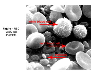
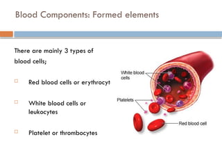
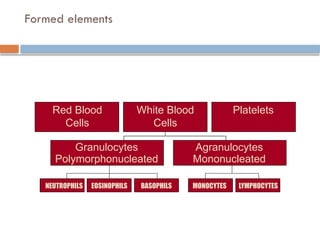
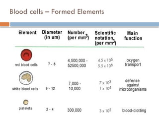
Blood Components: Formedelements
There are mainly 3 types of
blood cells;
Red blood cells or erythrocytes
White blood cells or
leukocytes
Platelet or thrombocytes
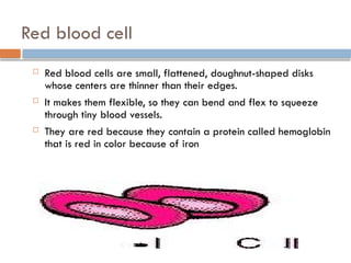
Red blood cell
Red blood cells are small, flattened, doughnut-shaped disks
whose centers are thinner than their edges.
It makes them flexible, so they can bend and flex to squeeze
through tiny blood vessels.
They are red because they contain a protein called hemoglobin
that is red in color because of iron
37.
Red blood cell
Red blood cells carry oxygen to body tissues and remove carbon
dioxide (which bound with hemoglobin).
The process by which red blood cells are produced is called
erythropoiesis.
Erythrocytes are continuously being produced in the bone marrow
of large bones, at a rate of about 2 million per second. (In the
embryo, the liver is the main site of red blood cell production).
In an adult, the total count of RBC is 4.5 to 5.5 million per mm3
of
blood.
38.
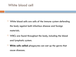
White blood cell
White blood cells are cells of the immune system defending
the body against both infectious disease and foreign
materials.
WBCs are found throughout the body, including the blood
and lymphatic system.
White cells called phagocytes can eat up the germs that
cause diseases.
39.
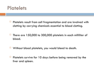
Platelets
Platelets resultfrom cell fragmentation and are involved with
clotting by carrying chemicals essential to blood clotting.
There are 150,000 to 300,000 platelets in each milliliter of
blood.
Without blood platelets, you would bleed to death.
Platelets survive for 10 days before being removed by the
liver and spleen.
40.
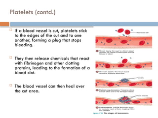
Platelets (contd.)
Ifa blood vessel is cut, platelets stick
to the edges of the cut and to one
another, forming a plug that stops
bleeding.
They then release chemicals that react
with fibrinogen and other clotting
proteins, leading to the formation of a
blood clot.
The blood vessel can then heal over
the cut area.
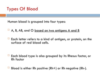
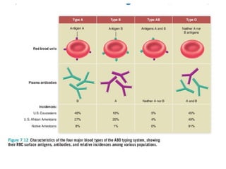
Types Of Blood
Humanblood is grouped into four types:
A, B, AB, and O based on two antigens A and B
Each letter refers to a kind of antigen, or protein, on the
surface of red blood cells.
Each blood type is also grouped by its Rhesus factor, or
Rh factor
Blood is either Rh positive (Rh+) or Rh negative (Rh-).
44.
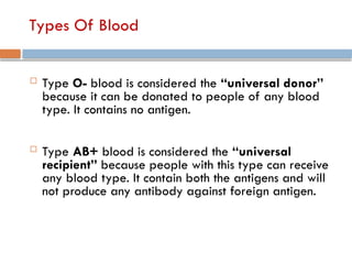
Types Of Blood
Type O- blood is considered the “universal donor”
because it can be donated to people of any blood
type. It contains no antigen.
Type AB+ blood is considered the “universal
recipient” because people with this type can receive
any blood type. It contain both the antigens and will
not produce any antibody against foreign antigen.
45.
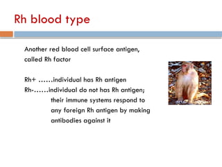
Rh blood type
Anotherred blood cell surface antigen,
called Rh factor
Rh+ ……individual has Rh antigen
Rh-……individual do not has Rh antigen;
their immune systems respond to
any foreign Rh antigen by making
antibodies against it
46.
Ques: A personwith blood group O- has antibodies against both A, B, and Rh antigens. Don't these
antibodies react with the antigens in the recipient's blood? Then how is O- blood a universal donor?
ANS: Why O- Blood is a Universal Donor
1.O- Blood Cells Lack A, B, and Rh Antigens:
1. Red blood cells (RBCs) from O- donors do not have A, B, or Rh (D) antigens on their surface.
2. Since antigens on donor RBCs trigger immune responses in recipients, the absence of these
antigens means the recipient's antibodies (against A, B, or Rh) will not react with O- blood.
2.Plasma (Containing Antibodies) is Typically Removed or Diluted:
1. Whole blood donations are often separated into components: RBCs, plasma, and platelets.
2. When O- blood is transfused as RBCs, the donor's plasma (which may contain A, B, and Rh
antibodies) is typically removed or significantly diluted in the recipient's blood. Therefore, the
antibodies from the O- donor are in such low concentrations that they don't cause a reaction in
the recipient.
Why O- Blood Does Not Harm Recipients
•The A, B, and Rh antibodies are primarily part of the plasma, not the red blood cells. During a
transfusion:
• If only RBCs are transfused (common practice), the antibodies in the donor plasma are
removed.
• In an emergency whole blood transfusion (less common), the volume of the donor's antibodies is
too small compared to the recipient's total blood volume to cause significant harm.
Key Takeaways
•Universal Donor refers to the red blood cells of O- blood, which lack antigens and are safe for any
recipient.
•The antibodies in the donor plasma are minimal after processing and do not typically pose a risk during
transfusion
#4 What are the major roles of circulatory system?
#5 What is the basic structure of circulatory system?
#7 Atrium (আর্ট্রিয়াম) → অলিন্দ
Ventricle (ভেন্ট্রিকল) → নিলয়
Capillary (ক্যাপিলারি) → কৈশিক
Vein (ভেইন) → শিরা
Artery (আর্টারি) → ধমনী
Superior Vena Cava (সুপিরিয়র ভেনা কাভা) → ঊর্ধ্ব প্রধান শিরা
The Superior Vena Cava (SVC) is a large vein that carries deoxygenated blood from the upper body (head, neck, arms, and upper chest) back to the right atrium of the heart.
#12 Venules are small blood vessels that collect deoxygenated blood from the capillaries and transport it to the larger veins, which then carry the blood back to the heart. They act as intermediaries between capillaries and veins, helping in the exchange of waste products and nutrients between blood and tissues.
![Blood Circulation in Heart:
Oxygenated blood from lungs to heart– Pulmonary
vein left Atrium [diastole] Left Ventricle [systole]
Aorta (to body)
Deoxygenated blood from body to heart – Superior and
Inferior Vena cava Right Atrium [diastole] Right
Ventricle [systole] Pulmonary Artery (to lungs)](https://image.slidesharecdn.com/topic-11lec-17circulatorysystem-250921182651-4a145320/85/Topic-11_Lec-17_Circulatory-System-pptx-14-320.jpg)


![Hypothalamus short notes on location, function and disorders by Dr. Neha [PT]...](https://cdn.slidesharecdn.com/ss_thumbnails/hypothalamusbydr-260124142231-2b48143d-thumbnail.jpg?width=640&height=640&fit=bounds)


