Download to read offline


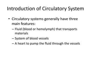
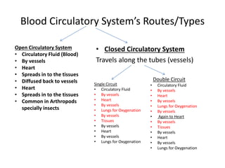
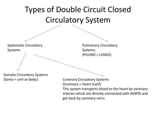

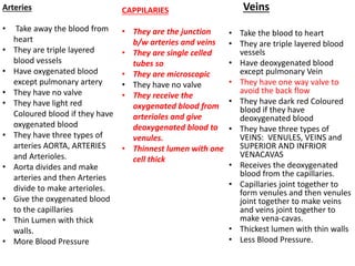
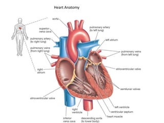
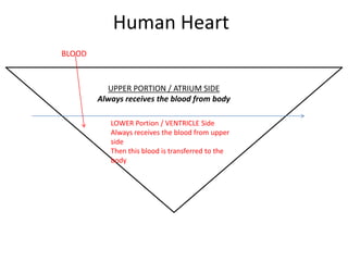
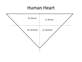
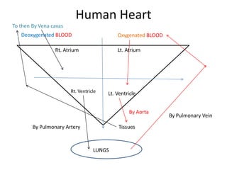
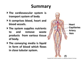
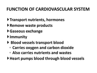
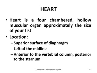
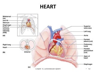
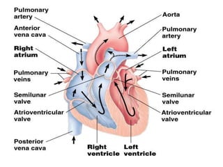
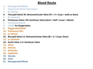


The circulatory system contains blood, blood vessels, and the heart. The heart pumps blood through a closed double circulatory system, where blood is pumped from the heart through arteries, then capillaries, then veins, and back to the heart. Blood carries oxygen, nutrients, waste, hormones, and other materials between tissues and organs through this circulatory loop. The main components include the heart, arteries and veins, and blood itself which contains plasma, red blood cells, white blood cells, and platelets.


![ANIMAL_CELL_,_TISSUE_AND_ORGAN_CULTURE[1].pptx](https://cdn.slidesharecdn.com/ss_thumbnails/animalcelltissueandorganculture1-260204172026-4462b440-thumbnail.jpg?width=640&height=640&fit=bounds)


