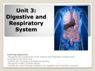
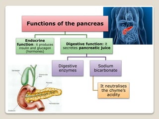
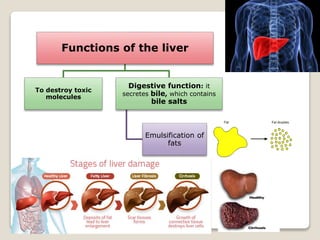
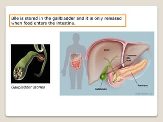
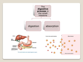
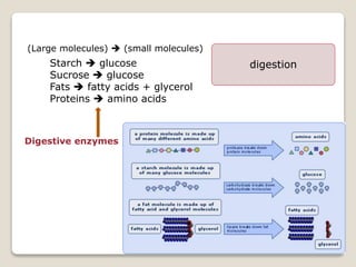
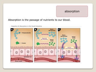
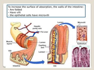
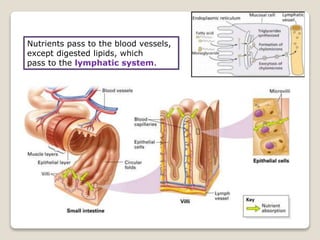
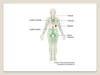
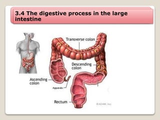
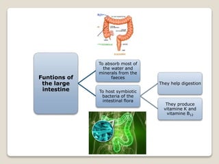
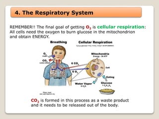
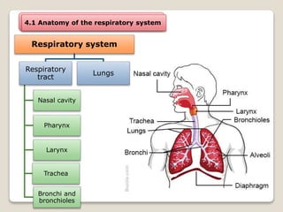
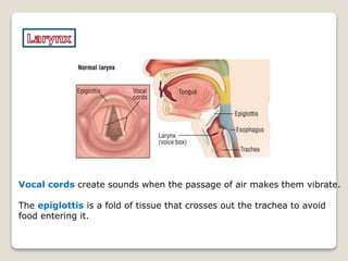
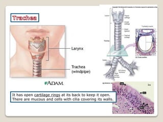
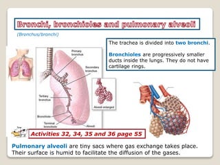
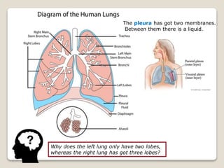
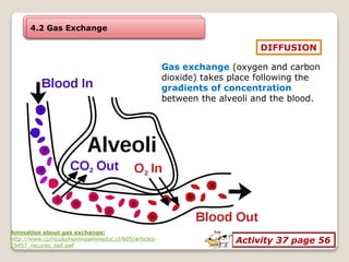
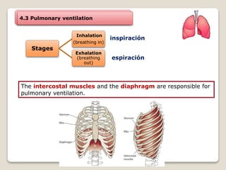
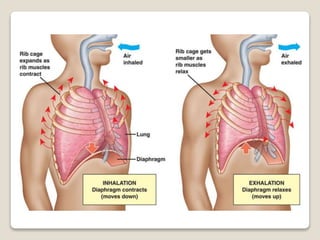
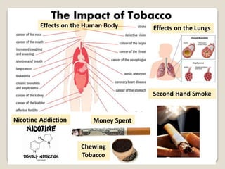
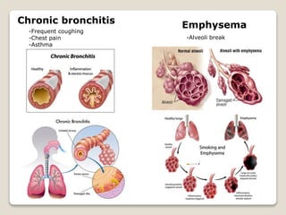
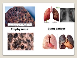
Unit 3:
Digestive and
Respiratory
System
Learning objectives:
• Recognise the components of the digetive and respiratory systems and
understand how they work.
• Explain gas exchange and digestive process.
• Recognise healthy livestyle habits.
• Identify the main illnesses related to the digestive and respiratory systems.
Instructions:
1. Write down the following definitions on your notebook
2. Listen to the word.
3. Match it with the corresponding definition.
Enzyme that starts protein digestion in the stomach. pepsin
Initial section of the small intestine. duodenum
Movement produced by the contraction
of the muscles found in the digestive tract.
peristaltic movements
Tooth used to tear. canine
Bag-like organ which stores bile. gallbladder
1. Systems involved in nutrition
Excretory system
Digestive system
Circulatory system
Respiratory system
 It gets the nutrients.
It gets oxygen
and eliminates carbon dioxide.
It transports nutrients,
O2 , CO2 and waste products.
It releases waste substances
outside the organism.
2. Digestive System
Parts of
the
digestive
system
Gastrointestinal
tract
Mouth
Pharynx
Esophagus
Stomach
Small
intestine
Large
intestine
Accessory glands
Salivary
glands
Liver
Pancreas
Activities 4, 5 and 6 page 45
3. Digestion
It is the process by which foods are transformed into nutrients.
Types of
processes
• Mechanical processes
•Chewing
•Muscular contractions
• Chemical processes
•Performed by digestive enzymes found in digestive
juices
3.1 The digestive process in the mouth
Functions
of saliva
It starts the
digestion of
starch molecules
(amylase)
It destroys some
bacteria
(lysozyme)
It lubricates the
bolus (mucin)
3.1.1 Salivation
3.1.2 Chewing (Mechanical processes)
To cut
To tear
To crush
3.2 The digestive process in the stomach
Cardia: valve which separates the esophagus from the stomach
Pylorus: valve which separates the stomach from the duodenum
Gastric juices
contain:
Pepsin:
enzyme that starts
protein digestion
Hydrochloric
acid (HCl):
•It activates and helps
pepsin.
•It destroys bacteria.
Food is transformed in the stomach
into a fluid substance called
chyme
Bolus  chyme
3.3 The digestive process in the small
intestine
Pylorus: valve which separates the stomach from the duodenum.
Ileocecal valve: valve which separates small intestine from
the large intestine. Sections of the
small intestine
Duodenum Jejunum Ileum
Pylorus
Ileocecal valve
The chyme mixes with the bile and the pancreatic and intestinal juices
and it is transformed into chyle.
Food Bolus Chyme Chyle
From the liver From the pancreas From the walls
of the intestine
Saliva
Gastric juices
Pancreatic juice
Bile
Intestinal juice
Functions of the pancreas
Endocrine
function: it produces
insulin and glucagon
(hormones)
Digestive function: it
secretes pancreatic juice
Digestive
enzymes
Sodium
bicarbonate
It neutralises
the chyme’s
acidity
Functions of the liver
To destroy toxic
molecules
Digestive function: it
secretes bile, which contains
bile salts
Emulsification of
fats
Bile is stored in the gallbladder and it is only released
when food enters the intestine.
Gallbladder stones
The
digestive
process in
the small
intestine
digestion absorption
Starch  glucose
Sucrose  glucose
Fats  fatty acids + glycerol
Proteins  amino acids
Digestive enzymes
(Large molecules)  (small molecules)
digestion
absorption
Absorption is the passage of nutrients to our blood.
To increase the surface of absorption, the walls of the intestine:
- Are folded
- Have villi
- the epithelial cells have microvilli
Nutrients pass to the blood vessels,
except digested lipids, which
pass to the lymphatic system.
3.4 The digestive process in the large
intestine
cecum
(ciego)
Funtions of
the large
intestine
To absorb most of
the water and
minerals from the
faeces
To host symbiotic
bacteria of the
intestinal flora
They help digestion
They produce
vitamine K and
vitamine B12
IN PAIRS
What does
each part of the drawing
represent?
4. The Respiratory System
REMEMBER!! The final goal of getting O2 is cellular respiration:
All cells need the oxygen to burn glucose in the mitochondrion
and obtain ENERGY.
CO2 is formed in this process as a waste product
and it needs to be released out of the body.
4.1 Anatomy of the respiratory system
Respiratory system
Respiratory
tract
Nasal cavity
Pharynx
Larynx
Trachea
Bronchi and
bronchioles
Lungs
The respiratory tract is covered by
a mucosa which purifies and humidifies
the air.
SinusitisAllergic rhinitis
Some illnesses
It is common to the digestive tract.
The function of the tonsils
is to produce white blood cells.
(amígdalas)
Vocal cords create sounds when the passage of air makes them vibrate.
The epiglottis is a fold of tissue that crosses out the trachea to avoid
food entering it.
It has open cartilage rings at its back to keep it open.
There are mucous and cells with cilia covering its walls.
(Bronchus/bronchi)
The trachea is divided into two bronchi.
Bronchioles are progressively smaller
ducts inside the lungs. They do not have
cartilage rings.
Pulmonary alveoli are tiny sacs where gas exchange takes place.
Their surface is humid to facilitate the diffusion of the gases.
Activities 32, 34, 35 and 36 page 55
Why does the left lung only have two lobes,
whereas the right lung has got three lobes?
The pleura has got two membranes.
Between them there is a liquid.
4.2 Gas Exchange
DIFFUSION
Gas exchange (oxygen and carbon
dioxide) takes place following the
gradients of concentration
between the alveoli and the blood.
Activity 37 page 56
Animation about gas exchange:
http://www.curriculumenlineamineduc.cl/605/articles-
25457_recurso_swf.swf
4.3 Pulmonary ventilation
Stages
Inhalation
(breathing in)
Exhalation
(breathing
out)
inspiración
espiración
The intercostal muscles and the diaphragm are responsible for
pulmonary ventilation.
Chronic bronchitis
-Frequent coughing
-Chest pain
-Asthma
Emphysema
-Alveoli break
Emphysema Lung cancer
Choose three of the healthy habits on page 58 and 60
that you already do.
Choose one that you will include in your life style.
WRITE THEM DOWN ON YOUR NOTEBOOK
Things which I already do
….
….
….
New habits which I will adquire
….
IN PAIRS
Activities 40, 41, 42, 43 pages 58 and 59
4 minutes
Discuss the following activities with your partner
HW: Activities 48, 54, 61 and READ AND UNDERSTAND SCIENCE

Unit 3 Digestive and Respiratory Systems

  • 1.
    Unit 3: Digestive and Respiratory System Learningobjectives: • Recognise the components of the digetive and respiratory systems and understand how they work. • Explain gas exchange and digestive process. • Recognise healthy livestyle habits. • Identify the main illnesses related to the digestive and respiratory systems.
  • 2.
    Instructions: 1. Write downthe following definitions on your notebook 2. Listen to the word. 3. Match it with the corresponding definition. Enzyme that starts protein digestion in the stomach. pepsin Initial section of the small intestine. duodenum Movement produced by the contraction of the muscles found in the digestive tract. peristaltic movements Tooth used to tear. canine Bag-like organ which stores bile. gallbladder
  • 3.
    1. Systems involvedin nutrition Excretory system Digestive system Circulatory system Respiratory system  It gets the nutrients. It gets oxygen and eliminates carbon dioxide. It transports nutrients, O2 , CO2 and waste products. It releases waste substances outside the organism.
  • 4.
    2. Digestive System Partsof the digestive system Gastrointestinal tract Mouth Pharynx Esophagus Stomach Small intestine Large intestine Accessory glands Salivary glands Liver Pancreas Activities 4, 5 and 6 page 45
  • 5.
    3. Digestion It isthe process by which foods are transformed into nutrients. Types of processes • Mechanical processes •Chewing •Muscular contractions • Chemical processes •Performed by digestive enzymes found in digestive juices
  • 6.
    3.1 The digestiveprocess in the mouth Functions of saliva It starts the digestion of starch molecules (amylase) It destroys some bacteria (lysozyme) It lubricates the bolus (mucin) 3.1.1 Salivation
  • 7.
    3.1.2 Chewing (Mechanicalprocesses) To cut To tear To crush
  • 8.
    3.2 The digestiveprocess in the stomach Cardia: valve which separates the esophagus from the stomach Pylorus: valve which separates the stomach from the duodenum
  • 9.
    Gastric juices contain: Pepsin: enzyme thatstarts protein digestion Hydrochloric acid (HCl): •It activates and helps pepsin. •It destroys bacteria.
  • 10.
    Food is transformedin the stomach into a fluid substance called chyme Bolus  chyme
  • 11.
    3.3 The digestiveprocess in the small intestine Pylorus: valve which separates the stomach from the duodenum. Ileocecal valve: valve which separates small intestine from the large intestine. Sections of the small intestine Duodenum Jejunum Ileum Pylorus Ileocecal valve
  • 12.
    The chyme mixeswith the bile and the pancreatic and intestinal juices and it is transformed into chyle. Food Bolus Chyme Chyle From the liver From the pancreas From the walls of the intestine Saliva Gastric juices Pancreatic juice Bile Intestinal juice
  • 13.
    Functions of thepancreas Endocrine function: it produces insulin and glucagon (hormones) Digestive function: it secretes pancreatic juice Digestive enzymes Sodium bicarbonate It neutralises the chyme’s acidity
  • 14.
    Functions of theliver To destroy toxic molecules Digestive function: it secretes bile, which contains bile salts Emulsification of fats
  • 15.
    Bile is storedin the gallbladder and it is only released when food enters the intestine. Gallbladder stones
  • 16.
  • 17.
    Starch  glucose Sucrose glucose Fats  fatty acids + glycerol Proteins  amino acids Digestive enzymes (Large molecules)  (small molecules) digestion
  • 18.
    absorption Absorption is thepassage of nutrients to our blood.
  • 19.
    To increase thesurface of absorption, the walls of the intestine: - Are folded - Have villi - the epithelial cells have microvilli
  • 20.
    Nutrients pass tothe blood vessels, except digested lipids, which pass to the lymphatic system.
  • 22.
    3.4 The digestiveprocess in the large intestine cecum (ciego)
  • 23.
    Funtions of the large intestine Toabsorb most of the water and minerals from the faeces To host symbiotic bacteria of the intestinal flora They help digestion They produce vitamine K and vitamine B12
  • 24.
    IN PAIRS What does eachpart of the drawing represent?
  • 25.
    4. The RespiratorySystem REMEMBER!! The final goal of getting O2 is cellular respiration: All cells need the oxygen to burn glucose in the mitochondrion and obtain ENERGY. CO2 is formed in this process as a waste product and it needs to be released out of the body.
  • 26.
    4.1 Anatomy ofthe respiratory system Respiratory system Respiratory tract Nasal cavity Pharynx Larynx Trachea Bronchi and bronchioles Lungs
  • 27.
    The respiratory tractis covered by a mucosa which purifies and humidifies the air. SinusitisAllergic rhinitis Some illnesses
  • 28.
    It is commonto the digestive tract. The function of the tonsils is to produce white blood cells. (amígdalas)
  • 29.
    Vocal cords createsounds when the passage of air makes them vibrate. The epiglottis is a fold of tissue that crosses out the trachea to avoid food entering it.
  • 30.
    It has opencartilage rings at its back to keep it open. There are mucous and cells with cilia covering its walls.
  • 31.
    (Bronchus/bronchi) The trachea isdivided into two bronchi. Bronchioles are progressively smaller ducts inside the lungs. They do not have cartilage rings. Pulmonary alveoli are tiny sacs where gas exchange takes place. Their surface is humid to facilitate the diffusion of the gases. Activities 32, 34, 35 and 36 page 55
  • 32.
    Why does theleft lung only have two lobes, whereas the right lung has got three lobes? The pleura has got two membranes. Between them there is a liquid.
  • 33.
    4.2 Gas Exchange DIFFUSION Gasexchange (oxygen and carbon dioxide) takes place following the gradients of concentration between the alveoli and the blood. Activity 37 page 56 Animation about gas exchange: http://www.curriculumenlineamineduc.cl/605/articles- 25457_recurso_swf.swf
  • 34.
    4.3 Pulmonary ventilation Stages Inhalation (breathingin) Exhalation (breathing out) inspiración espiración The intercostal muscles and the diaphragm are responsible for pulmonary ventilation.
  • 37.
    Chronic bronchitis -Frequent coughing -Chestpain -Asthma Emphysema -Alveoli break
  • 38.
  • 39.
    Choose three ofthe healthy habits on page 58 and 60 that you already do. Choose one that you will include in your life style. WRITE THEM DOWN ON YOUR NOTEBOOK Things which I already do …. …. …. New habits which I will adquire ….
  • 40.
    IN PAIRS Activities 40,41, 42, 43 pages 58 and 59 4 minutes Discuss the following activities with your partner HW: Activities 48, 54, 61 and READ AND UNDERSTAND SCIENCE

Editor's Notes

  • #3 Canine, duodenum, peristaltic movements, pepsin, gallbladder