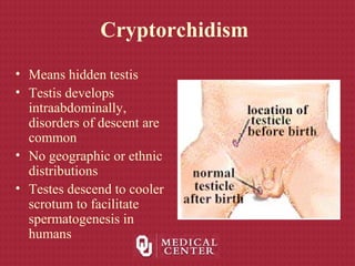
Cryptorchidism
Stephen Confer, MD
Ben O. Donovan, MD
Brad Kropp, MD
Dominic Frimberger, MD
University of Oklahoma
Department of Urology
Section of Pediatric Urology
Embryology of Gonads
• 5th
week - primordial germ
cells migrate from yolk
sac along dorsal
mesentary to the posterior
body wall
Migration of primordial
germ cells migrate from the
posterior extra-
embryonic mesoderm
through the mesenteries
and into the gonadal ridge
Embryology of Gonads
• 5 weeks- Sexually indifferent –
identical structures in male and
female
• Gonadal ridges develop on
dorsal abdominal wall week 5
– future ovaries or testes
• Wolffian ducts – future male
ducts
• Müllerian ducts – future
female ducts
Embryology of Gonads
• Mesenchymal proliferation occurs and genital ridges are formed
• Paramesonephric (Mullerian) ducts begin to form lateral to the
mesonephric ducts
• After the 6th
week genital ridges pursue fates as male or female.
• “Y” does this happen?
SRY acts on the indifferent gonad to
start the process of male sexual
development
Molecular Determination of Sex
Male Genital Development
• Sex cord cells differentiate into
Sertoli cells only if SRY protein
is present
• 7th
week – Sertoli cells organize
to form testis cords
– Will eventually form
seminiferous tubules
– Testis cord distally forms rete
testis
– Medially rete testis connect to
mesonephric ducts via 5-12
tubules (efferent ductules)
• Vas deferens develops from
mesonephric duct
Male Genital Development
• As Sertoli cells
differentiate in response to
SRY, secrete a
glycoprotein hormone
Mullerian-inhibiting
substance (MIS)
– MIS causes
paramesonephric
(mullerian) ducts to
regress between 8th
-10th
week
– Remnants may be
found
• Appendix testis
Male Genital Development• 9th
-10th
weeks – Leydig cells
differentiate
– Early testosterone regulated by
placental chorionic gonadotrophin –
eventually regulated by pituitary
gonadotrophins
• 8th
-12th
weeks – testosterone
secretion by Leydig cells stimulates
transformation of mesonephric duct
to the vas deferens
– Cranial portion – VD; ED
– Caudal portion -SV; Ejac. Duct
– Communication with rete testis is not
established until 3rd
month
Gonadal Descent
• Initial descent in both
sexes relies on the
gubernaculum
– Forms in the peritoneal
folds during 7th
week
– Superior end is attached to
gonad, inferiorly it is
attached to fascia between
the external and internal
obliques
• Processus vaginalis
– Evagination of peritoneum
Testicular Descent
Testicular Descent
• Layers encountered by processus
vaginalis
– Transversalis fascia
– Muscle fibers of internal
oblique (cremasteric)
– Thin layer of external oblique
muscle (external spermatic
fascia)
• Descent of testicle
– At level of internal ring by 3rd
month
– Complete descent to scrotum
between 7th
-9th
month
Normal Testicular Anatomy
Normal Anatomy
Duct System
Blood supply
Blood Testis
Barrier
Spermatic cord supplies
• Blood Supply
• Lymphatics
– Upper lumbar LN
• Nerve supply
– Sympathetic- T10-11
– Unable to differentiate
– Suggested to increase
or decrease dilation
Normal Anatomy
Embryologic Remnants
Normal Histology
Normal Seminiferous Tubule
Cryptorchidism
• Means hidden testis
• Testis develops
intraabdominally,
disorders of descent are
common
• No geographic or ethnic
distributions
• Testes descend to cooler
scrotum to facilitate
spermatogenesis in
humans
Embryology of Testis Descent
• Germ cells migrate from yolk sac to genital ridges at 6
weeks
• Testis differentiation begins to occur at 7 weeks as SRY
protein is produced
• Testosterone and MIF are secreted by testis during week 8
• Testosterone is controlled by maternal hCG, causes
wolffian differentiation into epididymis and vas
• MIF is produced by Sertoli cells and causes mullerian
regression=>appendix testis
Embryology of Testis Descent
(con’t)
• Growth of the gubernaculum is believed to be
controlled by testis paracrine function
• Testis is never more than 1.3 mm from internal
ring at any point in its development
• Testis remain in position but processus
vaginalis extends into scrotum from 12th
week
to 7th
month
Embryology of Testis Descent
(con’t)
• At 7 months, the vas and testicular vessels increase in
size, the gubernaculum swells, and the processus extends
into scrotum completely
• As the gubernaculum swells, the inguinal canal and
scrotum stretch=>testis rapidly descends
• The epididymis precedes the testis into the scrotum
• Canal portion of processus obliterates, testicular portion
persists as tunica vaginalis
• Gubernaculum atrophies
Incidence of Testicular Descent
• Of 1500 full term infants, 3.4% had true UDT
• Prematurity may predispose to UDT because birth occurs
before descent
• Of 142 premature infants, 30.3% had UDT
• Low birth weight also predisposes to UDT, as weight is
gained, testes descend
• At 1 y.o., 0.8-1.5% have UDT, 75% of full-term and
95% of premature UDT descend during first year
• Most descend during first 3 months after birth probably
due to elevated serum androgens during first 3 months
Incidence of Testicular Descent
(con’t)
• Cremasteric reflex is most active during 2-7 years
old=>retractile testes are misdiagnosed as UDT
– Estimates of UDT at 5 y.o. are about 10%
• Testes do not ascend postnatally
• Incidence of UDT in adulthood is < 1%
• Absence of one or both testes on exploration is 3-5%
• 10% of patients have bilateral UDT
• 14% of patients with UDT have a family member with
UDT
Theories Regarding the
Mechanisms of Testicular Descent
• Traction of the testis by gubernaculum and cremasters
– Severing the gubernaculum does not stop descent in animals
– Weak attachments between gubernaculum and scrotum
• Differential growth of the body wall in relation to
immobile gubernaculum
– Proximity to internal ring is maintained as body grows
– Gubernaculum swells and grows faster than the body
• Intraabdominal pressure pushes the testis through the
inguinal canal
– Increased pressure promotes descent supported by theoretical
and experimental data (Schechter 1960, Bergin 1970, Gier 1970)
Theories Regarding the
Mechanisms of Testicular Descent
(con’t)
• Development and maturation of the epididymis
contributing to descent
– Probably not as important as initially thought
• Effects of the genitofemoral nerve
– Severing the nerve prevents descent in rodents
Endocrine Aspects of UDT
• Exact mechanism of androgen induction of descent is
unknown
• No consistently discernable abnormalities have been
identified in patients w/ UDT
– Conflicting data on baseline LH, FSH, testosterone levels and
response to GnRH stimulation
• Endocrine factors probably play a major role in descent
– Testosterone induces testis descent in humans
– Androgens affect the nuclei of the genitofemoral nerve to
release modulating factors for gubernacular development
– Boys with spinal cord defects at or below nuclei of the nerve
have a higher incidence of UDT
– DHT binds to rat gubernaculum
H-P-T Axis Disorders and UDT
Hypothalamus LHRH Kallman’s Syndrome
Pituitary Gland LH (FSH) Anencephaly
Testis Testosterone
Spermatic cord
Gubernaculum
Processus Vaginalis
Organ Hormone DisorderOrgan Hormone Disorder
DHT Pseudovaginal
perineoscrotal hypospadias
Steroid-Receptor
Complex
Testicular Feminization
Reifenstein’s Syndrome
20,22-desmolase
3β-HSD, 17-hydroxylase
17,20-desmolase
17β-HSD
Classification of UDT
• Abdominal
– Impalpable by definition
– Usually “peeping”
• Canalicular
– Or “peeping” at external ring
• Ectopic
– 5 major sites corresponding to
gubernacular branches
– Superficial inguinal is most
common
• Rectractile
– Cremaster reflex
– Most common in 5-6 year olds
Whitaker/Kaplan Classification for
UDT
• Palpable
– Normal
– Retractile
– Ectopic
– Undescended
• Unpalpable
– Canalicular
– Intra-abdominal
– Emergent
– Absent
• Agenesis
• atrophy
Work Up of UDT
• Must determine if testes present at
birth
• Physical exam is very important to
evaluate retractile testes
– Non-palpable testis is
intraabdominal, intracanalicular,
absent
• Bilateral UDT requires hormonal
evaluation and challenge
– Elevated gonadotropins (FSH)
suggest bilateral anorchia
– Normal serum gonadotropins=>hCG
challenge (2000 IU x 3days)
– No testosterone response indicates
bilateral anorchia
Imaging of UDT
• Herniography-poor sensitivity and specificity
• U/S-good for inguinal testes, not reliable if higher
• CT-may be helpful for bilateral impalpable testes
– Difficult to perform in young children
• MRI-least invasive, most expensive
– Difficult to perform in young children
• Venography-invasive, pampiniform plexus
present=>testis present
– Non-visualized plexus or blindending does not eliminate testis
• Angiography-difficult to perform, high complications
• Overall accuracy of radiologic imaging for UDT = 44%
– PE is 53% accurate by PMD, 84% accuracy by peds GU
Laparoscopy
• Blind-ending vessels indicate
absent testis
• Vessels into the internal
ring=>inguinal exploration,
laparoscopic
orchiopexy/orchiectomy
• Intra-abdominal testis
– Laparoscopic orchiopexy
– 2 stage orchiopexy (Fowler-
Stephens)
– Abdominal exploration
Histology
• UDT contain atrophied
structures with thickened
basement membrane
• Epithelium contains atypical
germ cells
• Smaller seminiferous tubules
with fewer spermatogonia
• Higher level of UDT correlates
with severity of histologic
abnormalities
• Some of the changes can be
seen in the contralateral
normally descended testis
UDT histology
Normal testis histology
Neoplasm and UDT
• 10% of testis CA are in UDT
• UDT is 35-48x more likely to have malignancy
• Incidence of CA in UDT is 1:2550 testis tumors
• Abdominal UDT is 4x more likely than inguinal testis to develop
CA
• UDT tumors typically occur around puberty
• CIS occurs in 1.7% of UTD
• Orchiopexy should be performed between 1-1.5 years old
• 1/5 of testis CA in patients w/ hx of UDT occurs in contralateral
testis
Neoplasm and UDT
• For bilateral UDT, when CA is detected in one testis
there is a 15% chance of developing tumor in the
contralateral testis
– 30% if both were intra-abdominal UDT
• Seminoma is most common CA in UDT
– Embryonal is second most common
• Gonadoblastoma is most common CA in some intersex
disorders which present w/ UDT
– Often coexists with germ cell tumors
– Develop due to deletion on long arm of Y chromosome
Torsion and UDT
• Increased risk for torsion in
UDT due to anatomic
abnormality between testis
and mesentery
• Incidence is greatest after
puberty with increased testis
size
• Tumor that increases size also
increases risk of torsion
• 64% of adult torsion in UDT
patients had germ cell tumor
• Be aware of abdominal pain
and empty hemiscrotum=>
torsed intra-abdominal UDT
Hernia and UDT
• Processus vaginalis
should obliterate
between the 8th
month of gestation
and 1st
month of life
• UDT results in
patent processus
vaginalis
• Hernias are found in
90% of patients w/
UDT
Infertility and UDT
• Spermatogenesis is retarded by maldescent
• Bilateral UDT => poor fertility
• Higher UDT => more damage to seminiferous
tubules
• Earlier orchiopexy may improve chances for
recovery of spermatogenesis
• Sperm counts in unilateral UDT are much lower
than normal
– Contralateral testis may also be defective
Associated Anomalies with UDT
• Abnormalities with vas and
epididymis are associated
with UDT
– Extended length of epididymis,
partial atresia, total atresia,
dissociation from testis
• Cystic Fibrosis is associated
with vasal agenesis, UDT is
common
• DES exposure during
pregnancy results in
epdidymal defects and UDT
• Klinefelter’s syndrome
– Sterility the rule although
mosaicism may result in
fertility
• Noonan’s syndrome
– Male Turner’s syndrome
– Occasional fertility but not
passed to offspring
• Prader-Willi Syndrome
– Hypothalamic defect with
obesity, MR, hypotonia,
short stature
Hormonal Treatment
• hCG is given to stimulate Leydig cells to produce
testosterone=>descent of testes
– Success rate is 14-50% with doses ranging from 3K to 40K
IU for daily or weekly injections
– A dose of at least 10K is needed to have an effect, doses >
15K have significant side effects
– No change in testis histology or bone growth
• GnRH is given if basal LH is low and abnormality in
GnRH secretion is suspected
– Perinasal spray of 1.2mg/day is effective to stimulate LH
– 6%-70% success rate
Surgical Treatment
• Primary goal of pexation is to facilitate
examination
• Basic principles of orchipexy are: localization,
mobilization, cord dissection, isolation of
processus, tension-free relocation to scrotum
• Pexation does not reduce risk of cancer
• Orchiopexy should be performed before 2 y.o.
• Orchiectomy is an option for post-pubertal
males and dysgenetic testes
Orchiopexy
• 80% UDT are palpable
• Of non-palpable testes, 45% are absent, 30% are
abdominal, 25% are lower testes missed on
examination
• Orchiopexy is 92% successful for testes below external
ring and 87% for inguinal, 82% for peeping, 74% for
abdominal
• Success rates for different techniques are 89% for
inguinal exploration, 84% for microvascular repair,
81% for transabdominal, 73% for two-stage, 77% for
staged F-S repair, 67% for standard F-S repair
Standard Orchiopexy
• Transverse inguinal incision, watch for
testis
• Identify testis and divide gubernaculum
• Open tunics and evaluate testis
• Open external oblique fascia, avoid nerve
• Mobilize spermatic cord
• Finger dissection to enlarge scrotal cavity
medially
• Incise scrotal skin, create dartos pouch
• Pass clamp through pouch into inguinal
area and bring testis into pouch by
gubernaculum or tunica
• Pex testis with 4-0 vicryl sutures
• Complications: atrophy, retraction,
torsion, hematoma, nerve or vas injury
Fowler-Stephens Orchiopexy
• Testis has three sources of arterial blood flow
• Testicular artery and veins often limit mobilization
• < 1/3 of boys with intra-abdominal testes have long vasal loop
needed for vasal pedicle orchiopexy
• Testes located > 1cm above internal ring do poorly with this
technique
• Approach requires long inguinal incision and vasal loop must be
protected during inguinal dissection
• Must open hairpin turn of vas for length without disrupting blood
supply after dividing spermatic vessels
• Clamp testicular artery before ligation to ensure collateral blood
supply, incising tunica may be misleading
• Mobilization with wide peritoneal-vasal cuff, may need more
direct exit site
• Same scrotal fixation techniques
Two-Stage Fowler-Stephens
• 84% success rate with staged repair probably due to
improved collateral circulation
• Spermatic vessels are clipped > 1cm cranial to testis,
large clip facilitates identification
• Wait at least 6 months then perform orchiopexy open or
laparoscopically or combined
• Open: patient may not have external ring on affected
side, divide muscles in general area
• Divide vessels above clip, create wide peritoneal cuff
around testis and vas, straighten vasal loop
• Place in dartos pouch through medial incision in
abdominal wall
Laparoscopy
• 95% sensitive for identifying
and/or locating NPT
• Three likely findings:
– blind-ending vessels
– cord structures entering the ring
– intra-abdominal testis
• One-stage lap orchiopexy
brings testis medial to inferior
epigastric vessels into pouch
(Prentiss)
• If vessels and vas enter closed
internal ring, controversy about
groin exploration
• If internal ring is open, can
complete orchiopexy
laparoscopically or open
Laparoscopic view of intra-abdominal
testis at internal ring
Vanishing Testis
• Laparoscopic view of closed internal inguinal ring. A, vas deferens
and atretic spermatic vessels in vanishing testis syndrome. B,
ipsilateral normal vas deferens and spermatic vessels in contralateral
vanishing testis syndrome.
Algorithm for UDT

Undescended testicle

  • 1.
    Cryptorchidism Stephen Confer, MD BenO. Donovan, MD Brad Kropp, MD Dominic Frimberger, MD University of Oklahoma Department of Urology Section of Pediatric Urology
  • 2.
    Embryology of Gonads •5th week - primordial germ cells migrate from yolk sac along dorsal mesentary to the posterior body wall
  • 3.
    Migration of primordial germcells migrate from the posterior extra- embryonic mesoderm through the mesenteries and into the gonadal ridge Embryology of Gonads
  • 4.
    Embryology of Gonads •5 weeks- Sexually indifferent – identical structures in male and female • Gonadal ridges develop on dorsal abdominal wall week 5 – future ovaries or testes • Wolffian ducts – future male ducts • Müllerian ducts – future female ducts
  • 5.
    Embryology of Gonads •Mesenchymal proliferation occurs and genital ridges are formed • Paramesonephric (Mullerian) ducts begin to form lateral to the mesonephric ducts • After the 6th week genital ridges pursue fates as male or female. • “Y” does this happen?
  • 6.
    SRY acts onthe indifferent gonad to start the process of male sexual development Molecular Determination of Sex
  • 7.
    Male Genital Development •Sex cord cells differentiate into Sertoli cells only if SRY protein is present • 7th week – Sertoli cells organize to form testis cords – Will eventually form seminiferous tubules – Testis cord distally forms rete testis – Medially rete testis connect to mesonephric ducts via 5-12 tubules (efferent ductules) • Vas deferens develops from mesonephric duct
  • 8.
    Male Genital Development •As Sertoli cells differentiate in response to SRY, secrete a glycoprotein hormone Mullerian-inhibiting substance (MIS) – MIS causes paramesonephric (mullerian) ducts to regress between 8th -10th week – Remnants may be found • Appendix testis
  • 9.
    Male Genital Development•9th -10th weeks – Leydig cells differentiate – Early testosterone regulated by placental chorionic gonadotrophin – eventually regulated by pituitary gonadotrophins • 8th -12th weeks – testosterone secretion by Leydig cells stimulates transformation of mesonephric duct to the vas deferens – Cranial portion – VD; ED – Caudal portion -SV; Ejac. Duct – Communication with rete testis is not established until 3rd month
  • 10.
    Gonadal Descent • Initialdescent in both sexes relies on the gubernaculum – Forms in the peritoneal folds during 7th week – Superior end is attached to gonad, inferiorly it is attached to fascia between the external and internal obliques • Processus vaginalis – Evagination of peritoneum
  • 11.
  • 12.
    Testicular Descent • Layersencountered by processus vaginalis – Transversalis fascia – Muscle fibers of internal oblique (cremasteric) – Thin layer of external oblique muscle (external spermatic fascia) • Descent of testicle – At level of internal ring by 3rd month – Complete descent to scrotum between 7th -9th month
  • 13.
  • 14.
    Normal Anatomy Duct System Bloodsupply Blood Testis Barrier
  • 15.
    Spermatic cord supplies •Blood Supply • Lymphatics – Upper lumbar LN • Nerve supply – Sympathetic- T10-11 – Unable to differentiate – Suggested to increase or decrease dilation
  • 16.
  • 17.
  • 18.
  • 19.
  • 20.
    Cryptorchidism • Means hiddentestis • Testis develops intraabdominally, disorders of descent are common • No geographic or ethnic distributions • Testes descend to cooler scrotum to facilitate spermatogenesis in humans
  • 21.
    Embryology of TestisDescent • Germ cells migrate from yolk sac to genital ridges at 6 weeks • Testis differentiation begins to occur at 7 weeks as SRY protein is produced • Testosterone and MIF are secreted by testis during week 8 • Testosterone is controlled by maternal hCG, causes wolffian differentiation into epididymis and vas • MIF is produced by Sertoli cells and causes mullerian regression=>appendix testis
  • 22.
    Embryology of TestisDescent (con’t) • Growth of the gubernaculum is believed to be controlled by testis paracrine function • Testis is never more than 1.3 mm from internal ring at any point in its development • Testis remain in position but processus vaginalis extends into scrotum from 12th week to 7th month
  • 23.
    Embryology of TestisDescent (con’t) • At 7 months, the vas and testicular vessels increase in size, the gubernaculum swells, and the processus extends into scrotum completely • As the gubernaculum swells, the inguinal canal and scrotum stretch=>testis rapidly descends • The epididymis precedes the testis into the scrotum • Canal portion of processus obliterates, testicular portion persists as tunica vaginalis • Gubernaculum atrophies
  • 24.
    Incidence of TesticularDescent • Of 1500 full term infants, 3.4% had true UDT • Prematurity may predispose to UDT because birth occurs before descent • Of 142 premature infants, 30.3% had UDT • Low birth weight also predisposes to UDT, as weight is gained, testes descend • At 1 y.o., 0.8-1.5% have UDT, 75% of full-term and 95% of premature UDT descend during first year • Most descend during first 3 months after birth probably due to elevated serum androgens during first 3 months
  • 25.
    Incidence of TesticularDescent (con’t) • Cremasteric reflex is most active during 2-7 years old=>retractile testes are misdiagnosed as UDT – Estimates of UDT at 5 y.o. are about 10% • Testes do not ascend postnatally • Incidence of UDT in adulthood is < 1% • Absence of one or both testes on exploration is 3-5% • 10% of patients have bilateral UDT • 14% of patients with UDT have a family member with UDT
  • 26.
    Theories Regarding the Mechanismsof Testicular Descent • Traction of the testis by gubernaculum and cremasters – Severing the gubernaculum does not stop descent in animals – Weak attachments between gubernaculum and scrotum • Differential growth of the body wall in relation to immobile gubernaculum – Proximity to internal ring is maintained as body grows – Gubernaculum swells and grows faster than the body • Intraabdominal pressure pushes the testis through the inguinal canal – Increased pressure promotes descent supported by theoretical and experimental data (Schechter 1960, Bergin 1970, Gier 1970)
  • 27.
    Theories Regarding the Mechanismsof Testicular Descent (con’t) • Development and maturation of the epididymis contributing to descent – Probably not as important as initially thought • Effects of the genitofemoral nerve – Severing the nerve prevents descent in rodents
  • 28.
    Endocrine Aspects ofUDT • Exact mechanism of androgen induction of descent is unknown • No consistently discernable abnormalities have been identified in patients w/ UDT – Conflicting data on baseline LH, FSH, testosterone levels and response to GnRH stimulation • Endocrine factors probably play a major role in descent – Testosterone induces testis descent in humans – Androgens affect the nuclei of the genitofemoral nerve to release modulating factors for gubernacular development – Boys with spinal cord defects at or below nuclei of the nerve have a higher incidence of UDT – DHT binds to rat gubernaculum
  • 29.
    H-P-T Axis Disordersand UDT Hypothalamus LHRH Kallman’s Syndrome Pituitary Gland LH (FSH) Anencephaly Testis Testosterone Spermatic cord Gubernaculum Processus Vaginalis Organ Hormone DisorderOrgan Hormone Disorder DHT Pseudovaginal perineoscrotal hypospadias Steroid-Receptor Complex Testicular Feminization Reifenstein’s Syndrome 20,22-desmolase 3β-HSD, 17-hydroxylase 17,20-desmolase 17β-HSD
  • 30.
    Classification of UDT •Abdominal – Impalpable by definition – Usually “peeping” • Canalicular – Or “peeping” at external ring • Ectopic – 5 major sites corresponding to gubernacular branches – Superficial inguinal is most common • Rectractile – Cremaster reflex – Most common in 5-6 year olds
  • 31.
    Whitaker/Kaplan Classification for UDT •Palpable – Normal – Retractile – Ectopic – Undescended • Unpalpable – Canalicular – Intra-abdominal – Emergent – Absent • Agenesis • atrophy
  • 32.
    Work Up ofUDT • Must determine if testes present at birth • Physical exam is very important to evaluate retractile testes – Non-palpable testis is intraabdominal, intracanalicular, absent • Bilateral UDT requires hormonal evaluation and challenge – Elevated gonadotropins (FSH) suggest bilateral anorchia – Normal serum gonadotropins=>hCG challenge (2000 IU x 3days) – No testosterone response indicates bilateral anorchia
  • 33.
    Imaging of UDT •Herniography-poor sensitivity and specificity • U/S-good for inguinal testes, not reliable if higher • CT-may be helpful for bilateral impalpable testes – Difficult to perform in young children • MRI-least invasive, most expensive – Difficult to perform in young children • Venography-invasive, pampiniform plexus present=>testis present – Non-visualized plexus or blindending does not eliminate testis • Angiography-difficult to perform, high complications • Overall accuracy of radiologic imaging for UDT = 44% – PE is 53% accurate by PMD, 84% accuracy by peds GU
  • 34.
    Laparoscopy • Blind-ending vesselsindicate absent testis • Vessels into the internal ring=>inguinal exploration, laparoscopic orchiopexy/orchiectomy • Intra-abdominal testis – Laparoscopic orchiopexy – 2 stage orchiopexy (Fowler- Stephens) – Abdominal exploration
  • 35.
    Histology • UDT containatrophied structures with thickened basement membrane • Epithelium contains atypical germ cells • Smaller seminiferous tubules with fewer spermatogonia • Higher level of UDT correlates with severity of histologic abnormalities • Some of the changes can be seen in the contralateral normally descended testis UDT histology Normal testis histology
  • 36.
    Neoplasm and UDT •10% of testis CA are in UDT • UDT is 35-48x more likely to have malignancy • Incidence of CA in UDT is 1:2550 testis tumors • Abdominal UDT is 4x more likely than inguinal testis to develop CA • UDT tumors typically occur around puberty • CIS occurs in 1.7% of UTD • Orchiopexy should be performed between 1-1.5 years old • 1/5 of testis CA in patients w/ hx of UDT occurs in contralateral testis
  • 37.
    Neoplasm and UDT •For bilateral UDT, when CA is detected in one testis there is a 15% chance of developing tumor in the contralateral testis – 30% if both were intra-abdominal UDT • Seminoma is most common CA in UDT – Embryonal is second most common • Gonadoblastoma is most common CA in some intersex disorders which present w/ UDT – Often coexists with germ cell tumors – Develop due to deletion on long arm of Y chromosome
  • 38.
    Torsion and UDT •Increased risk for torsion in UDT due to anatomic abnormality between testis and mesentery • Incidence is greatest after puberty with increased testis size • Tumor that increases size also increases risk of torsion • 64% of adult torsion in UDT patients had germ cell tumor • Be aware of abdominal pain and empty hemiscrotum=> torsed intra-abdominal UDT
  • 39.
    Hernia and UDT •Processus vaginalis should obliterate between the 8th month of gestation and 1st month of life • UDT results in patent processus vaginalis • Hernias are found in 90% of patients w/ UDT
  • 40.
    Infertility and UDT •Spermatogenesis is retarded by maldescent • Bilateral UDT => poor fertility • Higher UDT => more damage to seminiferous tubules • Earlier orchiopexy may improve chances for recovery of spermatogenesis • Sperm counts in unilateral UDT are much lower than normal – Contralateral testis may also be defective
  • 41.
    Associated Anomalies withUDT • Abnormalities with vas and epididymis are associated with UDT – Extended length of epididymis, partial atresia, total atresia, dissociation from testis • Cystic Fibrosis is associated with vasal agenesis, UDT is common • DES exposure during pregnancy results in epdidymal defects and UDT • Klinefelter’s syndrome – Sterility the rule although mosaicism may result in fertility • Noonan’s syndrome – Male Turner’s syndrome – Occasional fertility but not passed to offspring • Prader-Willi Syndrome – Hypothalamic defect with obesity, MR, hypotonia, short stature
  • 42.
    Hormonal Treatment • hCGis given to stimulate Leydig cells to produce testosterone=>descent of testes – Success rate is 14-50% with doses ranging from 3K to 40K IU for daily or weekly injections – A dose of at least 10K is needed to have an effect, doses > 15K have significant side effects – No change in testis histology or bone growth • GnRH is given if basal LH is low and abnormality in GnRH secretion is suspected – Perinasal spray of 1.2mg/day is effective to stimulate LH – 6%-70% success rate
  • 43.
    Surgical Treatment • Primarygoal of pexation is to facilitate examination • Basic principles of orchipexy are: localization, mobilization, cord dissection, isolation of processus, tension-free relocation to scrotum • Pexation does not reduce risk of cancer • Orchiopexy should be performed before 2 y.o. • Orchiectomy is an option for post-pubertal males and dysgenetic testes
  • 44.
    Orchiopexy • 80% UDTare palpable • Of non-palpable testes, 45% are absent, 30% are abdominal, 25% are lower testes missed on examination • Orchiopexy is 92% successful for testes below external ring and 87% for inguinal, 82% for peeping, 74% for abdominal • Success rates for different techniques are 89% for inguinal exploration, 84% for microvascular repair, 81% for transabdominal, 73% for two-stage, 77% for staged F-S repair, 67% for standard F-S repair
  • 45.
    Standard Orchiopexy • Transverseinguinal incision, watch for testis • Identify testis and divide gubernaculum • Open tunics and evaluate testis • Open external oblique fascia, avoid nerve • Mobilize spermatic cord • Finger dissection to enlarge scrotal cavity medially • Incise scrotal skin, create dartos pouch • Pass clamp through pouch into inguinal area and bring testis into pouch by gubernaculum or tunica • Pex testis with 4-0 vicryl sutures • Complications: atrophy, retraction, torsion, hematoma, nerve or vas injury
  • 46.
    Fowler-Stephens Orchiopexy • Testishas three sources of arterial blood flow • Testicular artery and veins often limit mobilization • < 1/3 of boys with intra-abdominal testes have long vasal loop needed for vasal pedicle orchiopexy • Testes located > 1cm above internal ring do poorly with this technique • Approach requires long inguinal incision and vasal loop must be protected during inguinal dissection • Must open hairpin turn of vas for length without disrupting blood supply after dividing spermatic vessels • Clamp testicular artery before ligation to ensure collateral blood supply, incising tunica may be misleading • Mobilization with wide peritoneal-vasal cuff, may need more direct exit site • Same scrotal fixation techniques
  • 47.
    Two-Stage Fowler-Stephens • 84%success rate with staged repair probably due to improved collateral circulation • Spermatic vessels are clipped > 1cm cranial to testis, large clip facilitates identification • Wait at least 6 months then perform orchiopexy open or laparoscopically or combined • Open: patient may not have external ring on affected side, divide muscles in general area • Divide vessels above clip, create wide peritoneal cuff around testis and vas, straighten vasal loop • Place in dartos pouch through medial incision in abdominal wall
  • 48.
    Laparoscopy • 95% sensitivefor identifying and/or locating NPT • Three likely findings: – blind-ending vessels – cord structures entering the ring – intra-abdominal testis • One-stage lap orchiopexy brings testis medial to inferior epigastric vessels into pouch (Prentiss) • If vessels and vas enter closed internal ring, controversy about groin exploration • If internal ring is open, can complete orchiopexy laparoscopically or open Laparoscopic view of intra-abdominal testis at internal ring
  • 49.
    Vanishing Testis • Laparoscopicview of closed internal inguinal ring. A, vas deferens and atretic spermatic vessels in vanishing testis syndrome. B, ipsilateral normal vas deferens and spermatic vessels in contralateral vanishing testis syndrome.
  • 50.