Downloaded 419 times
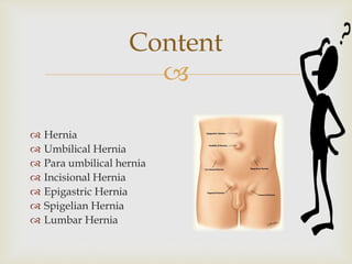


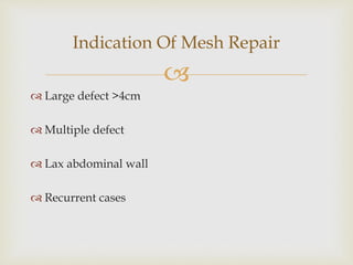
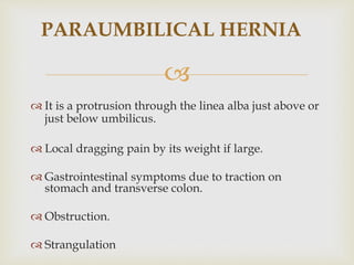


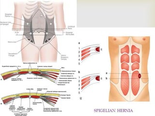


Dr. Shirish Silwal provides a summary of different types of hernias including inguinal, umbilical, paraumbilical, incisional, epigastric, spigelian, and lumbar hernias. The document discusses the history, anatomy, causes, presentations, complications, and management approaches for each hernia type. Meshes are recommended for repair when there is a large defect size, multiple defects, or lax abdominal walls to create a tension-free repair and reduce recurrence rates.