Primary surway
A: spontaneousbreathing
B: Normal breath sound, trachea at midline
C: BP 100/62 mmHg PR 94 /min
Capillary refill < 2 second
D: E4V5M6, pupil 3 mm RTLBE
E:- Left arm deformities(volar), tenderness,
swelling at left wrist
- ecchymosis
- No active bleeding
8.
Physical examination
GA:A youngThai female, good conscious, crying
Vital sign: BP 100/62 mmHg PR 94 /min
RR 18/min BT 36.3 ⁰C
HEENT : not pale conjunctivae, anicteric sclerae
CVS : normal s1s2, no murmur
Lung: clear both lungs, no adventitious sound
Abdomen: solf, not tender, normoactive bowel sound
9.
Physical Examination
Extremities(Affected part)
Left wrist -Deformity (volar displacement&dorsal
angulation)
–marked tenderness, swelling
-limit ROM due to pain
-Capillary refill < 2 sec
-Sensation intact
-No external wound
-No active bleeding
Smith’s Fracture Etiology
Smith’s Fracture is a distal radius fracture with
forward displacement of the distal fragment.
Considered a reverse Colle’s fracture
Caused by falling backwards which causes forced
pronation on the wrist.
Most commonly age 60-70 and young male.
Smith fractures account for less than 3% of all
fractures of the radius and ulna
Clinical Evaluation
Painand swelling in wrist generally after a fall
backwards onto the outstretched hand. Often
gross deformity in wrist.
Document neurovascular exam
Evaluated for carpal tunnel syndrome
19.
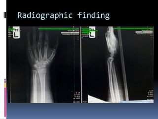
X-Ray Finding
Fracturesof the distal radius with associated
palmar angulation of the distal fracture
fragment. Classically, these fractures are extra-
articular transverse fractures and can be thought
of as a reverse Colles fracture.
21.
Radiographic features
The fracturecan be split into three types
-Type I
extra-articular transverse fracture through the distal radius
most common: -85%
-Type II
Intra-articular oblique fracture
equivalent to a reverse Barton fracture
~13%
-Type III
juxta-articular oblique fracture
uncommon: <2%
23.
Smith’s Fracture Associatedinjury
*Scapholunate ligament tear:
21.5% with intraarticular fracture
6.7% with extraarticular fracture
*Median nerve injury
*Triangular Fibrocartilage Complex injury (TFCC) up to 50%
when ulnar styloid fx present
*Carpal ligament injury
*Tendon injury, attritional EPL rupture
*Compartment syndrome
*Ulnar styloid fracture
*Distal radial ulnar joint (DRUJ) instability
24.
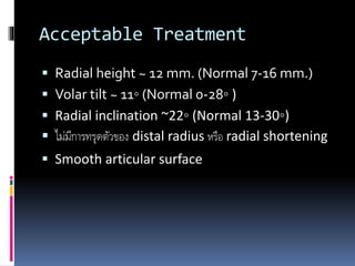
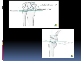
Treatment
Distal radius fractureAcceptableReduction
<2 mm articular stepoff
<5 mm shortening
<10⁰ dorsal tilt
Surgical indication
Radial shortening > 3mm,
dorsal tilt>10
Intra-articular displacement or step-off>2 mm.
(AAOS Clinical PracticeGuideline,2011)
25.
Treatment
ดึง tractionในท่า supination และดัน distal fragment
จากด้าน volar ไปด้าน dorsal ข้อสาคัญคือ ต้องใส่เฝือก long
arm cast ให้ข้อศอกงอ 90⁰ supination และ dorsiflex
ส่วนใหญ่ในปัจจุบันนิยมผ่าตัดด้าน volar และใส่ volar
buttress plate ป้องกันไม่ให้distal fragment เคลื่อนหลุด ซึ่ง
ได้ผลการรักษาดีกว่าการใส่เฝือก