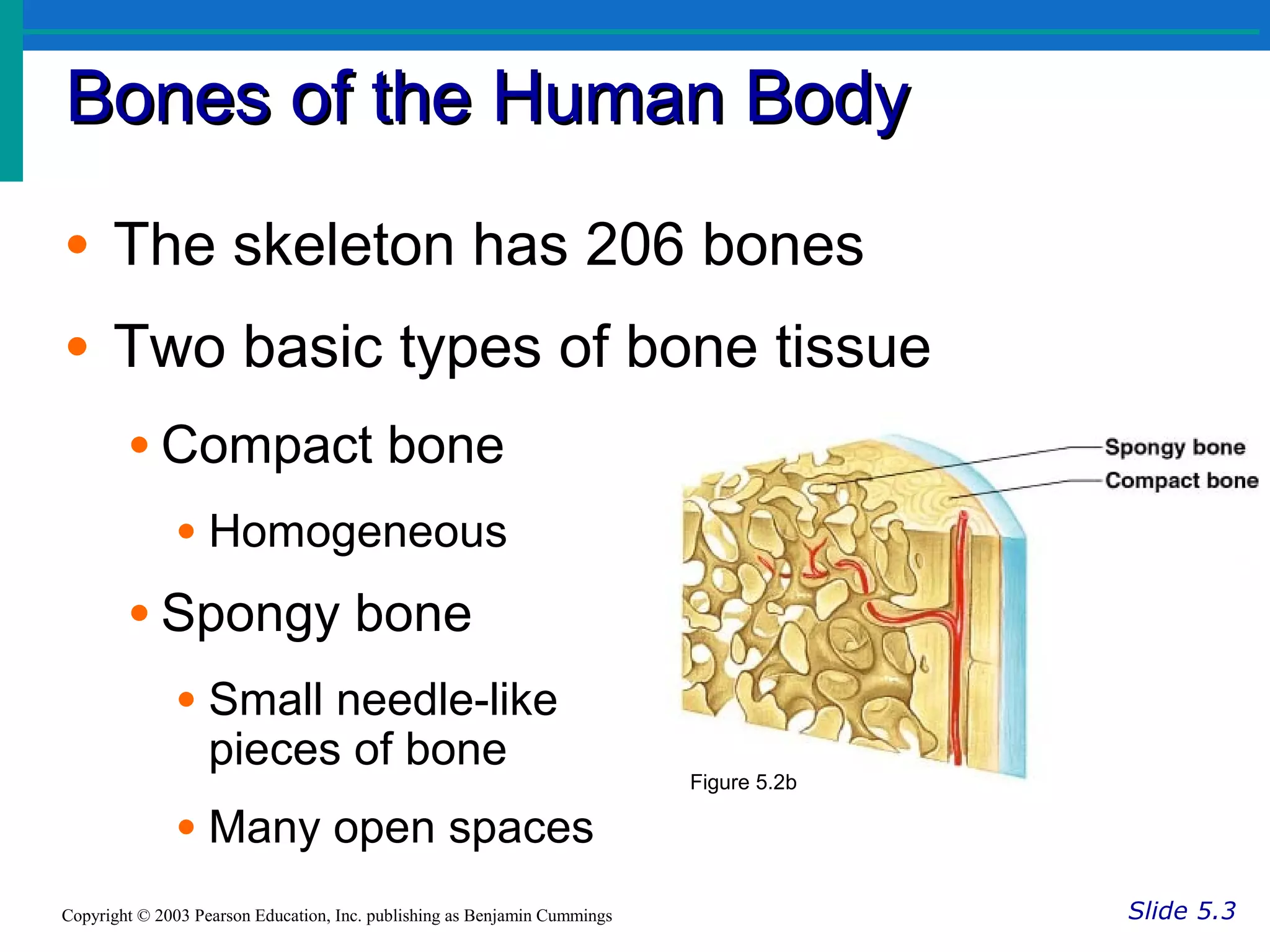
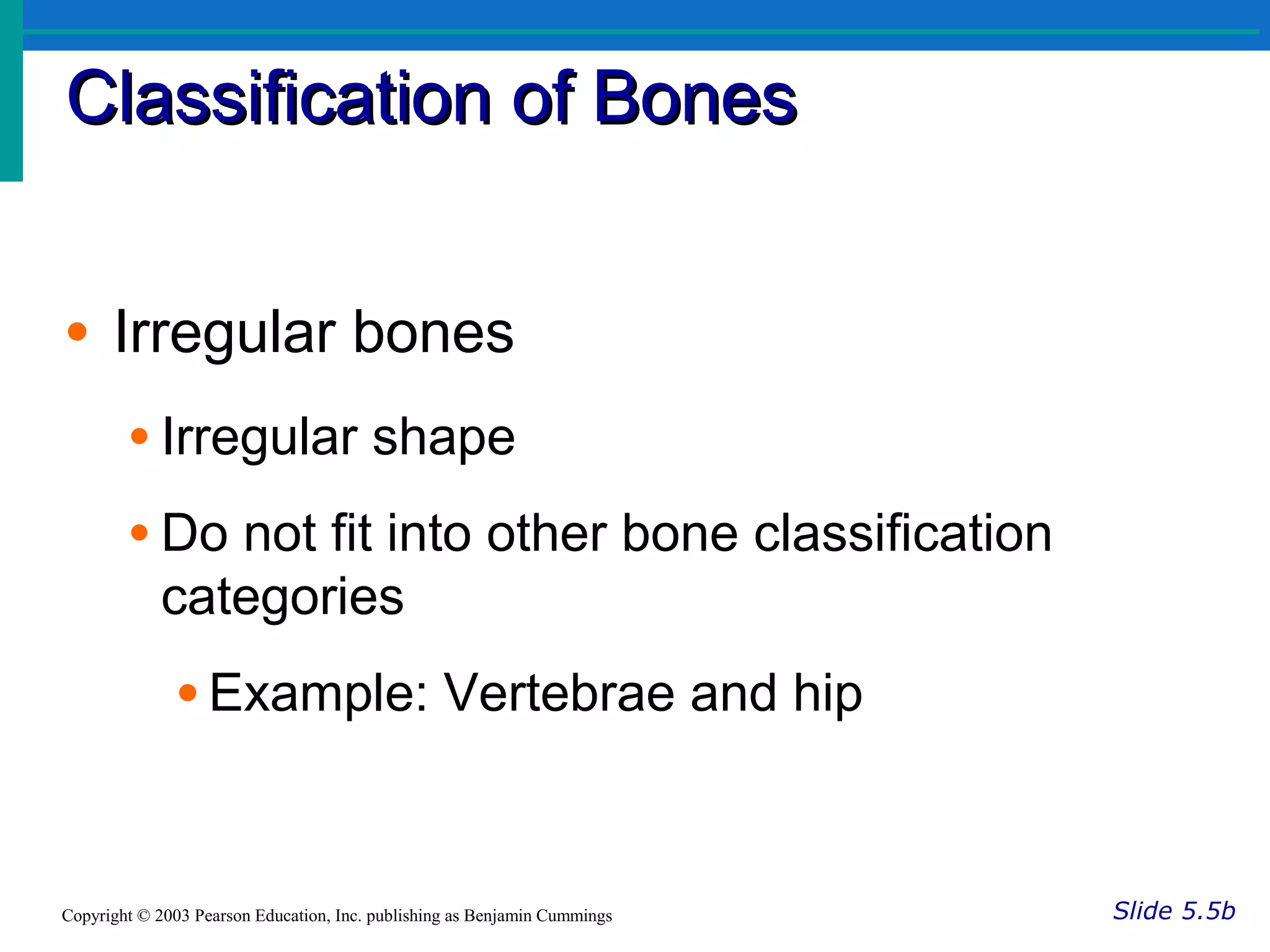
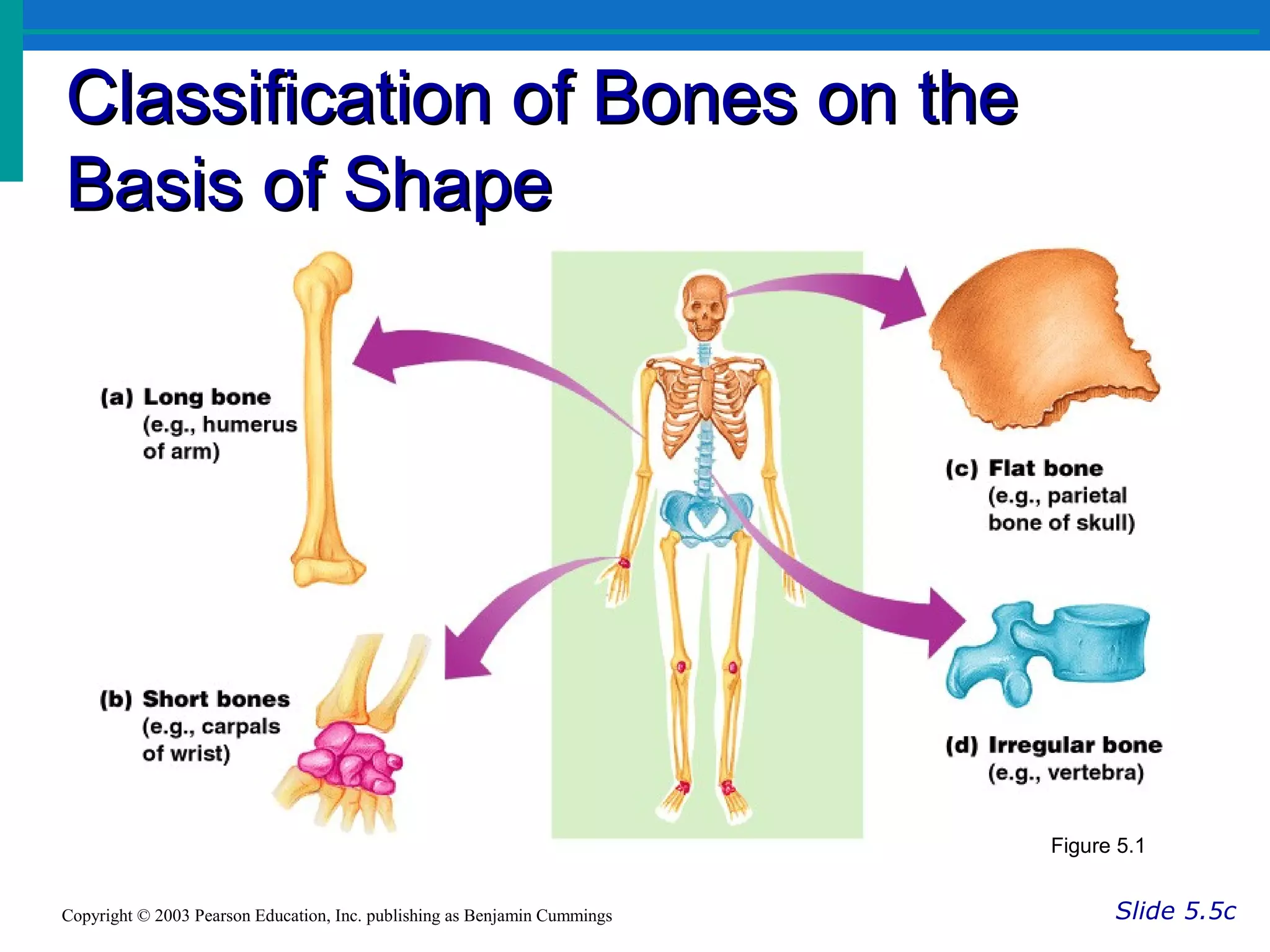
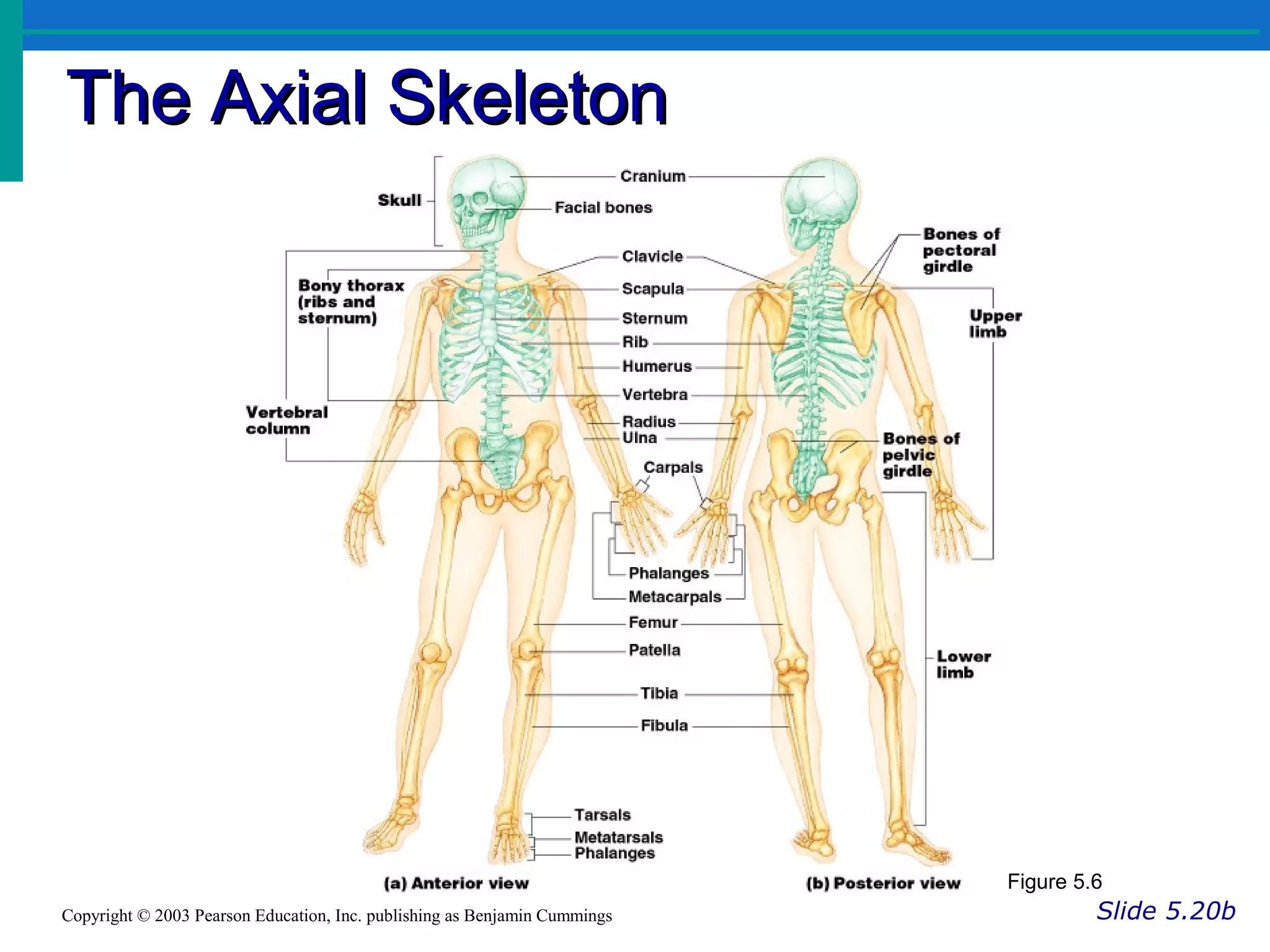
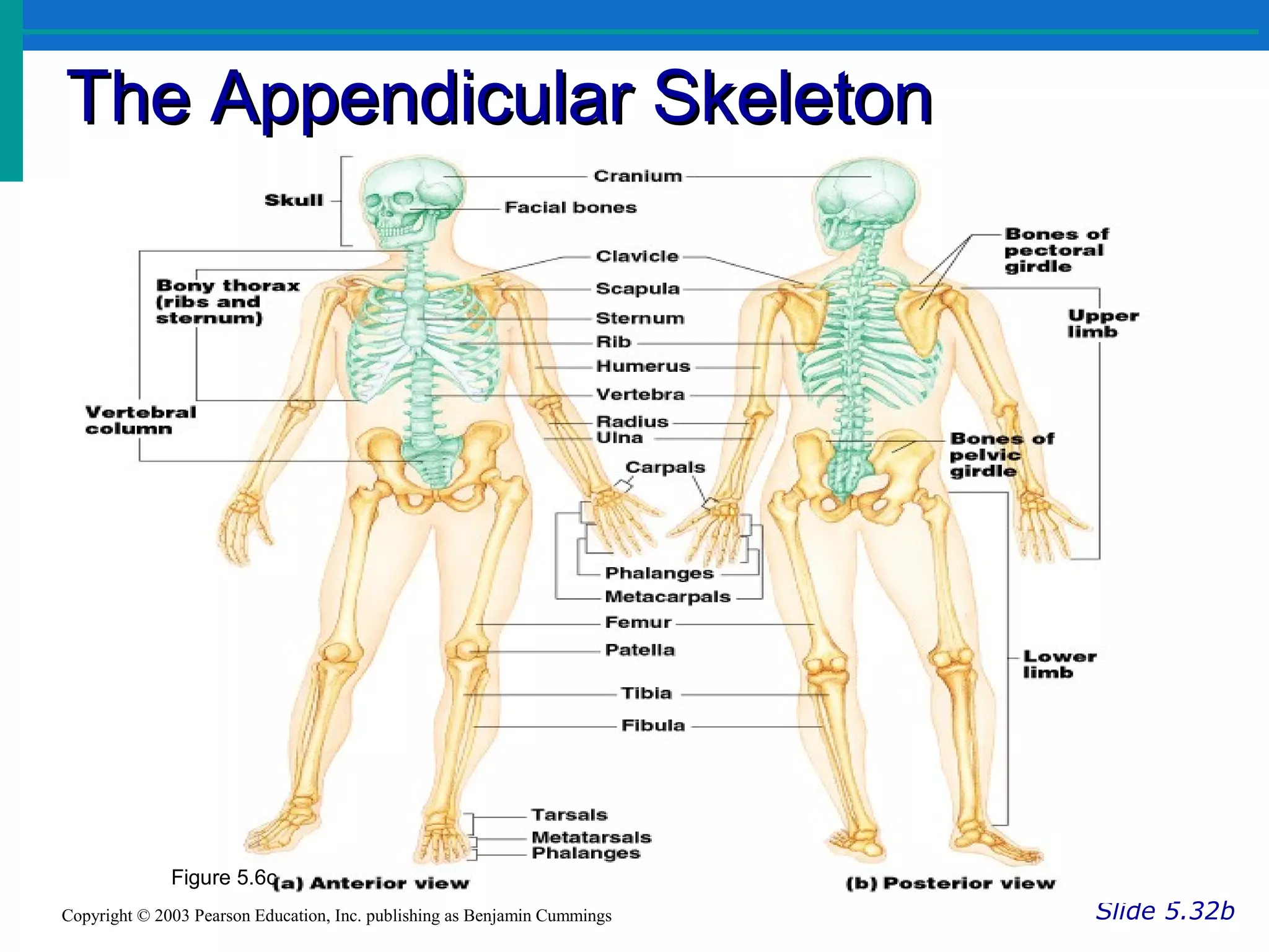
Anatomy of Skeletal System
The Skeletal SystemThe Skeletal System
Slide 5.1Copyright © 2003 Pearson Education, Inc. publishing as Benjamin Cummings
• Parts of the skeletal system
• Bones (skeleton)
• Joints
• Cartilages
• Ligaments (bone to bone)(tendon=bone to
muscle)
• Divided into two divisions
• Axial skeleton
• Appendicular skeleton – limbs and girdle
Functions of Skeletal System
• SUPPORT: Hard framework that supports and anchors
the soft organs of the body.
• PROTECTION: Surrounds organs such as the brain and
spinal cord.
• MOVEMENT: Allows for muscle attachment therefore the
bones are used as levers.
• STORAGE: Minerals and lipids are stored within bone
material.
• BLOOD CELL FORMATION: The bone marrow is
responsible for blood cell production.
Bones of the Human BodyBones of the Human Body
Slide 5.3Copyright © 2003 Pearson Education, Inc. publishing as Benjamin Cummings
• The skeleton has 206 bones
• Two basic types of bone tissue
• Compact bone
• Homogeneous
• Spongy bone
• Small needle-like
pieces of bone
• Many open spaces
Figure 5.2b
Classification of BonesClassification of Bones
Slide 5.4aCopyright © 2003 Pearson Education, Inc. publishing as Benjamin Cummings
• Long bones
• Typically longer than wide
• Have a shaft with heads at both ends
• Contain mostly compact bone
• Examples: Femur, humerus
Classification of BonesClassification of Bones
Slide 5.4bCopyright © 2003 Pearson Education, Inc. publishing as Benjamin Cummings
• Short bones
• Generally cube-shape
• Contain mostly spongy bone
• Examples: Carpals, tarsals
Classification of Bones on theClassification of Bones on the
Basis of ShapeBasis of Shape
Slide 5.4cCopyright © 2003 Pearson Education, Inc. publishing as Benjamin Cummings
Figure 5.1
Classification of BonesClassification of Bones
Slide 5.5aCopyright © 2003 Pearson Education, Inc. publishing as Benjamin Cummings
• Flat bones
• Thin and flattened
• Usually curved
• Thin layers of compact bone around a layer
of spongy bone
• Examples: Skull, ribs, sternum
Classification of BonesClassification of Bones
Slide 5.5bCopyright © 2003 Pearson Education, Inc. publishing as Benjamin Cummings
• Irregular bones
• Irregular shape
• Do not fit into other bone classification
categories
• Example: Vertebrae and hip
Classification of BonesClassification of Bones
• Sesamoid bones:
• Small bone
• It can change the direction of tendon
• Eg. Knee caps(patellae)
Classification of Bones on theClassification of Bones on the
Basis of ShapeBasis of Shape
Slide 5.5cCopyright © 2003 Pearson Education, Inc. publishing as Benjamin Cummings
Figure 5.1
Gross Anatomy of a Long BoneGross Anatomy of a Long Bone
Slide 5.6Copyright © 2003 Pearson Education, Inc. publishing as Benjamin Cummings
• Diaphysis
• Shaft
• Composed of
compact bone
• Epiphysis
• Ends of the bone
• Composed mostly of
spongy bone Figure 5.2a
Structures of a Long BoneStructures of a Long Bone
Slide 5.7Copyright © 2003 Pearson Education, Inc. publishing as Benjamin Cummings
• Periosteum
• Outside covering of
the diaphysis
• Fibrous connective
tissue membrane
• Sharpey’s fibers
• Secure periosteum to
underlying bone
• Arteries
• Supply bone cells
with nutrients
Figure 5.2c
Structures of a Long BoneStructures of a Long Bone
Slide 5.8aCopyright © 2003 Pearson Education, Inc. publishing as Benjamin Cummings
• Articular cartilage
• Covers the
external surface of
the epiphyses
• Made of hyaline
cartilage
• Decreases friction
at joint surfaces Figure 5.2a
Structures of a Long BoneStructures of a Long Bone
Slide 5.8bCopyright © 2003 Pearson Education, Inc. publishing as Benjamin Cummings
• Medullary cavity
• Cavity of the shaft
• Contains yellow
marrow (mostly fat)
in adults
• Contains red marrow
(for blood cell
formation) in infants Figure 5.2a
Bone Markings - Page 119Bone Markings - Page 119
Slide 5.9Copyright © 2003 Pearson Education, Inc. publishing as Benjamin Cummings
• Surface features of bones
• Sites of attachments for muscles, tendons,
and ligaments
• Passages for nerves and blood vessels
• Categories of bone markings
• Projections and processes – grow out from the
bone surface
• Depressions or cavities – indentations
Microscopic Anatomy of BoneMicroscopic Anatomy of Bone
Slide 5.10aCopyright © 2003 Pearson Education, Inc. publishing as Benjamin Cummings
• Osteon (Haversian System)
• A unit of bone
• Central (Haversian) canal
• Opening in the center of an osteon
• Carries blood vessels and nerves
• Perforating (Volkman’s) canal
• Canal perpendicular to the central canal
• Carries blood vessels and nerves
Microscopic Anatomy of BoneMicroscopic Anatomy of Bone
Slide 5.10bCopyright © 2003 Pearson Education, Inc. publishing as Benjamin Cummings
Figure 5.3
Microscopic Anatomy of BoneMicroscopic Anatomy of Bone
Slide 5.11aCopyright © 2003 Pearson Education, Inc. publishing as Benjamin Cummings
• Lacunae
• Cavities containing
bone cells
(osteocytes)
• Arranged in
concentric rings
• Lamellae
• Rings around the
central canal
• Sites of lacunae Figure 5.3
Microscopic Anatomy of BoneMicroscopic Anatomy of Bone
Slide 5.11bCopyright © 2003 Pearson Education, Inc. publishing as Benjamin Cummings
• Canaliculi
• Tiny canals
• Radiate from the
central canal to
lacunae
• Form a transport
system
Figure 5.3
Types of Bone CellsTypes of Bone Cells
Slide 5.15Copyright © 2003 Pearson Education, Inc. publishing as Benjamin Cummings
• Osteocytes
• Mature bone cells
• Osteoblasts
• Bone-forming cells
• Osteoclasts
• Bone-destroying cells
• Break down bone matrix for remodeling and
release of calcium
• Bone remodeling is a process by both
osteoblasts and osteoclasts
Bone formation(osteogenesis)
• Osification- two process
1.Intramembranous
2.Endochondral ossification
Bone formation(osteogenesis)
• Intramembranous: -within membrane
• Stepa of process-mesenchymal cell-osteoprogenitor cell-
osteoblast, the cluster called center of ossification.
• Osteoblast –secrets matrix
• Osteocytes –stop matrix secretion
• Deposition of calcium and other minerals on matrix to
harden it.
• Bone matrix form tuberculae
• It appears like spongy bone
Endochondral phase – with in
cartilage
Changes in the Human SkeletonChanges in the Human Skeleton
Slide 5.12Copyright © 2003 Pearson Education, Inc. publishing as Benjamin Cummings
• In embryos, the skeleton is primarily hyaline
cartilage
• During development, much of this cartilage
is replaced by bone
• Cartilage remains in isolated areas
• Bridge of the nose
• Parts of ribs
• Joints
Bone GrowthBone Growth
Slide 5.13aCopyright © 2003 Pearson Education, Inc. publishing as Benjamin Cummings
• Epiphyseal plates allow for growth of long
bone during childhood
• New cartilage is continuously formed
• Older cartilage becomes ossified
• Cartilage is broken down
• Bone replaces cartilage
Bone GrowthBone Growth
Slide 5.13bCopyright © 2003 Pearson Education, Inc. publishing as Benjamin Cummings
• Bones are remodeled and lengthened
until growth stops
• Bones change shape somewhat
• Bones grow in width
Long Bone Formation and GrowthLong Bone Formation and Growth
Slide 5.14aCopyright © 2003 Pearson Education, Inc. publishing as Benjamin Cummings
Figure 5.4a
Bone FracturesBone Fractures
Slide 5.16Copyright © 2003 Pearson Education, Inc. publishing as Benjamin Cummings
• A break in a bone
• Types of bone fractures
• Closed (simple) fracture – break that does not
penetrate the skin
• Open (compound) fracture – broken bone
penetrates through the skin
• Bone fractures are treated by reduction
and immobilization
• Realignment of the bone
Common Types of FracturesCommon Types of Fractures
Slide 5.17Copyright © 2003 Pearson Education, Inc. publishing as Benjamin Cummings
Table 5.2
Repair of Bone FracturesRepair of Bone Fractures
Slide 5.18Copyright © 2003 Pearson Education, Inc. publishing as Benjamin Cummings
• Hematoma (blood-filled swelling) is
formed
• Break is splinted by fibrocartilage to
form a callus
• Fibrocartilage callus is replaced by a
bony callus
• Bony callus is remodeled to form a
permanent patch
Stages in the Healing of a BoneStages in the Healing of a Bone
FractureFracture
Slide 5.19Copyright © 2003 Pearson Education, Inc. publishing as Benjamin Cummings
Figure 5.5
The Axial SkeletonThe Axial Skeleton
Slide 5.20aCopyright © 2003 Pearson Education, Inc. publishing as Benjamin Cummings
• Forms the longitudinal part of the body
• Divided into three parts
• Skull
• Vertebral column
• Bony thorax
The Axial SkeletonThe Axial Skeleton
Slide 5.20bCopyright © 2003 Pearson Education, Inc. publishing as Benjamin Cummings
Figure 5.6
The SkullThe Skull
Slide 5.21aCopyright © 2003 Pearson Education, Inc. publishing as Benjamin Cummings
• Two sets of bones
• Cranium
• Facial bones
• Bones are joined by sutures
• Only the mandible is attached by a
freely movable joint
SuturesSutures
• Sutures – Immovable joints that join skull bones together
• Form boundaries between skull bones
• Four sutures:
• Coronal – between parietal and frontal
• Sagittal– between parietal bones
• Lambdoid – between the parietal and occipital
• Squamous – between the parietal and temporal
Fontanels – usually ossify by 2 years of age
Anterior fontanel- closed at 18 month
Posterior fontanel- closed at 2 month
Frontal
(Coronal)
Sagittal
Squamous
Lambdoid
Sutures
•skull = 22 bones
•cranium = 8 bones: frontal, occipital, 2 temporals, 2 parietals, sphenoid and
ethmoid
•facial bones = 14 bones: nasals, maxillae, zygomatics, mandible, lacrimals,
palatines, inferior nasal conchae, vomer.
•skull forms a larger cranial cavity
-also forms the nasal cavity, the orbits, paranasal sinuses
mandible and auditory ossicles are the only movable skull bones
•cranial bones also: attach to membranes called meninges
-stabilize positions of the brain, blood vessels
-outer surface provides large areas for muscle attachment that
move the head or provide facial expressions
The Adult SkullThe Adult Skull
Frontal
Frontal View
Frontal bone
• Bone of fore head
• Forms orbital cavity and supraorbital margins
• Prominent ridges above the eyes and supraorbital
foramen.
Parietal
Lateral View
Parietal bone
• Two bone
• Greater portion of sides and roof of cranial cavity.
• Inner surface is concave and grooved by blood and brain
vessels
Temporal bone
• Two bone
• Present on lateral side
• Squamous part is flat
• Articulate with parietal bone
• Zygomatic process articulates with zygomatic bone
• Petrous portion – floor of cranium
• Temporal articulates with mandible –temporomandibular
joints
• Posterior inferior portion called – mastoid portion
• The process called mastoid process
• Mandibular fossa- between squamous and petrous portion
Occipital
Lateral View
Occipital bone
• Forms back of the head
• Foramen magnum (large hole)inferior part of the brain-
spinal cord passes to cranial cavity.
• Occipital condyles are oval process with convex surface,
present on each side of foramen magnum – articulates
with first cervical vertebra.
Sphenoid bone
• Wedge shape, at middle part of the base of skull
• Articulate with all bones of cranium
• Middle portion called hypophysial fossa
• Greater and lesser wings projects laterally on anterior and
posterior side
• Pterygoid process on inferior surface.
Nasal
Frontal View
Nasal bone
• Two bones
• Flat bones
• It forms lateral and superior surface of the bridge of nose
Vomer
Frontal View
Vomer bone
• Triangular bone
• It forms the inferior and posterior part of the nasal septum.
• Plate of ethmoid, septal cartilage, vomer and parts of
palatine bone, maxilla forms nasal septum
Zygoma
Frontal View
Zygomatic or cheek bone
• It forms the prominence of cheeks and part of the floor
and lateral walls of orbital cavities.
Maxilla
Frontal View
Maxilla or upper jaw bone
• Paired maxillae jointed to form the maxillary joints
• It forms floor of orbits, part of the lateral wall, floor of nasal
cavity, most of the hard palate.
• Forms anterior three quarters of hard palate
Mandible
Frontal View
Mandible
• Largest, strongest, movable joint
• Helps for chewing
• Curved bone, consist of body and rami(branch).
• Ramus devides into the chydylar process to
temporal bone and make temporomendibular
joint, other is coronoid process attach with
muscles and ligaments
• Angle and mandibular notch between processes
Lacrimal bone
• Two in number
• Finger nail shaped
• Its surface is thin and rough
• Posterior lateral to the nasal bone
• Lacrimal bone contains a foramen, through which
nasolacrimal duct /tear duct pass.
Palatine bone
• Two in number
• L-shaped
• Horizontal part joins to hard palate
• Perpendicular part projects upward to form lateral wall of
nasal cavity
Inferior conchae
• Two in number- scroll-like bone
• A part of lateral wall
• Filters and warm the air
The Hyoid BoneThe Hyoid Bone
Slide 5.26Copyright © 2003 Pearson Education, Inc. publishing as Benjamin Cummings
• U shaped /hourse shoe
shaped
• The only bone that
does not articulate with
another bone
• Serves as a moveable
base for the tongue
Figure 5.12
Figure 6.4 Sectional Anatomy of the
Skull, Part I
14 Facial Bones
The SkullThe Skull
Slide 5.21bCopyright © 2003 Pearson Education, Inc. publishing as Benjamin Cummings
Figure 5.7
Bones of the SkullBones of the Skull
Slide 5.22Copyright © 2003 Pearson Education, Inc. publishing as Benjamin Cummings
Figure 5.11
Human Skull, Superior ViewHuman Skull, Superior View
Slide 5.23Copyright © 2003 Pearson Education, Inc. publishing as Benjamin Cummings
Figure 5.8
Human Skull, Inferior ViewHuman Skull, Inferior View
Slide 5.24Copyright © 2003 Pearson Education, Inc. publishing as Benjamin Cummings
Figure 5.9
Paranasal Sinuses
• part of the nasal complex
• Paired cavities in ethmoid,
sphenoid, frontal and maxillary
• Lined with mucous
membranes and open into
nasal cavity though openings
called ostia
• Resonating chambers for
voice, lighten the skull
• Sinusitis is inflammation of the
membrane (allergy)
• infection can easily spread
from one sinus to the other
through the nasal cavity
• can also spread to other
tissues
 frontal sinusesfrontal sinuses
 sphenoid sinusessphenoid sinuses
 ethmoid sinusesethmoid sinuses
 maxillarymaxillary
Paranasal SinusesParanasal Sinuses
Slide 5.25aCopyright © 2003 Pearson Education, Inc. publishing as Benjamin Cummings
• Hollow portions of bones surrounding
the nasal cavity
Figure 5.10
Paranasal SinusesParanasal Sinuses
Slide 5.25bCopyright © 2003 Pearson Education, Inc. publishing as Benjamin Cummings
• Functions of paranasal sinuses
• Lighten the skull
• Give resonance and amplification to voice
Figure 5.10
The Fetal SkullThe Fetal Skull
Slide 5.27aCopyright © 2003 Pearson Education, Inc. publishing as Benjamin Cummings
• The fetal skull is
large compared
to the infants
total body length
Figure 5.13
The Fetal SkullThe Fetal Skull
Slide 5.27bCopyright © 2003 Pearson Education, Inc. publishing as Benjamin Cummings
• Fontanelles –
fibrous membranes
connecting the
cranial bones
• Allow the brain
to grow
• Convert to bone
within 24 months
after birth
Figure 5.13
The Vertebral ColumnThe Vertebral Column
Slide 5.28Copyright © 2003 Pearson Education, Inc. publishing as Benjamin Cummings
• Vertebrae
separated by
intervertebral
discs
• The spine has
a normal
curvature
• Each vertebrae
is given a name
according to its
location
Figure 5.14
• Length of column 71cm (male) and 61cm (female)
• Strong flexible rod that can bend.
• Serve as point of attachment for the ribs
• Intervertebral foramina is the opening between vertebra
• Adult contains 26 vertebra.
Characteristics of a typical
vertebrae
• The body- situated anteriorly
• Size change as per the sites
• They are smallest in the cervical region and become larger
towards lumber region.
• Vertebral arch- encloses large vertebral foramen.
• The ring projects 2 pedicle at backward, 2 lamina which
attach to the pedicle.
• Transverse processes project laterally, where two lamina
meets at midline posteriorly.
• They form spinous process.
• Neural arch has four articulating surfaces. Two articulate
with above vertebra, and two blow vertebra.
Structure of a Typical VertebraeStructure of a Typical Vertebrae
Slide 5.29Copyright © 2003 Pearson Education, Inc. publishing as Benjamin Cummings
Figure 5.16
Typical Vertebrae • Body
• weight bearing
• Vertebral arch
• pedicles
• laminae
• Vertebral foramen
• Seven processes
• 2 transverse
• 1 spinous
• 4 articular
Cervical vertebra
• Seven- C1 to C7
• Atlas is the first vertebra
• It consist ring bone with two transvers process.
• The axis is the second vertebra, the body is small.
Thoracic vertebra
• 12 in number
• Large strong than cervical
• The bodies and transverse processes have facets for
articulation of ribs
Lumber Region
• Five lumber vertebra L1 –L5
• Largest strongest, their projections are short and thick.
Sacrum
Figure 7.18a, b
Sacrum
• Triangular bone
• Formed by union of five sacral vertebrae.
• Anterior side is concave.
• Lateral portion contains sacral ala(wing)
• The posterior surface is convex.
• Upper side sacral promontory present.
• Sacral tuberosity is a rough surface
• Female sacrum is shorter, wider and more curved
compare to male.
Coccyx
• Triangular shaped bone
• Fusion of 4 coccygeal vertebra
• Transverse process are present laterally
Movement of vertebral colums
Flexion - forward
Extension- backward
Lateral flexion- bend on side
Rotation -
Functions of vertebral colums
• It supplies blood, nerve, lymph supply on each side of
body.
• Supports skull
• Flexible body movement possible
• Protective cover for spinal cord
The Bony ThoraxThe Bony Thorax
Slide 5.31aCopyright © 2003 Pearson Education, Inc. publishing as Benjamin Cummings
• Forms a cage to protect major organs
• Cone shaped
• Superiorly narrow, inferiorly broad
• Made-up of three parts
• 1 Sternum
• 12 pairs of ribs
• 12 Thoracic vertebrae Figure 5.19a
The Bony ThoraxThe Bony Thorax
Slide 5.31bCopyright © 2003 Pearson Education, Inc. publishing as Benjamin Cummings
Figure 5.19a
Sternum
• Flat, narrow,
• Length 15cm
• Three parts
• Manubrium-triangular, superior portion
• Body-the middle largest portion
• Xiphoid process-inferior smallest portion
• Jugular notch/ Suprasternal notch– depression
on superior surface of manubrium.
• Clavicular notch- each side of jugular notch.
Sternum
• Formed from three parts :
• Manubrium – superior part
• Articulates with medial end of clavicles
• Body – bulk of sternum
• Sides are articulate for costal cartilage of ribs 2–7
• Xiphoid process – inferior end of sternum
• Ossifies around age 40
Ribs
• All ribs attach to vertebral column posteriorly
• True ribs - superior seven pairs of ribs
• Attach to sternum by costal cartilage
• False ribs – inferior five pairs of ribs ,attach indirectly to the
sternum
•floating ribs ribs 11–12 are short and free anteriorly.
Ribs
• 12 pairs of ribs
• Articulate posteriorly to thoracic vertebra
• Anteriorly to sternum with costal cartilage
• Last two pairs have no anterior attachment
• First seven –true ribs
• Five pairs – false ribs
• Head of typical rib project posteriorly, wedge shaped.
The Appendicular SkeletonThe Appendicular Skeleton
Slide 5.32aCopyright © 2003 Pearson Education, Inc. publishing as Benjamin Cummings
• Limbs (appendages)
• Pectoral girdle
• Pelvic girdle
The Appendicular SkeletonThe Appendicular Skeleton
Slide 5.32bCopyright © 2003 Pearson Education, Inc. publishing as Benjamin Cummings
Figure 5.6c
The Pectoral (Shoulder) GirdleThe Pectoral (Shoulder) Girdle
Slide 5.33Copyright © 2003 Pearson Education, Inc. publishing as Benjamin Cummings
• Composed of two bones
• Clavicle – collarbone
• Scapula – shoulder blade
• These bones allow the upper limb to
have exceptionally free movement
Bones of the Shoulder GirdleBones of the Shoulder Girdle
Slide 5.34aCopyright © 2003 Pearson Education, Inc. publishing as Benjamin Cummings
Figure 5.20a, b
The Clavicles
Figure 8–2b, c
Clavicle
• Collar bone
• Long slender s- shaped bone
• 2 curve- concave and convex
• It articulates with manubrium of sternum by
sternoclavicular joint
• Also attach with acromion process of scapula and form
acromio-clavicular joint
Anatomy of
The
scapula
Figure 8–3a
Scapula
• Large triangular flat bone
• Present on posterior part of the thorax between
second and seventh ribs
• Two angle- superior angle, inferior angle
• Three border –superior, medial, lateral
• Two surfaces- anterior and posterior
• Lateral end of spine is flattened, expanded process
called acromion.
• Glenoid cavity-is depression of inferior surface of
acromion. At the lateral end of superior border is a
projection at anterior surface called coracoid
process. Muscles are attached at this part.
Bones of the Upper LimbBones of the Upper Limb
Slide 5.35aCopyright © 2003 Pearson Education, Inc. publishing as Benjamin Cummings
• The arm
is
formed
by a
single
bone
• Humerus
Figure 5.21a, b
ANATOMY OF The Humerus
Figure 8–4
Humerus
• Largest longest bone
• Articulates proximally with the scapula distally at elbow
with radius and ulna.
• Head articulate with glenoid cavity of scapula.
• Greater and lesser tubercle are rough projection at distal
of head.
• Intertubercular salcus present between two tubercles. It is
deep groove
• At middle portion –shaft, roughened v shaped area called
deltoid tuberosity.
• Medial and lateral epicondyle are rough projection.
Bones of the Upper LimbBones of the Upper Limb
Slide 5.35bCopyright © 2003 Pearson Education, Inc. publishing as Benjamin Cummings
• The
forearm
has two
bones
• Ulna
• Radius
Figure 5.21c
The Forearm
Figure 8–5
Ulna and radius
• Bones of forearm
• Ulna is longer than radius
• Olecranon process present at proximal end of ulna
• Trochlear notch is a large curve area, present between
olecranon and coronooid process.
• In anatomical position-two bones are parallel.
• Articulate with humerus by elbow joint
• Carpal bone at wrist joint
• Proximal and distal radioulnar joint.
Bones of the Upper LimbBones of the Upper Limb
Slide 5.36Copyright © 2003 Pearson Education, Inc. publishing as Benjamin Cummings
• The hand
• Carpals –
wrist
• Metacarpal
s – palm
• Phalanges
– fingers
Figure 5.22
Carpals or wrist bones
• Eight small bones – arranged in two rows of four.
a)Proximal row: scaphoid, lunate, triquetral, pisiform,
attached to wrist joint
b)Distal row:trapezium. Trapezoid, capitate, hamate- joints
with metacarpal bone.
Metacarpal bones
• Five bones- form the palm of hand
• Numbered as I to V from thumb side to inward.
• Proximal end articulates with carpal bones
• Distal ends with phalanges.
Phalanges or finger bones
• 14 in numbers-
• Referred as thumb, index, middle, ring, little finger.
• Articulate with metacarpals.
Bones of the Pelvic GirdleBones of the Pelvic Girdle
Slide 5.37Copyright © 2003 Pearson Education, Inc. publishing as Benjamin Cummings
• Hip bones
• Composed of three pair of fused bones
• Ilium
• Ischium
• Pubic bone
• The total weight of the upper body rests on the
pelvis
• Protects several organs
• Reproductive organs
• Urinary bladder
• Part of the large intestine
The Pelvic Girdle
Figure 8–7
The Pelvis
Figure 8–8
Innominate bones/hip bones
• 2 in number, formed by fusion of three bones
• Illium, ischium, pubic
• Ilium- superior border called iliac crest,
• Ends of border called iliac spine- 4 in number
• Anterior superior iliac spine
• Anterior inferior iliac spine
• Posterior superior iliac spine
• Posterior inferior iliac spine
• Greater sciatic notch is present
Ischium
• Posterior and inferior part of hip bone
• It consist lesser sciatic notch, ischial spine, ishial
tuberosity
Pubis
• Anterior – inferior part of hip bone
• It consist superior and inferior rami
• Body between rami called symphysis pubis
• Anterior border called pubic crest
• On lateral end projection called pubic tubercle
The PelvisThe Pelvis
Slide 5.38aCopyright © 2003 Pearson Education, Inc. publishing as Benjamin Cummings
Figure 5.23a
Gender Differences of the PelvisGender Differences of the Pelvis
Slide 5.39Copyright © 2003 Pearson Education, Inc. publishing as Benjamin Cummings
Figure 5.23c
Lower limb
• Composed of 60 bones
• At each side
1 femur
1 tibia
1 fibula
1 patella
7 tarsal bone
5 metatarsal bone
14 phalanges
Bones of the Lower LimbsBones of the Lower Limbs
Slide 5.40aCopyright © 2003 Pearson Education, Inc. publishing as Benjamin Cummings
• The thigh
has one
bone
Femur – thigh
bone
Figure 5.35a, b
Femur
• Longest, heaviest, strongest bone
• Proximally articulates with hip bone at
acetabulum cavity.
• Distal articulates tibia and fibula
• Greater and lesser trochanter are the projections.
• Posterior one third triangular flat part called
popliteal surface
• At distal end there are two articulars –medial and
lateral condyle
Patella /knee cap
• Small triangular bone
• Located anterior of knee joint
Function: protects knee joint
maintain position of the tendon
Bones of the Lower LimbsBones of the Lower Limbs
Slide 5.40bCopyright © 2003 Pearson Education, Inc. publishing as Benjamin Cummings
• The leg has
two bones
• Tibia
• Fibula
Figure 5.35c
Tibia and fibula
• Tibia is longer than fibula.
• There are medial condyle and lateral condyle.
• Lateral condyle extended on fibula.
• At distal part medial malleolus on tibia and lateral
malleolus on fibula
• Tibial tuberosity is point of attachment of muscle and
ligaments
The Tibia and Fibula
Figure 8–13
Bones of the Lower LimbsBones of the Lower Limbs
Slide 5.41Copyright © 2003 Pearson Education, Inc. publishing as Benjamin Cummings
• The foot
• Tarsus – ankle
• Metatarsals –
sole
• Phalanges –
toes
Figure 5.25
Tarsals
• Seven in number
• Posterior part of foot
1 talus
1 calcaneus
1 navicular
3 cuneiform
1 cuboid
Talus articulates with tibia and fibula at ankle joint
Calcaneus forms heel of foot
The Ankle-Also called the tarsus:
consists of 7 tarsal bones
Figure 8–14a
Metatarsal
• Five bones
• Numbered I to V from medial to lateral
• At proximal ends articulates with tarsal bones and
at distal articulates with phalanges
• Phalanges
• 14 in numbers.
• Arranged in same manner like fingers
• Big toe has 2 large, and four toes have three
phalanges
Joints
Key words
• Articulation- joint
• Bursa –cushion like sac
• Diathrosis- freely movable joint
• Suture – immovable joints between skull bone
• Symphasis- joint formed by fibrocartilage.
JointsJoints
Slide 5.43Copyright © 2003 Pearson Education, Inc. publishing as Benjamin Cummings
• Articulations of bones
• Functions of joints
• Hold bones together
• Allow for mobility
• Ways joints are classified
• Functionally
• Structurally
Functional Classification of JointsFunctional Classification of Joints
Slide 5.44Copyright © 2003 Pearson Education, Inc. publishing as Benjamin Cummings
• Synarthroses – immovable joints
• Amphiarthroses – slightly moveable
joints
• Diarthroses – freely moveable joints
Types
• Synarthroses
• No movements
• Primarily axial
skeleton
• Bones connected
with fibrous tissue
ligament
• Examples: Skull
sutures and distal
Tibia/Fibula
Types
• Amphiarthroses
• Slightly movable
• Axial skeleton
• Connected by
cartilage
• Intervertebral
joints, pubic
symphysis
Types
• Diarthroses – freely movable
• Also called synovial (fluid filled joint cavity)
• Primarily found in the limbs
• Plane of movement depends on the joint
Structural Classification of JointsStructural Classification of Joints
Slide 5.45Copyright © 2003 Pearson Education, Inc. publishing as Benjamin Cummings
• Fibrous joints
• Generally immovable
• Cartilaginous joints
• Immovable or slightly moveable
• Synovial joints
• Freely moveable
Fibrous JointsFibrous Joints
Slide 5.46Copyright © 2003 Pearson Education, Inc. publishing as Benjamin Cummings
• Bones united by fibrous tissue –
synarthrosis or largely immovable.
Figure 5.27d, e
Cartilaginous Joints – mostlyCartilaginous Joints – mostly
amphiarthrosisamphiarthrosis
Slide 5.47Copyright © 2003 Pearson Education, Inc. publishing as Benjamin Cummings
• Bones connected by cartilage
• Examples
• Pubic
symphysis
• Intervertebral
joints
Figure 5.27b, c
Synovial JointsSynovial Joints
Slide 5.48Copyright © 2003 Pearson Education, Inc. publishing as Benjamin Cummings
• Articulating
bones are
separated by a
joint cavity
• Synovial fluid
is found in the
joint cavity
Figure 5.27f–h
The Synovial JointThe Synovial Joint
Slide 5.51Copyright © 2003 Pearson Education, Inc. publishing as Benjamin Cummings
Figure 5.28
Synovial Joints - Structure
1. Articular cartilage: hyaline
2. Joint Cavity: space filled with lubricating fluid
3. Fibrous Capsule: fibrous CT lined with a smooth
synovial membrane
4. Reinforcing Ligament: can be inside or outside
the joint capsule
5. Synovial Fluid: viscous and lubricating
6. Tendons sheath an elongated bursa that rapes
around a tendon subjected to friction.
7. Menisci: cartilaginous discs
Synovial Joint
Shoulder joint
• Ball and socket joint, multiaxial synovial joint
• Humerus fit into glenoid cavity of scapula
• Greater rate of movement
• Articulating capsule is more loose, completely envelops
the articulation
• Strength and stability is depend on four muscle: supra
spinatus, infra spinatus, tarrous, rotator
• Three ligament: Cora cohumeral ligament, Glenohumeral
ligament, Transverse humeral ligament
Muscles in shoulder joint
• Coracobrachialis muscle – upper arm, at coracoid
process to mid arm
• Deltoid muscle- clevicle, acromion process and spine of
scapula to deltoid tuberosity
• Pectoralis muscle – at anterior thoracic wall, clevicle and
humerus
• Latissimus dorsi – iliac crest, spinous process of lumber
and thoracic vertebra to upward at bicipital groove of
humerus
Movements
• Flexion
• Extension
• Hyper extension
• Abduction
• Adduction
• Circumduction
• Rotation – longitudinal axis
• Inversion –inward
• Eversion –outward
• Dorsiflexion-bending foot, upper surface
• Planter flexion-bending foot in direction of sole
• Protraction- mandible and shoulder girdle forward
• Retraction –mandible and shoulder girdle backward
• Elevation –body upward
• Depression –body down ward
• Opposition – angular of thumb to finger
• Reposition –thumb to anatomical position
Elbow joint
• Hinge joint – between humerus and radius
• Extracapsular stuctures- anterior, posterior, medial ,
lateral strengthening ligaments
• Muscles-brachialis and triceps
Hip joint
• Ball and socket joint, multi axial joint
• Head of femur articulates with acetabulum cavity of hip
joint
• Deepened by acetabulo labrum- hold the socket secure
by synovial membrane
• Three ligaments:
• Iliofemoral
• Pubofemoral
• ischiofemora
Muscles and movement
• Muscles:
• Psoas muscle
• Iliacus muscle
• Quadriceps femoris
• Gluteal muscles
• Sartorius muscle
Movements:
• Flexion
• Extension
• Adduction
• Abduction
• Medial rotation
• Lateral rotation
Knee joint
• Called tibio-femoral joint
• Largest most complex joint
• Synovial with modified hinge joint
• Small degree of rotation movement
• Synovial membrane covers ligaments and pads of fat
• Bursae- cushion the knee joint
• Supra patellar- above patella
• Prepatellar- between skin and patella
• Infra patellar- between skin and tubular tuberosity
Muscles and movement
• Flexion
• extension
• slight degree of rotation- laterally medially
• Muscles :
• Hamstring muscles
• Gastrocnemius muscle

Skeletal system in a Brief

  • 1.
  • 2.
    The Skeletal SystemTheSkeletal System Slide 5.1Copyright © 2003 Pearson Education, Inc. publishing as Benjamin Cummings • Parts of the skeletal system • Bones (skeleton) • Joints • Cartilages • Ligaments (bone to bone)(tendon=bone to muscle) • Divided into two divisions • Axial skeleton • Appendicular skeleton – limbs and girdle
  • 4.
    Functions of SkeletalSystem • SUPPORT: Hard framework that supports and anchors the soft organs of the body. • PROTECTION: Surrounds organs such as the brain and spinal cord. • MOVEMENT: Allows for muscle attachment therefore the bones are used as levers. • STORAGE: Minerals and lipids are stored within bone material. • BLOOD CELL FORMATION: The bone marrow is responsible for blood cell production.
  • 5.
    Bones of theHuman BodyBones of the Human Body Slide 5.3Copyright © 2003 Pearson Education, Inc. publishing as Benjamin Cummings • The skeleton has 206 bones • Two basic types of bone tissue • Compact bone • Homogeneous • Spongy bone • Small needle-like pieces of bone • Many open spaces Figure 5.2b
  • 6.
    Classification of BonesClassificationof Bones Slide 5.4aCopyright © 2003 Pearson Education, Inc. publishing as Benjamin Cummings • Long bones • Typically longer than wide • Have a shaft with heads at both ends • Contain mostly compact bone • Examples: Femur, humerus
  • 7.
    Classification of BonesClassificationof Bones Slide 5.4bCopyright © 2003 Pearson Education, Inc. publishing as Benjamin Cummings • Short bones • Generally cube-shape • Contain mostly spongy bone • Examples: Carpals, tarsals
  • 8.
    Classification of Boneson theClassification of Bones on the Basis of ShapeBasis of Shape Slide 5.4cCopyright © 2003 Pearson Education, Inc. publishing as Benjamin Cummings Figure 5.1
  • 9.
    Classification of BonesClassificationof Bones Slide 5.5aCopyright © 2003 Pearson Education, Inc. publishing as Benjamin Cummings • Flat bones • Thin and flattened • Usually curved • Thin layers of compact bone around a layer of spongy bone • Examples: Skull, ribs, sternum
  • 10.
    Classification of BonesClassificationof Bones Slide 5.5bCopyright © 2003 Pearson Education, Inc. publishing as Benjamin Cummings • Irregular bones • Irregular shape • Do not fit into other bone classification categories • Example: Vertebrae and hip
  • 11.
    Classification of BonesClassificationof Bones • Sesamoid bones: • Small bone • It can change the direction of tendon • Eg. Knee caps(patellae)
  • 12.
    Classification of Boneson theClassification of Bones on the Basis of ShapeBasis of Shape Slide 5.5cCopyright © 2003 Pearson Education, Inc. publishing as Benjamin Cummings Figure 5.1
  • 13.
    Gross Anatomy ofa Long BoneGross Anatomy of a Long Bone Slide 5.6Copyright © 2003 Pearson Education, Inc. publishing as Benjamin Cummings • Diaphysis • Shaft • Composed of compact bone • Epiphysis • Ends of the bone • Composed mostly of spongy bone Figure 5.2a
  • 14.
    Structures of aLong BoneStructures of a Long Bone Slide 5.7Copyright © 2003 Pearson Education, Inc. publishing as Benjamin Cummings • Periosteum • Outside covering of the diaphysis • Fibrous connective tissue membrane • Sharpey’s fibers • Secure periosteum to underlying bone • Arteries • Supply bone cells with nutrients Figure 5.2c
  • 15.
    Structures of aLong BoneStructures of a Long Bone Slide 5.8aCopyright © 2003 Pearson Education, Inc. publishing as Benjamin Cummings • Articular cartilage • Covers the external surface of the epiphyses • Made of hyaline cartilage • Decreases friction at joint surfaces Figure 5.2a
  • 16.
    Structures of aLong BoneStructures of a Long Bone Slide 5.8bCopyright © 2003 Pearson Education, Inc. publishing as Benjamin Cummings • Medullary cavity • Cavity of the shaft • Contains yellow marrow (mostly fat) in adults • Contains red marrow (for blood cell formation) in infants Figure 5.2a
  • 17.
    Bone Markings -Page 119Bone Markings - Page 119 Slide 5.9Copyright © 2003 Pearson Education, Inc. publishing as Benjamin Cummings • Surface features of bones • Sites of attachments for muscles, tendons, and ligaments • Passages for nerves and blood vessels • Categories of bone markings • Projections and processes – grow out from the bone surface • Depressions or cavities – indentations
  • 18.
    Microscopic Anatomy ofBoneMicroscopic Anatomy of Bone Slide 5.10aCopyright © 2003 Pearson Education, Inc. publishing as Benjamin Cummings • Osteon (Haversian System) • A unit of bone • Central (Haversian) canal • Opening in the center of an osteon • Carries blood vessels and nerves • Perforating (Volkman’s) canal • Canal perpendicular to the central canal • Carries blood vessels and nerves
  • 19.
    Microscopic Anatomy ofBoneMicroscopic Anatomy of Bone Slide 5.10bCopyright © 2003 Pearson Education, Inc. publishing as Benjamin Cummings Figure 5.3
  • 20.
    Microscopic Anatomy ofBoneMicroscopic Anatomy of Bone Slide 5.11aCopyright © 2003 Pearson Education, Inc. publishing as Benjamin Cummings • Lacunae • Cavities containing bone cells (osteocytes) • Arranged in concentric rings • Lamellae • Rings around the central canal • Sites of lacunae Figure 5.3
  • 21.
    Microscopic Anatomy ofBoneMicroscopic Anatomy of Bone Slide 5.11bCopyright © 2003 Pearson Education, Inc. publishing as Benjamin Cummings • Canaliculi • Tiny canals • Radiate from the central canal to lacunae • Form a transport system Figure 5.3
  • 22.
    Types of BoneCellsTypes of Bone Cells Slide 5.15Copyright © 2003 Pearson Education, Inc. publishing as Benjamin Cummings • Osteocytes • Mature bone cells • Osteoblasts • Bone-forming cells • Osteoclasts • Bone-destroying cells • Break down bone matrix for remodeling and release of calcium • Bone remodeling is a process by both osteoblasts and osteoclasts
  • 23.
    Bone formation(osteogenesis) • Osification-two process 1.Intramembranous 2.Endochondral ossification
  • 24.
    Bone formation(osteogenesis) • Intramembranous:-within membrane • Stepa of process-mesenchymal cell-osteoprogenitor cell- osteoblast, the cluster called center of ossification. • Osteoblast –secrets matrix • Osteocytes –stop matrix secretion • Deposition of calcium and other minerals on matrix to harden it. • Bone matrix form tuberculae • It appears like spongy bone
  • 25.
    Endochondral phase –with in cartilage
  • 26.
    Changes in theHuman SkeletonChanges in the Human Skeleton Slide 5.12Copyright © 2003 Pearson Education, Inc. publishing as Benjamin Cummings • In embryos, the skeleton is primarily hyaline cartilage • During development, much of this cartilage is replaced by bone • Cartilage remains in isolated areas • Bridge of the nose • Parts of ribs • Joints
  • 27.
    Bone GrowthBone Growth Slide5.13aCopyright © 2003 Pearson Education, Inc. publishing as Benjamin Cummings • Epiphyseal plates allow for growth of long bone during childhood • New cartilage is continuously formed • Older cartilage becomes ossified • Cartilage is broken down • Bone replaces cartilage
  • 28.
    Bone GrowthBone Growth Slide5.13bCopyright © 2003 Pearson Education, Inc. publishing as Benjamin Cummings • Bones are remodeled and lengthened until growth stops • Bones change shape somewhat • Bones grow in width
  • 29.
    Long Bone Formationand GrowthLong Bone Formation and Growth Slide 5.14aCopyright © 2003 Pearson Education, Inc. publishing as Benjamin Cummings Figure 5.4a
  • 30.
    Bone FracturesBone Fractures Slide5.16Copyright © 2003 Pearson Education, Inc. publishing as Benjamin Cummings • A break in a bone • Types of bone fractures • Closed (simple) fracture – break that does not penetrate the skin • Open (compound) fracture – broken bone penetrates through the skin • Bone fractures are treated by reduction and immobilization • Realignment of the bone
  • 31.
    Common Types ofFracturesCommon Types of Fractures Slide 5.17Copyright © 2003 Pearson Education, Inc. publishing as Benjamin Cummings Table 5.2
  • 32.
    Repair of BoneFracturesRepair of Bone Fractures Slide 5.18Copyright © 2003 Pearson Education, Inc. publishing as Benjamin Cummings • Hematoma (blood-filled swelling) is formed • Break is splinted by fibrocartilage to form a callus • Fibrocartilage callus is replaced by a bony callus • Bony callus is remodeled to form a permanent patch
  • 33.
    Stages in theHealing of a BoneStages in the Healing of a Bone FractureFracture Slide 5.19Copyright © 2003 Pearson Education, Inc. publishing as Benjamin Cummings Figure 5.5
  • 34.
    The Axial SkeletonTheAxial Skeleton Slide 5.20aCopyright © 2003 Pearson Education, Inc. publishing as Benjamin Cummings • Forms the longitudinal part of the body • Divided into three parts • Skull • Vertebral column • Bony thorax
  • 35.
    The Axial SkeletonTheAxial Skeleton Slide 5.20bCopyright © 2003 Pearson Education, Inc. publishing as Benjamin Cummings Figure 5.6
  • 36.
    The SkullThe Skull Slide5.21aCopyright © 2003 Pearson Education, Inc. publishing as Benjamin Cummings • Two sets of bones • Cranium • Facial bones • Bones are joined by sutures • Only the mandible is attached by a freely movable joint
  • 38.
    SuturesSutures • Sutures –Immovable joints that join skull bones together • Form boundaries between skull bones • Four sutures: • Coronal – between parietal and frontal • Sagittal– between parietal bones • Lambdoid – between the parietal and occipital • Squamous – between the parietal and temporal Fontanels – usually ossify by 2 years of age Anterior fontanel- closed at 18 month Posterior fontanel- closed at 2 month
  • 39.
  • 40.
    •skull = 22bones •cranium = 8 bones: frontal, occipital, 2 temporals, 2 parietals, sphenoid and ethmoid •facial bones = 14 bones: nasals, maxillae, zygomatics, mandible, lacrimals, palatines, inferior nasal conchae, vomer. •skull forms a larger cranial cavity -also forms the nasal cavity, the orbits, paranasal sinuses mandible and auditory ossicles are the only movable skull bones •cranial bones also: attach to membranes called meninges -stabilize positions of the brain, blood vessels -outer surface provides large areas for muscle attachment that move the head or provide facial expressions The Adult SkullThe Adult Skull
  • 41.
  • 42.
    Frontal bone • Boneof fore head • Forms orbital cavity and supraorbital margins • Prominent ridges above the eyes and supraorbital foramen.
  • 43.
  • 44.
    Parietal bone • Twobone • Greater portion of sides and roof of cranial cavity. • Inner surface is concave and grooved by blood and brain vessels
  • 45.
    Temporal bone • Twobone • Present on lateral side • Squamous part is flat • Articulate with parietal bone • Zygomatic process articulates with zygomatic bone • Petrous portion – floor of cranium • Temporal articulates with mandible –temporomandibular joints • Posterior inferior portion called – mastoid portion • The process called mastoid process • Mandibular fossa- between squamous and petrous portion
  • 46.
  • 47.
    Occipital bone • Formsback of the head • Foramen magnum (large hole)inferior part of the brain- spinal cord passes to cranial cavity. • Occipital condyles are oval process with convex surface, present on each side of foramen magnum – articulates with first cervical vertebra.
  • 48.
    Sphenoid bone • Wedgeshape, at middle part of the base of skull • Articulate with all bones of cranium • Middle portion called hypophysial fossa • Greater and lesser wings projects laterally on anterior and posterior side • Pterygoid process on inferior surface.
  • 49.
  • 50.
    Nasal bone • Twobones • Flat bones • It forms lateral and superior surface of the bridge of nose
  • 51.
  • 52.
    Vomer bone • Triangularbone • It forms the inferior and posterior part of the nasal septum. • Plate of ethmoid, septal cartilage, vomer and parts of palatine bone, maxilla forms nasal septum
  • 53.
  • 54.
    Zygomatic or cheekbone • It forms the prominence of cheeks and part of the floor and lateral walls of orbital cavities.
  • 55.
  • 56.
    Maxilla or upperjaw bone • Paired maxillae jointed to form the maxillary joints • It forms floor of orbits, part of the lateral wall, floor of nasal cavity, most of the hard palate. • Forms anterior three quarters of hard palate
  • 57.
  • 58.
    Mandible • Largest, strongest,movable joint • Helps for chewing • Curved bone, consist of body and rami(branch). • Ramus devides into the chydylar process to temporal bone and make temporomendibular joint, other is coronoid process attach with muscles and ligaments • Angle and mandibular notch between processes
  • 59.
    Lacrimal bone • Twoin number • Finger nail shaped • Its surface is thin and rough • Posterior lateral to the nasal bone • Lacrimal bone contains a foramen, through which nasolacrimal duct /tear duct pass.
  • 60.
    Palatine bone • Twoin number • L-shaped • Horizontal part joins to hard palate • Perpendicular part projects upward to form lateral wall of nasal cavity
  • 61.
    Inferior conchae • Twoin number- scroll-like bone • A part of lateral wall • Filters and warm the air
  • 62.
    The Hyoid BoneTheHyoid Bone Slide 5.26Copyright © 2003 Pearson Education, Inc. publishing as Benjamin Cummings • U shaped /hourse shoe shaped • The only bone that does not articulate with another bone • Serves as a moveable base for the tongue Figure 5.12
  • 65.
    Figure 6.4 SectionalAnatomy of the Skull, Part I
  • 67.
  • 68.
    The SkullThe Skull Slide5.21bCopyright © 2003 Pearson Education, Inc. publishing as Benjamin Cummings Figure 5.7
  • 69.
    Bones of theSkullBones of the Skull Slide 5.22Copyright © 2003 Pearson Education, Inc. publishing as Benjamin Cummings Figure 5.11
  • 70.
    Human Skull, SuperiorViewHuman Skull, Superior View Slide 5.23Copyright © 2003 Pearson Education, Inc. publishing as Benjamin Cummings Figure 5.8
  • 71.
    Human Skull, InferiorViewHuman Skull, Inferior View Slide 5.24Copyright © 2003 Pearson Education, Inc. publishing as Benjamin Cummings Figure 5.9
  • 72.
    Paranasal Sinuses • partof the nasal complex • Paired cavities in ethmoid, sphenoid, frontal and maxillary • Lined with mucous membranes and open into nasal cavity though openings called ostia • Resonating chambers for voice, lighten the skull • Sinusitis is inflammation of the membrane (allergy) • infection can easily spread from one sinus to the other through the nasal cavity • can also spread to other tissues  frontal sinusesfrontal sinuses  sphenoid sinusessphenoid sinuses  ethmoid sinusesethmoid sinuses  maxillarymaxillary
  • 73.
    Paranasal SinusesParanasal Sinuses Slide5.25aCopyright © 2003 Pearson Education, Inc. publishing as Benjamin Cummings • Hollow portions of bones surrounding the nasal cavity Figure 5.10
  • 74.
    Paranasal SinusesParanasal Sinuses Slide5.25bCopyright © 2003 Pearson Education, Inc. publishing as Benjamin Cummings • Functions of paranasal sinuses • Lighten the skull • Give resonance and amplification to voice Figure 5.10
  • 75.
    The Fetal SkullTheFetal Skull Slide 5.27aCopyright © 2003 Pearson Education, Inc. publishing as Benjamin Cummings • The fetal skull is large compared to the infants total body length Figure 5.13
  • 76.
    The Fetal SkullTheFetal Skull Slide 5.27bCopyright © 2003 Pearson Education, Inc. publishing as Benjamin Cummings • Fontanelles – fibrous membranes connecting the cranial bones • Allow the brain to grow • Convert to bone within 24 months after birth Figure 5.13
  • 77.
    The Vertebral ColumnTheVertebral Column Slide 5.28Copyright © 2003 Pearson Education, Inc. publishing as Benjamin Cummings • Vertebrae separated by intervertebral discs • The spine has a normal curvature • Each vertebrae is given a name according to its location Figure 5.14
  • 78.
    • Length ofcolumn 71cm (male) and 61cm (female) • Strong flexible rod that can bend. • Serve as point of attachment for the ribs • Intervertebral foramina is the opening between vertebra • Adult contains 26 vertebra.
  • 79.
    Characteristics of atypical vertebrae • The body- situated anteriorly • Size change as per the sites • They are smallest in the cervical region and become larger towards lumber region. • Vertebral arch- encloses large vertebral foramen. • The ring projects 2 pedicle at backward, 2 lamina which attach to the pedicle. • Transverse processes project laterally, where two lamina meets at midline posteriorly. • They form spinous process. • Neural arch has four articulating surfaces. Two articulate with above vertebra, and two blow vertebra.
  • 80.
    Structure of aTypical VertebraeStructure of a Typical Vertebrae Slide 5.29Copyright © 2003 Pearson Education, Inc. publishing as Benjamin Cummings Figure 5.16
  • 81.
    Typical Vertebrae •Body • weight bearing • Vertebral arch • pedicles • laminae • Vertebral foramen • Seven processes • 2 transverse • 1 spinous • 4 articular
  • 82.
    Cervical vertebra • Seven-C1 to C7 • Atlas is the first vertebra • It consist ring bone with two transvers process. • The axis is the second vertebra, the body is small.
  • 83.
    Thoracic vertebra • 12in number • Large strong than cervical • The bodies and transverse processes have facets for articulation of ribs Lumber Region • Five lumber vertebra L1 –L5 • Largest strongest, their projections are short and thick.
  • 84.
  • 85.
    Sacrum • Triangular bone •Formed by union of five sacral vertebrae. • Anterior side is concave. • Lateral portion contains sacral ala(wing) • The posterior surface is convex. • Upper side sacral promontory present. • Sacral tuberosity is a rough surface • Female sacrum is shorter, wider and more curved compare to male.
  • 86.
    Coccyx • Triangular shapedbone • Fusion of 4 coccygeal vertebra • Transverse process are present laterally Movement of vertebral colums Flexion - forward Extension- backward Lateral flexion- bend on side Rotation -
  • 87.
    Functions of vertebralcolums • It supplies blood, nerve, lymph supply on each side of body. • Supports skull • Flexible body movement possible • Protective cover for spinal cord
  • 88.
    The Bony ThoraxTheBony Thorax Slide 5.31aCopyright © 2003 Pearson Education, Inc. publishing as Benjamin Cummings • Forms a cage to protect major organs • Cone shaped • Superiorly narrow, inferiorly broad • Made-up of three parts • 1 Sternum • 12 pairs of ribs • 12 Thoracic vertebrae Figure 5.19a
  • 89.
    The Bony ThoraxTheBony Thorax Slide 5.31bCopyright © 2003 Pearson Education, Inc. publishing as Benjamin Cummings Figure 5.19a
  • 90.
    Sternum • Flat, narrow, •Length 15cm • Three parts • Manubrium-triangular, superior portion • Body-the middle largest portion • Xiphoid process-inferior smallest portion • Jugular notch/ Suprasternal notch– depression on superior surface of manubrium. • Clavicular notch- each side of jugular notch.
  • 91.
    Sternum • Formed fromthree parts : • Manubrium – superior part • Articulates with medial end of clavicles • Body – bulk of sternum • Sides are articulate for costal cartilage of ribs 2–7 • Xiphoid process – inferior end of sternum • Ossifies around age 40
  • 92.
    Ribs • All ribsattach to vertebral column posteriorly • True ribs - superior seven pairs of ribs • Attach to sternum by costal cartilage • False ribs – inferior five pairs of ribs ,attach indirectly to the sternum •floating ribs ribs 11–12 are short and free anteriorly.
  • 93.
    Ribs • 12 pairsof ribs • Articulate posteriorly to thoracic vertebra • Anteriorly to sternum with costal cartilage • Last two pairs have no anterior attachment • First seven –true ribs • Five pairs – false ribs • Head of typical rib project posteriorly, wedge shaped.
  • 94.
    The Appendicular SkeletonTheAppendicular Skeleton Slide 5.32aCopyright © 2003 Pearson Education, Inc. publishing as Benjamin Cummings • Limbs (appendages) • Pectoral girdle • Pelvic girdle
  • 95.
    The Appendicular SkeletonTheAppendicular Skeleton Slide 5.32bCopyright © 2003 Pearson Education, Inc. publishing as Benjamin Cummings Figure 5.6c
  • 96.
    The Pectoral (Shoulder)GirdleThe Pectoral (Shoulder) Girdle Slide 5.33Copyright © 2003 Pearson Education, Inc. publishing as Benjamin Cummings • Composed of two bones • Clavicle – collarbone • Scapula – shoulder blade • These bones allow the upper limb to have exceptionally free movement
  • 97.
    Bones of theShoulder GirdleBones of the Shoulder Girdle Slide 5.34aCopyright © 2003 Pearson Education, Inc. publishing as Benjamin Cummings Figure 5.20a, b
  • 98.
  • 99.
    Clavicle • Collar bone •Long slender s- shaped bone • 2 curve- concave and convex • It articulates with manubrium of sternum by sternoclavicular joint • Also attach with acromion process of scapula and form acromio-clavicular joint
  • 100.
  • 101.
    Scapula • Large triangularflat bone • Present on posterior part of the thorax between second and seventh ribs • Two angle- superior angle, inferior angle • Three border –superior, medial, lateral • Two surfaces- anterior and posterior • Lateral end of spine is flattened, expanded process called acromion. • Glenoid cavity-is depression of inferior surface of acromion. At the lateral end of superior border is a projection at anterior surface called coracoid process. Muscles are attached at this part.
  • 102.
    Bones of theUpper LimbBones of the Upper Limb Slide 5.35aCopyright © 2003 Pearson Education, Inc. publishing as Benjamin Cummings • The arm is formed by a single bone • Humerus Figure 5.21a, b
  • 103.
    ANATOMY OF TheHumerus Figure 8–4
  • 104.
    Humerus • Largest longestbone • Articulates proximally with the scapula distally at elbow with radius and ulna. • Head articulate with glenoid cavity of scapula. • Greater and lesser tubercle are rough projection at distal of head. • Intertubercular salcus present between two tubercles. It is deep groove • At middle portion –shaft, roughened v shaped area called deltoid tuberosity. • Medial and lateral epicondyle are rough projection.
  • 105.
    Bones of theUpper LimbBones of the Upper Limb Slide 5.35bCopyright © 2003 Pearson Education, Inc. publishing as Benjamin Cummings • The forearm has two bones • Ulna • Radius Figure 5.21c
  • 106.
  • 107.
    Ulna and radius •Bones of forearm • Ulna is longer than radius • Olecranon process present at proximal end of ulna • Trochlear notch is a large curve area, present between olecranon and coronooid process. • In anatomical position-two bones are parallel. • Articulate with humerus by elbow joint • Carpal bone at wrist joint • Proximal and distal radioulnar joint.
  • 108.
    Bones of theUpper LimbBones of the Upper Limb Slide 5.36Copyright © 2003 Pearson Education, Inc. publishing as Benjamin Cummings • The hand • Carpals – wrist • Metacarpal s – palm • Phalanges – fingers Figure 5.22
  • 109.
    Carpals or wristbones • Eight small bones – arranged in two rows of four. a)Proximal row: scaphoid, lunate, triquetral, pisiform, attached to wrist joint b)Distal row:trapezium. Trapezoid, capitate, hamate- joints with metacarpal bone.
  • 110.
    Metacarpal bones • Fivebones- form the palm of hand • Numbered as I to V from thumb side to inward. • Proximal end articulates with carpal bones • Distal ends with phalanges.
  • 111.
    Phalanges or fingerbones • 14 in numbers- • Referred as thumb, index, middle, ring, little finger. • Articulate with metacarpals.
  • 112.
    Bones of thePelvic GirdleBones of the Pelvic Girdle Slide 5.37Copyright © 2003 Pearson Education, Inc. publishing as Benjamin Cummings • Hip bones • Composed of three pair of fused bones • Ilium • Ischium • Pubic bone • The total weight of the upper body rests on the pelvis • Protects several organs • Reproductive organs • Urinary bladder • Part of the large intestine
  • 113.
  • 114.
  • 115.
    Innominate bones/hip bones •2 in number, formed by fusion of three bones • Illium, ischium, pubic • Ilium- superior border called iliac crest, • Ends of border called iliac spine- 4 in number • Anterior superior iliac spine • Anterior inferior iliac spine • Posterior superior iliac spine • Posterior inferior iliac spine • Greater sciatic notch is present
  • 116.
    Ischium • Posterior andinferior part of hip bone • It consist lesser sciatic notch, ischial spine, ishial tuberosity
  • 117.
    Pubis • Anterior –inferior part of hip bone • It consist superior and inferior rami • Body between rami called symphysis pubis • Anterior border called pubic crest • On lateral end projection called pubic tubercle
  • 118.
    The PelvisThe Pelvis Slide5.38aCopyright © 2003 Pearson Education, Inc. publishing as Benjamin Cummings Figure 5.23a
  • 119.
    Gender Differences ofthe PelvisGender Differences of the Pelvis Slide 5.39Copyright © 2003 Pearson Education, Inc. publishing as Benjamin Cummings Figure 5.23c
  • 120.
    Lower limb • Composedof 60 bones • At each side 1 femur 1 tibia 1 fibula 1 patella 7 tarsal bone 5 metatarsal bone 14 phalanges
  • 121.
    Bones of theLower LimbsBones of the Lower Limbs Slide 5.40aCopyright © 2003 Pearson Education, Inc. publishing as Benjamin Cummings • The thigh has one bone Femur – thigh bone Figure 5.35a, b
  • 122.
    Femur • Longest, heaviest,strongest bone • Proximally articulates with hip bone at acetabulum cavity. • Distal articulates tibia and fibula • Greater and lesser trochanter are the projections. • Posterior one third triangular flat part called popliteal surface • At distal end there are two articulars –medial and lateral condyle
  • 123.
    Patella /knee cap •Small triangular bone • Located anterior of knee joint Function: protects knee joint maintain position of the tendon
  • 124.
    Bones of theLower LimbsBones of the Lower Limbs Slide 5.40bCopyright © 2003 Pearson Education, Inc. publishing as Benjamin Cummings • The leg has two bones • Tibia • Fibula Figure 5.35c
  • 125.
    Tibia and fibula •Tibia is longer than fibula. • There are medial condyle and lateral condyle. • Lateral condyle extended on fibula. • At distal part medial malleolus on tibia and lateral malleolus on fibula • Tibial tuberosity is point of attachment of muscle and ligaments
  • 126.
    The Tibia andFibula Figure 8–13
  • 127.
    Bones of theLower LimbsBones of the Lower Limbs Slide 5.41Copyright © 2003 Pearson Education, Inc. publishing as Benjamin Cummings • The foot • Tarsus – ankle • Metatarsals – sole • Phalanges – toes Figure 5.25
  • 128.
    Tarsals • Seven innumber • Posterior part of foot 1 talus 1 calcaneus 1 navicular 3 cuneiform 1 cuboid Talus articulates with tibia and fibula at ankle joint Calcaneus forms heel of foot
  • 130.
    The Ankle-Also calledthe tarsus: consists of 7 tarsal bones Figure 8–14a
  • 131.
    Metatarsal • Five bones •Numbered I to V from medial to lateral • At proximal ends articulates with tarsal bones and at distal articulates with phalanges • Phalanges • 14 in numbers. • Arranged in same manner like fingers • Big toe has 2 large, and four toes have three phalanges
  • 132.
    Joints Key words • Articulation-joint • Bursa –cushion like sac • Diathrosis- freely movable joint • Suture – immovable joints between skull bone • Symphasis- joint formed by fibrocartilage.
  • 133.
    JointsJoints Slide 5.43Copyright ©2003 Pearson Education, Inc. publishing as Benjamin Cummings • Articulations of bones • Functions of joints • Hold bones together • Allow for mobility • Ways joints are classified • Functionally • Structurally
  • 134.
    Functional Classification ofJointsFunctional Classification of Joints Slide 5.44Copyright © 2003 Pearson Education, Inc. publishing as Benjamin Cummings • Synarthroses – immovable joints • Amphiarthroses – slightly moveable joints • Diarthroses – freely moveable joints
  • 135.
    Types • Synarthroses • Nomovements • Primarily axial skeleton • Bones connected with fibrous tissue ligament • Examples: Skull sutures and distal Tibia/Fibula
  • 136.
    Types • Amphiarthroses • Slightlymovable • Axial skeleton • Connected by cartilage • Intervertebral joints, pubic symphysis
  • 137.
    Types • Diarthroses –freely movable • Also called synovial (fluid filled joint cavity) • Primarily found in the limbs • Plane of movement depends on the joint
  • 138.
    Structural Classification ofJointsStructural Classification of Joints Slide 5.45Copyright © 2003 Pearson Education, Inc. publishing as Benjamin Cummings • Fibrous joints • Generally immovable • Cartilaginous joints • Immovable or slightly moveable • Synovial joints • Freely moveable
  • 139.
    Fibrous JointsFibrous Joints Slide5.46Copyright © 2003 Pearson Education, Inc. publishing as Benjamin Cummings • Bones united by fibrous tissue – synarthrosis or largely immovable. Figure 5.27d, e
  • 140.
    Cartilaginous Joints –mostlyCartilaginous Joints – mostly amphiarthrosisamphiarthrosis Slide 5.47Copyright © 2003 Pearson Education, Inc. publishing as Benjamin Cummings • Bones connected by cartilage • Examples • Pubic symphysis • Intervertebral joints Figure 5.27b, c
  • 141.
    Synovial JointsSynovial Joints Slide5.48Copyright © 2003 Pearson Education, Inc. publishing as Benjamin Cummings • Articulating bones are separated by a joint cavity • Synovial fluid is found in the joint cavity Figure 5.27f–h
  • 142.
    The Synovial JointTheSynovial Joint Slide 5.51Copyright © 2003 Pearson Education, Inc. publishing as Benjamin Cummings Figure 5.28
  • 143.
    Synovial Joints -Structure 1. Articular cartilage: hyaline 2. Joint Cavity: space filled with lubricating fluid 3. Fibrous Capsule: fibrous CT lined with a smooth synovial membrane 4. Reinforcing Ligament: can be inside or outside the joint capsule 5. Synovial Fluid: viscous and lubricating 6. Tendons sheath an elongated bursa that rapes around a tendon subjected to friction. 7. Menisci: cartilaginous discs
  • 144.
  • 145.
    Shoulder joint • Balland socket joint, multiaxial synovial joint • Humerus fit into glenoid cavity of scapula • Greater rate of movement • Articulating capsule is more loose, completely envelops the articulation • Strength and stability is depend on four muscle: supra spinatus, infra spinatus, tarrous, rotator • Three ligament: Cora cohumeral ligament, Glenohumeral ligament, Transverse humeral ligament
  • 146.
    Muscles in shoulderjoint • Coracobrachialis muscle – upper arm, at coracoid process to mid arm • Deltoid muscle- clevicle, acromion process and spine of scapula to deltoid tuberosity • Pectoralis muscle – at anterior thoracic wall, clevicle and humerus • Latissimus dorsi – iliac crest, spinous process of lumber and thoracic vertebra to upward at bicipital groove of humerus
  • 147.
    Movements • Flexion • Extension •Hyper extension • Abduction • Adduction • Circumduction • Rotation – longitudinal axis • Inversion –inward • Eversion –outward • Dorsiflexion-bending foot, upper surface • Planter flexion-bending foot in direction of sole
  • 148.
    • Protraction- mandibleand shoulder girdle forward • Retraction –mandible and shoulder girdle backward • Elevation –body upward • Depression –body down ward • Opposition – angular of thumb to finger • Reposition –thumb to anatomical position
  • 149.
    Elbow joint • Hingejoint – between humerus and radius • Extracapsular stuctures- anterior, posterior, medial , lateral strengthening ligaments • Muscles-brachialis and triceps
  • 150.
    Hip joint • Balland socket joint, multi axial joint • Head of femur articulates with acetabulum cavity of hip joint • Deepened by acetabulo labrum- hold the socket secure by synovial membrane • Three ligaments: • Iliofemoral • Pubofemoral • ischiofemora
  • 151.
    Muscles and movement •Muscles: • Psoas muscle • Iliacus muscle • Quadriceps femoris • Gluteal muscles • Sartorius muscle
  • 152.
    Movements: • Flexion • Extension •Adduction • Abduction • Medial rotation • Lateral rotation
  • 153.
    Knee joint • Calledtibio-femoral joint • Largest most complex joint • Synovial with modified hinge joint • Small degree of rotation movement • Synovial membrane covers ligaments and pads of fat • Bursae- cushion the knee joint • Supra patellar- above patella • Prepatellar- between skin and patella • Infra patellar- between skin and tubular tuberosity
  • 154.
    Muscles and movement •Flexion • extension • slight degree of rotation- laterally medially • Muscles : • Hamstring muscles • Gastrocnemius muscle