Lecture on General Oteology: study of the structure and function of the skeleton and bony structures
Plan of the lecture
1. General concepts about skeleton
2. Bone as an organ
3. Functions of the skeleton
4. Classification of bones
5. Types of bone ossification
6. Development of bones
Plan of thelecture
1. General concepts about skeleton
2. Bone as an organ
3. Functions of the skeleton
4. Classification of bones
5. Types of bone ossification
6. Development of bones
3.
THE LOCOMOTOR APPARATUS– ITS
COMPONENETS AND FUNCTIONAL ROLE
The skeleton is a complex of hard
structures that is of mesenchymal origin
and possesses a mechanical significance.
The term skeleton comes from a Greek word
meaning “dried up”.
NB: All the bones and articulations of the
body make up the passive part of the
locomotor apparatus.
4.
The skeleton
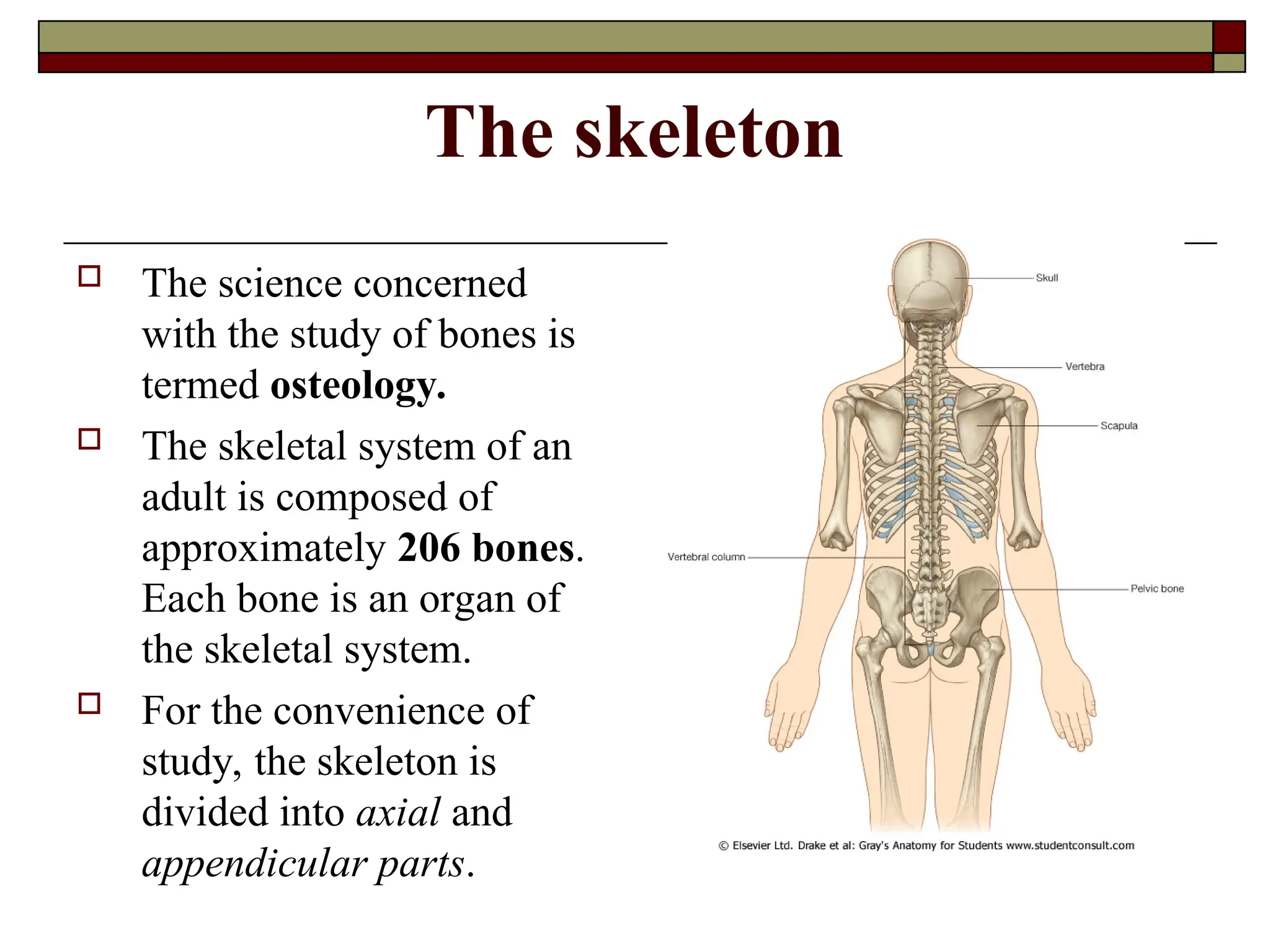
Thescience concerned
with the study of bones is
termed osteology.
The skeletal system of an
adult is composed of
approximately 206 bones.
Each bone is an organ of
the skeletal system.
For the convenience of
study, the skeleton is
divided into axial and
appendicular parts.
5.
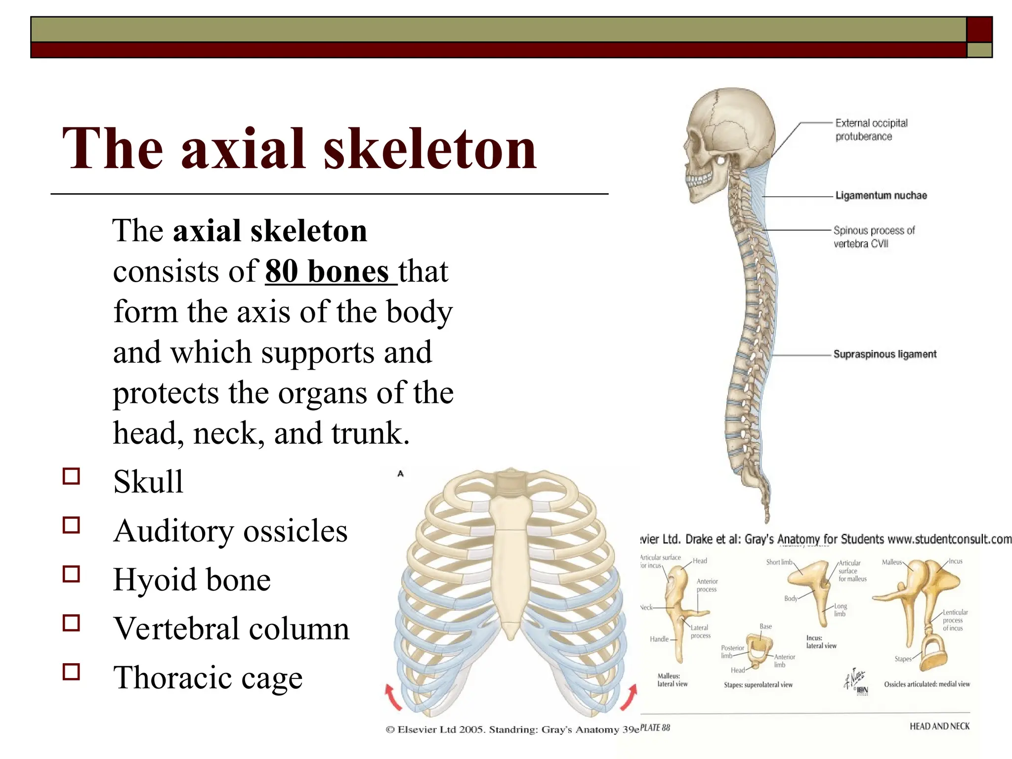
The axial skeleton
Theaxial skeleton
consists of 80 bones that
form the axis of the body
and which supports and
protects the organs of the
head, neck, and trunk.
Skull
Auditory ossicles
Hyoid bone
Vertebral column
Thoracic cage
6.
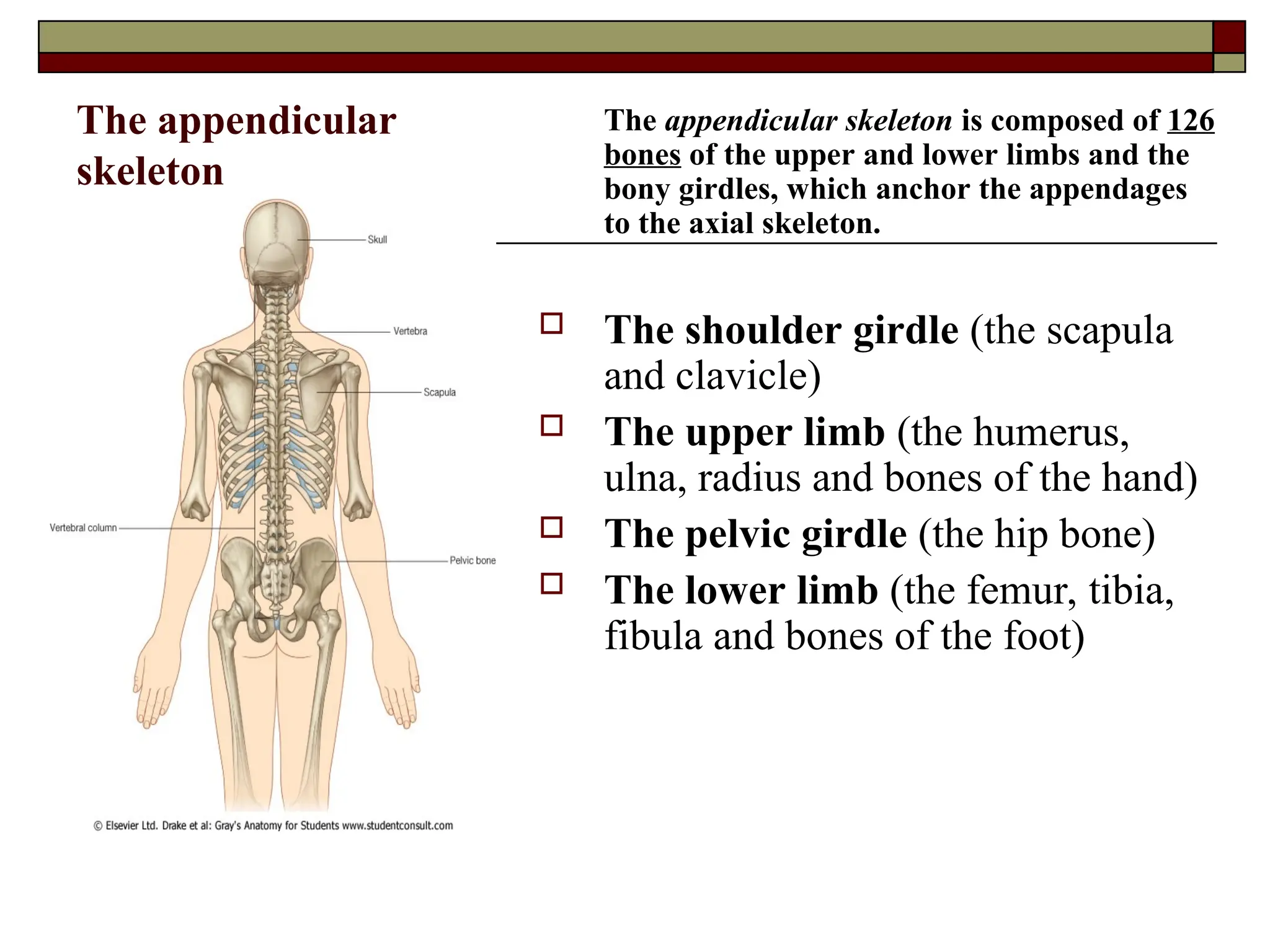
The appendicular
skeleton
The appendicularskeleton is composed of 126
bones of the upper and lower limbs and the
bony girdles, which anchor the appendages
to the axial skeleton.
The shoulder girdle (the scapula
and clavicle)
The upper limb (the humerus,
ulna, radius and bones of the hand)
The pelvic girdle (the hip bone)
The lower limb (the femur, tibia,
fibula and bones of the foot)
7.
BONE AS ANORGAN
STRUCTURE OF A BONE AND STRUCTURE OF THE
PERIOSTEUM
Bone (osis) is one of the hardest structures of
the body. It possesses also a certain degree of
toughness and elasticity. Its color, in a fresh
state, is pinkish-white externally, and red
within.
8.
Types of bonetissue
There are two types of bone tissue:
a) compact bony tissue
b) spongy bony tissue
The names imply that the two types differ in density, or how tightly the
tissue is packed together.
There are three types of cells that contribute to bone homeostasis.
a) osteoblasts are bone-forming cell
b) osteoclasts resorb or break down the bone
c) osteocytes are mature bone cells.
An equilibrium between osteoblasts and osteoclasts maintains the bone tissue.
9.
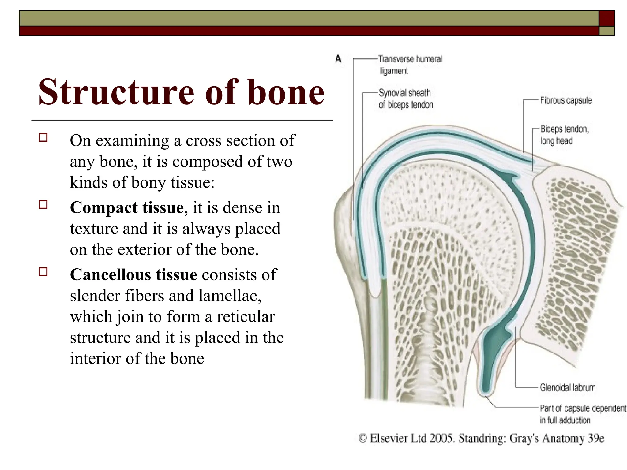
Structure of bone
On examining a cross section of
any bone, it is composed of two
kinds of bony tissue:
Compact tissue, it is dense in
texture and it is always placed
on the exterior of the bone.
Cancellous tissue consists of
slender fibers and lamellae,
which join to form a reticular
structure and it is placed in the
interior of the bone
10.
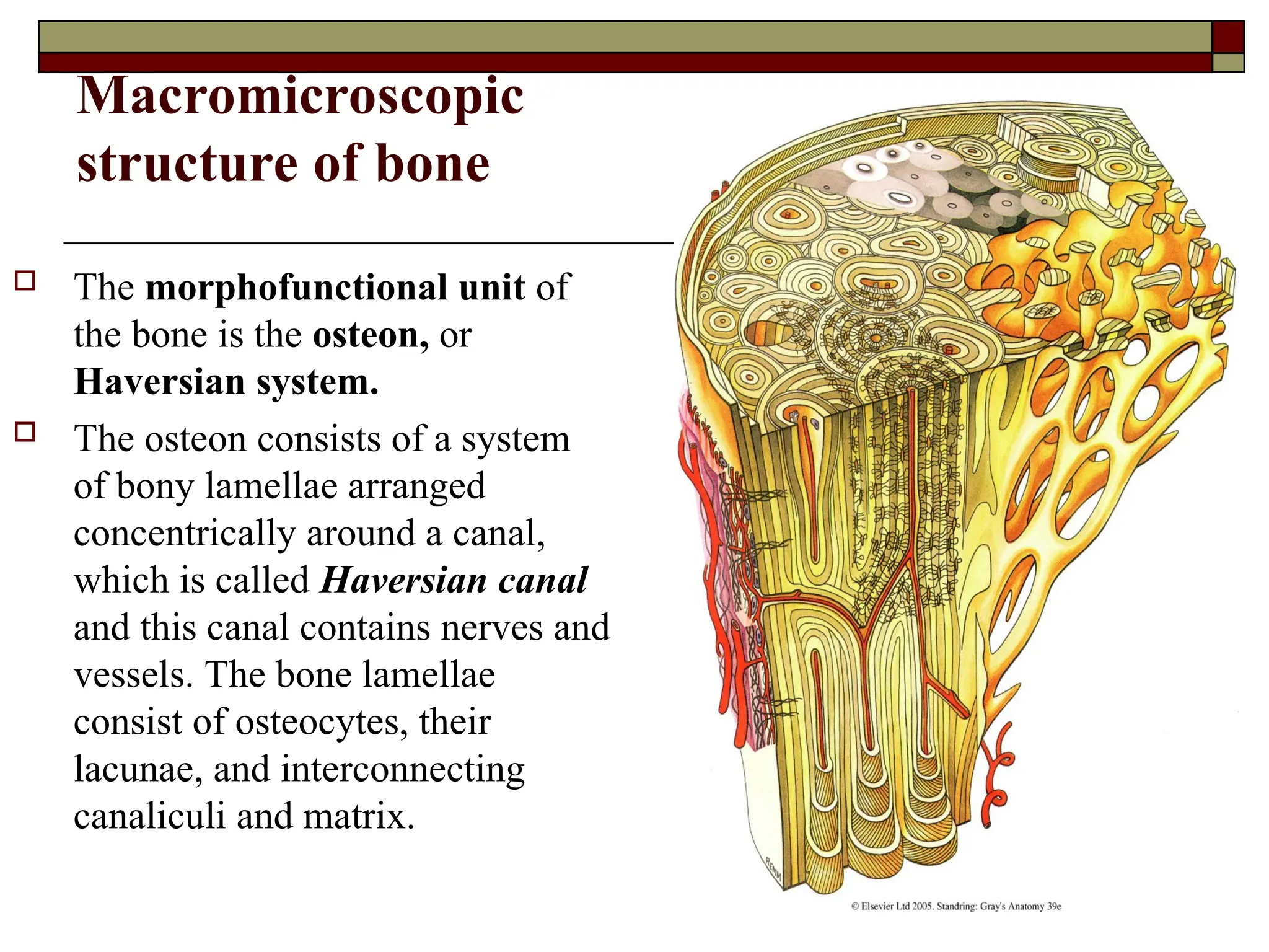
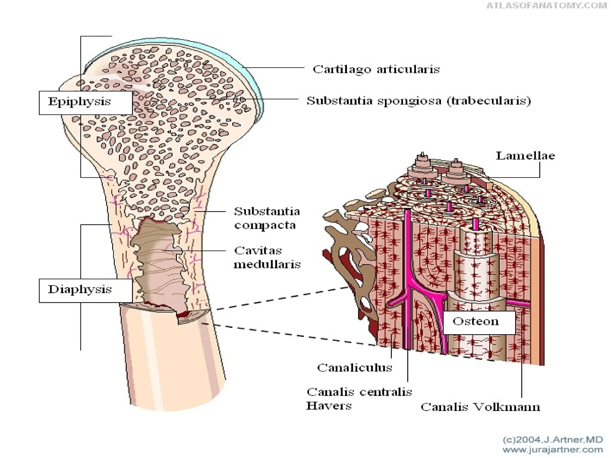
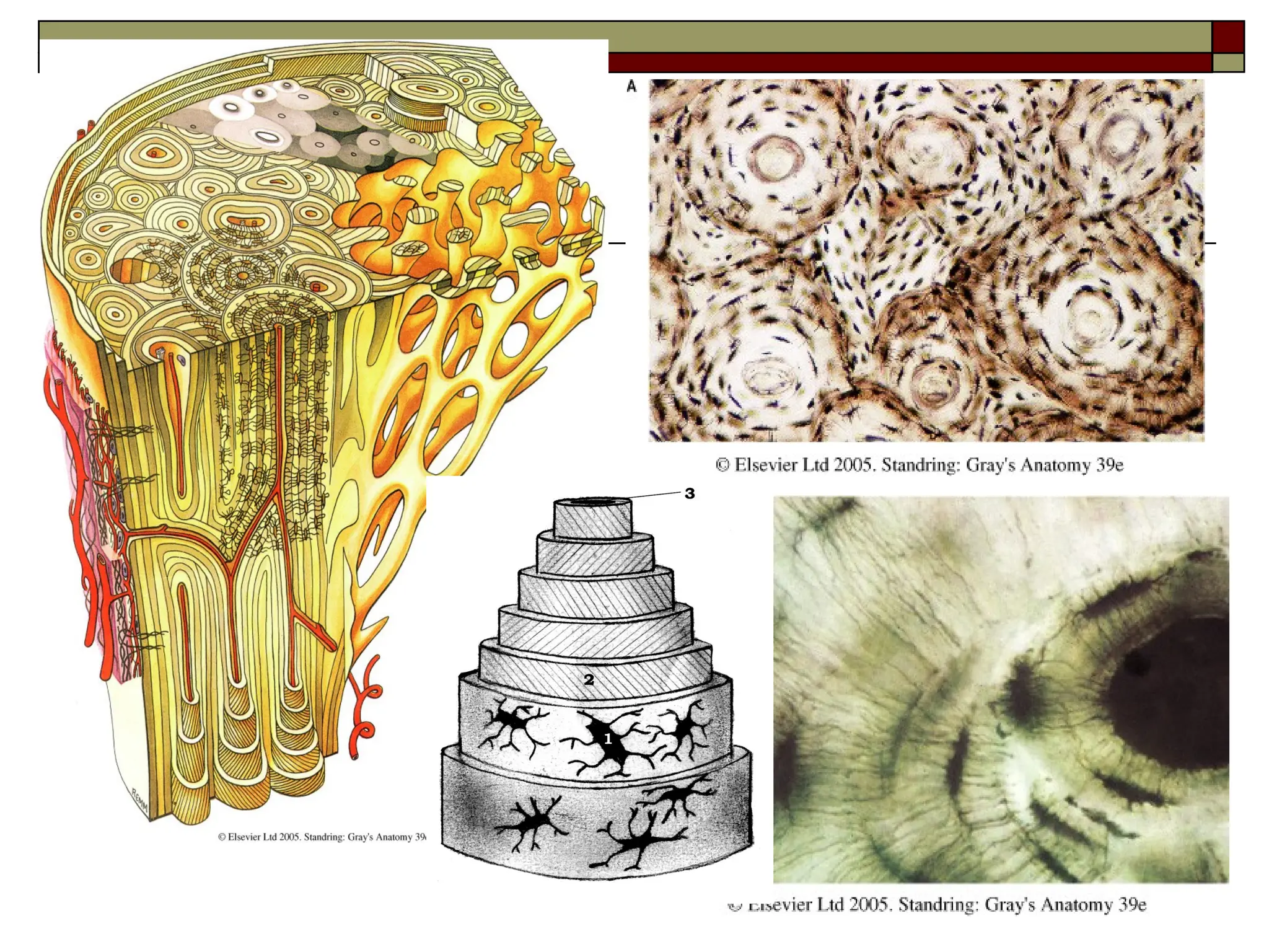
Macromicroscopic
structure of bone
The morphofunctional unit of
the bone is the osteon, or
Haversian system.
The osteon consists of a system
of bony lamellae arranged
concentrically around a canal,
which is called Haversian canal
and this canal contains nerves and
vessels. The bone lamellae
consist of osteocytes, their
lacunae, and interconnecting
canaliculi and matrix.
13.
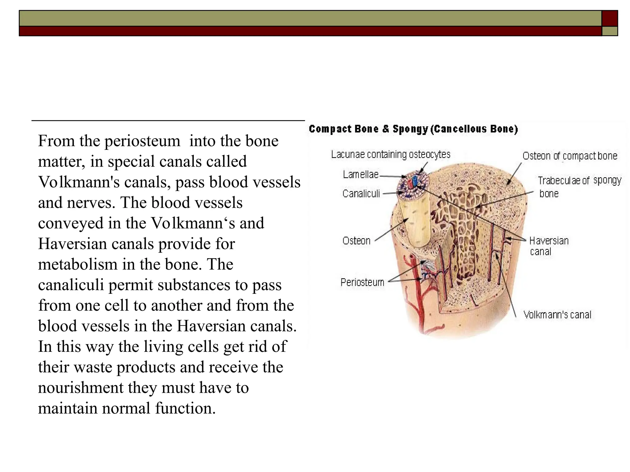
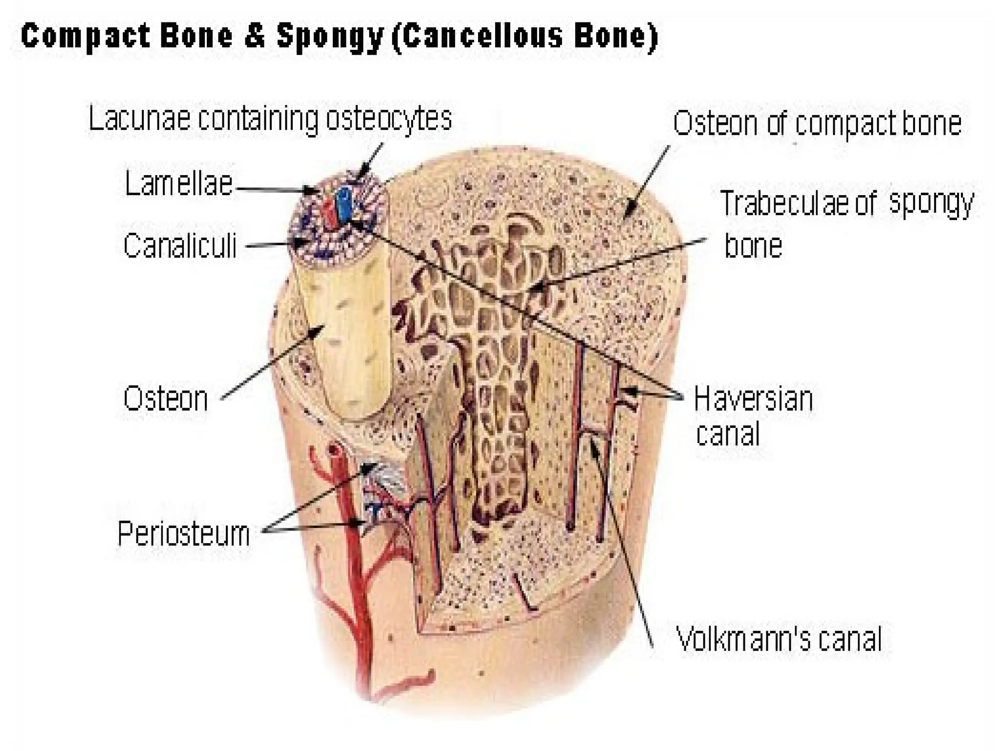
From the periosteuminto the bone
matter, in special canals called
Volkmann's canals, pass blood vessels
and nerves. The blood vessels
conveyed in the Volkmann‘s and
Haversian canals provide for
metabolism in the bone. The
canaliculi permit substances to pass
from one cell to another and from the
blood vessels in the Haversian canals.
In this way the living cells get rid of
their waste products and receive the
nourishment they must have to
maintain normal function.
15.
The spongy bonetissue
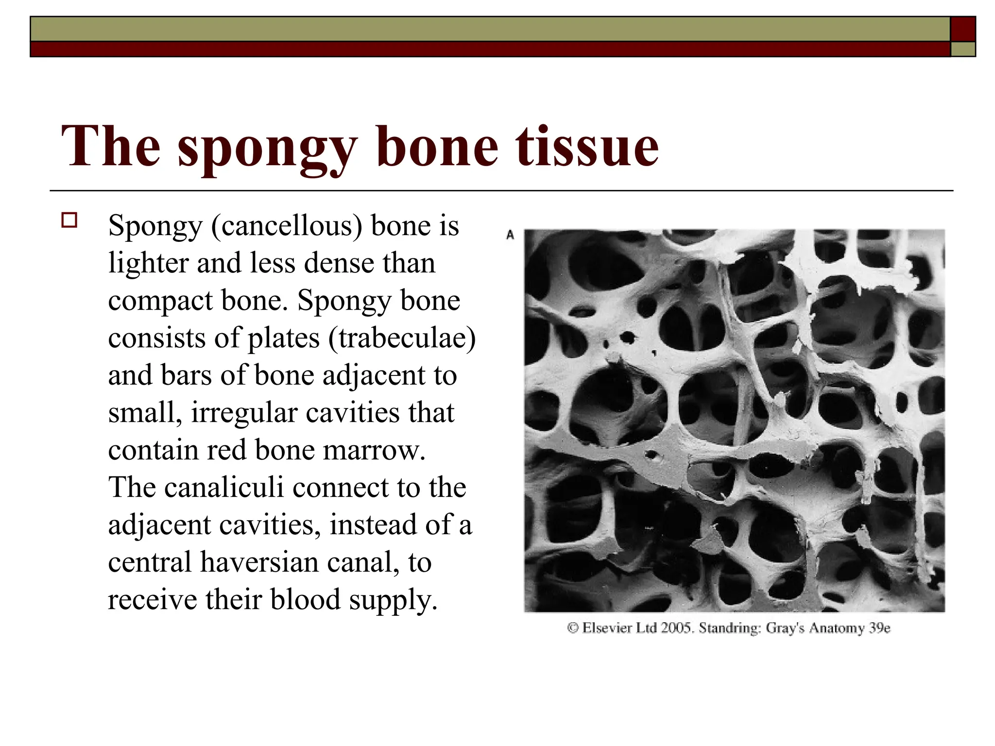
Spongy (cancellous) bone is
lighter and less dense than
compact bone. Spongy bone
consists of plates (trabeculae)
and bars of bone adjacent to
small, irregular cavities that
contain red bone marrow.
The canaliculi connect to the
adjacent cavities, instead of a
central haversian canal, to
receive their blood supply.
16.
The spongy bonetissue
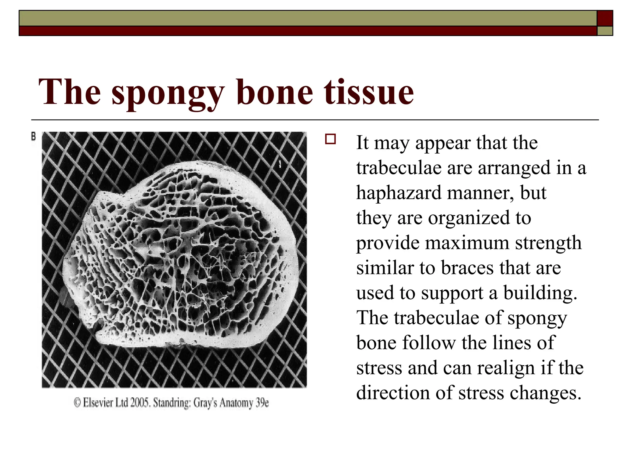
It may appear that the
trabeculae are arranged in a
haphazard manner, but
they are organized to
provide maximum strength
similar to braces that are
used to support a building.
The trabeculae of spongy
bone follow the lines of
stress and can realign if the
direction of stress changes.
17.
The periosteum
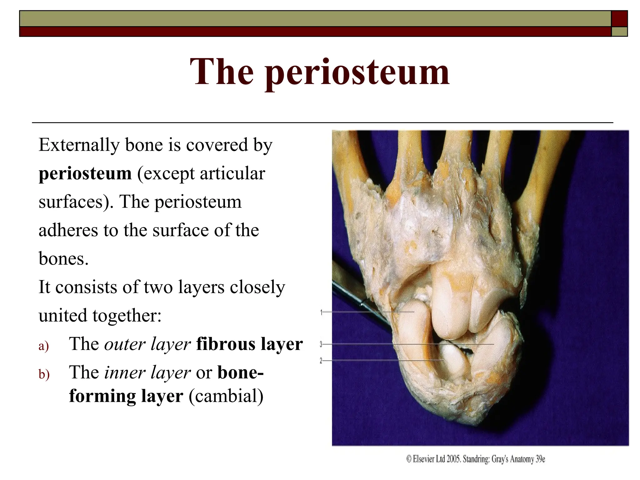
Externally boneis covered by
periosteum (except articular
surfaces). The periosteum
adheres to the surface of the
bones.
It consists of two layers closely
united together:
a) The outer layer fibrous layer
b) The inner layer or bone-
forming layer (cambial)
18.
Structure of
the periosteum
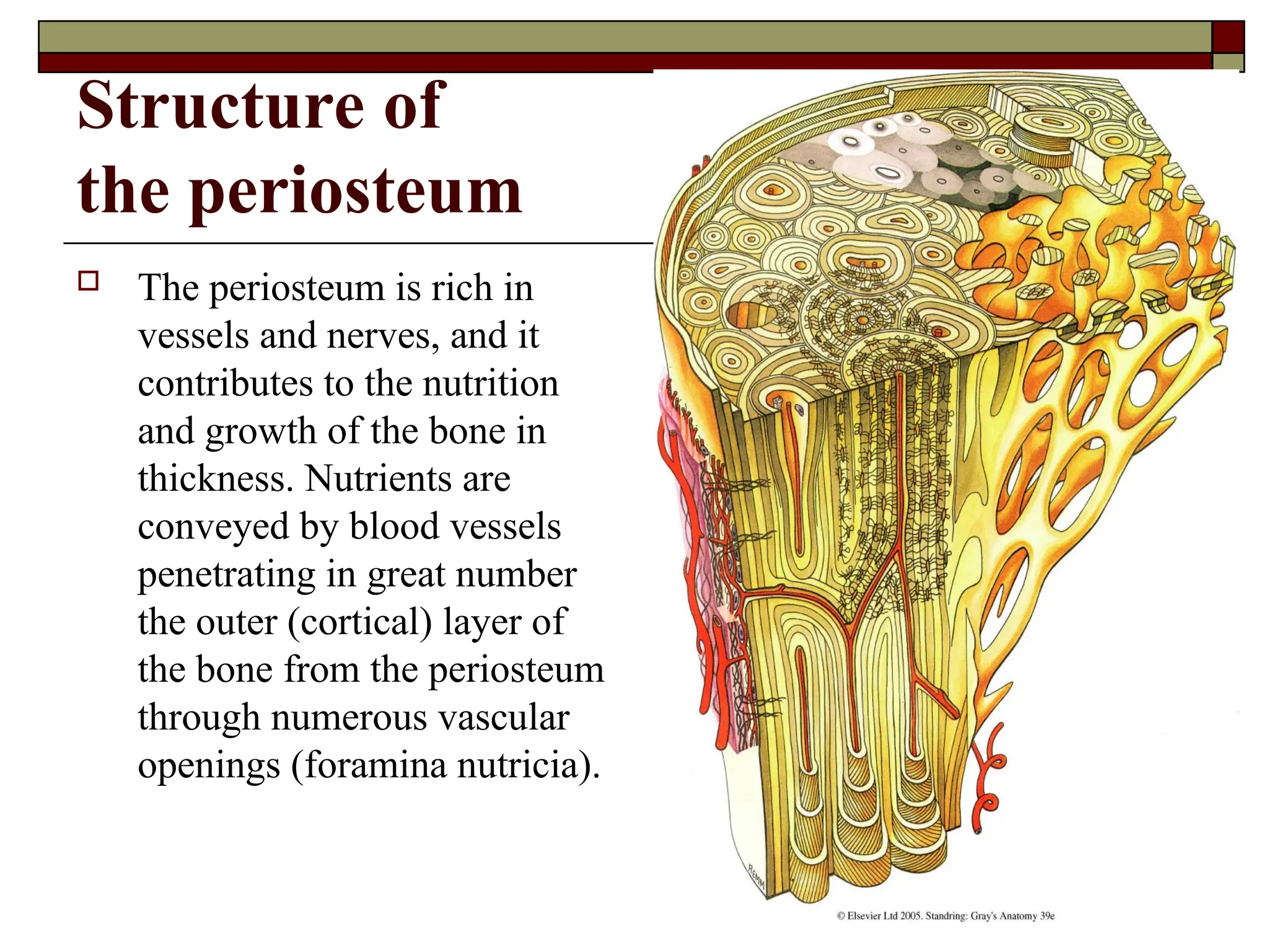
The periosteum is rich in
vessels and nerves, and it
contributes to the nutrition
and growth of the bone in
thickness. Nutrients are
conveyed by blood vessels
penetrating in great number
the outer (cortical) layer of
the bone from the periosteum
through numerous vascular
openings (foramina nutricia).
19.
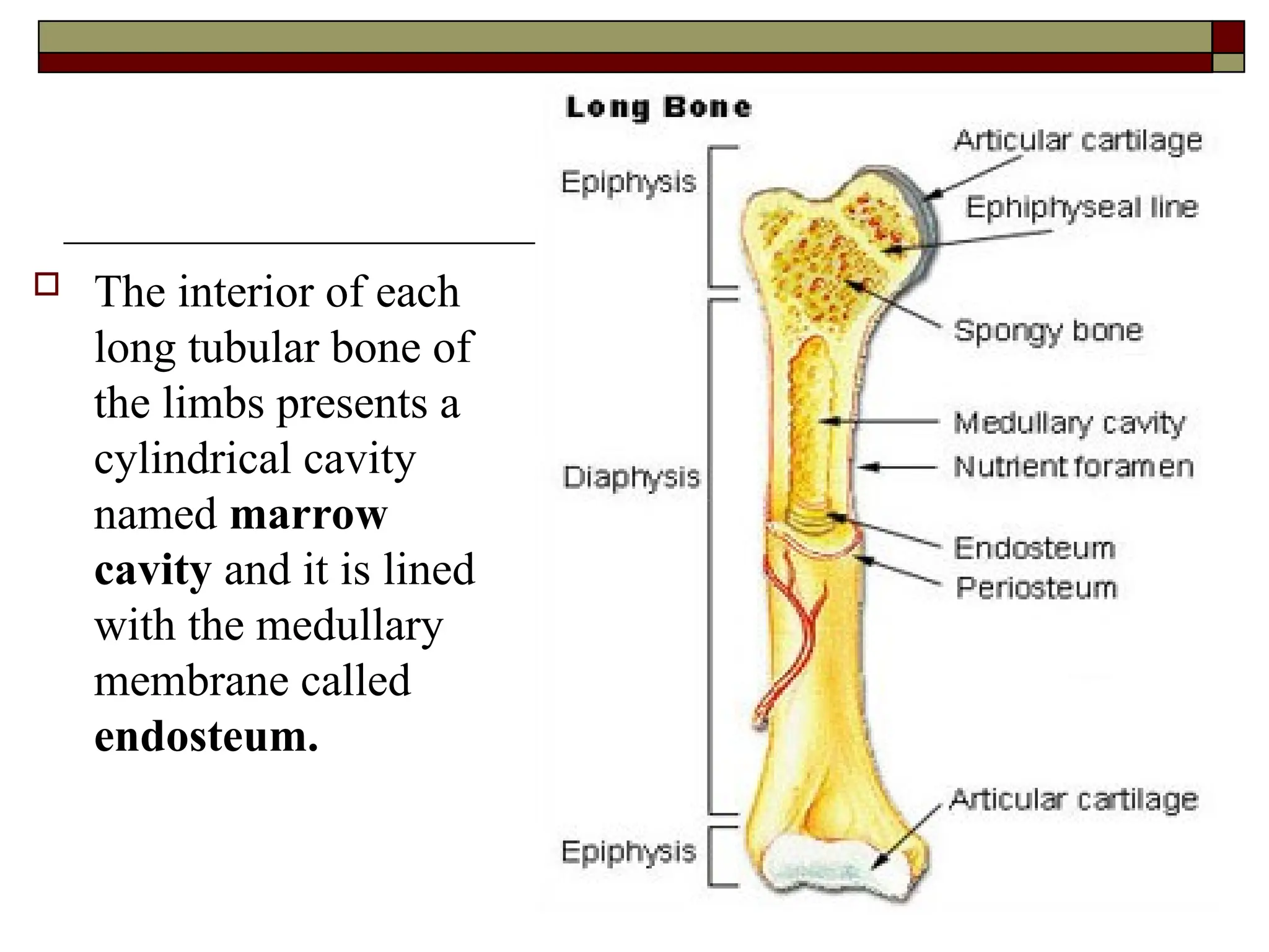
The interiorof each
long tubular bone of
the limbs presents a
cylindrical cavity
named marrow
cavity and it is lined
with the medullary
membrane called
endosteum.
20.
CHEMICAL COMPOSITION ANDPHYSICAL
PROPERTIES OF BONE
Bone matter consists of two types of chemical material:
Organic – 1/3, mainly ossein (it provides elasticity
to the bone).
Inorganic – 2/3, mainly calcium phosphate in
particular 51.04% (provides hardness to the bone).
The bone contains vitamins A, D and C. A lack of
salts or vitamin D in the period of growth reduces
bone hardness and causes deformities of bones
(rickets) in children. Vitamin A deficiency leads to
abnormal thickness of bones, and the bone cavities
and canals become empty.
21.
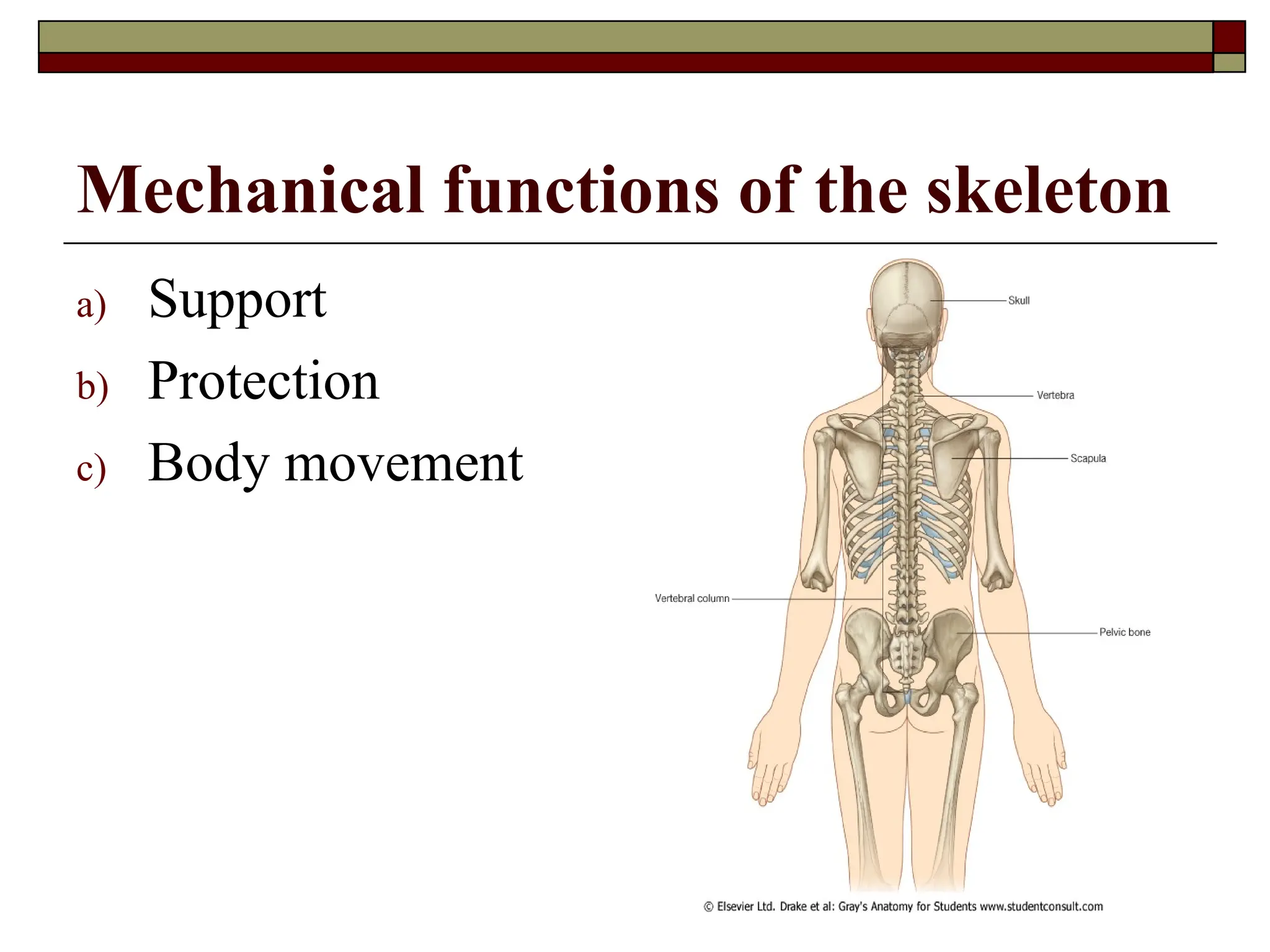
Functions of theskeleton
Biological functions
Mechanical functions
Bone marrow
Thebony compartments contain bony marrow,
medulla ossium. Two types of bone marrow can be
distinguished:
red bone marrow
white bone marrow
The white or yellow marrow fills up the medullary
cavities of the shafts of the long tubular bones.
The red marrow is located within the cancellous
tissue and extends into the larger bony canals
(Haversian canals) that contain blood vessels.
24.
Haemopoiesis function
The bonemarrow provides
haemopoiesis function and biological
protection of the organism. It takes part
in nutrition, development and growth of
the bone. The red marrow concerned
with haemopoiesis and bone formation,
has an active role in the healing of
fractures. Red marrow predominates in
infants and in children, with growth of
child the red marrow is gradually
replaced by yellow marrow.
NB: The
bones of the
embryo and
new-born
contain only
red marrow.
25.
Haemopoiesis function
Thered bone marrow of an adult produces white blood
cells, red blood cells, and platelets.
In an infant, the spleen and liver produce red blood
cells, but as the bones mature, the bone marrow
performs this task.
It is estimated that an average of 1 million blood cells
are produced every second by the bone marrow to
replace those that are worn out and destroyed by the
liver.
26.
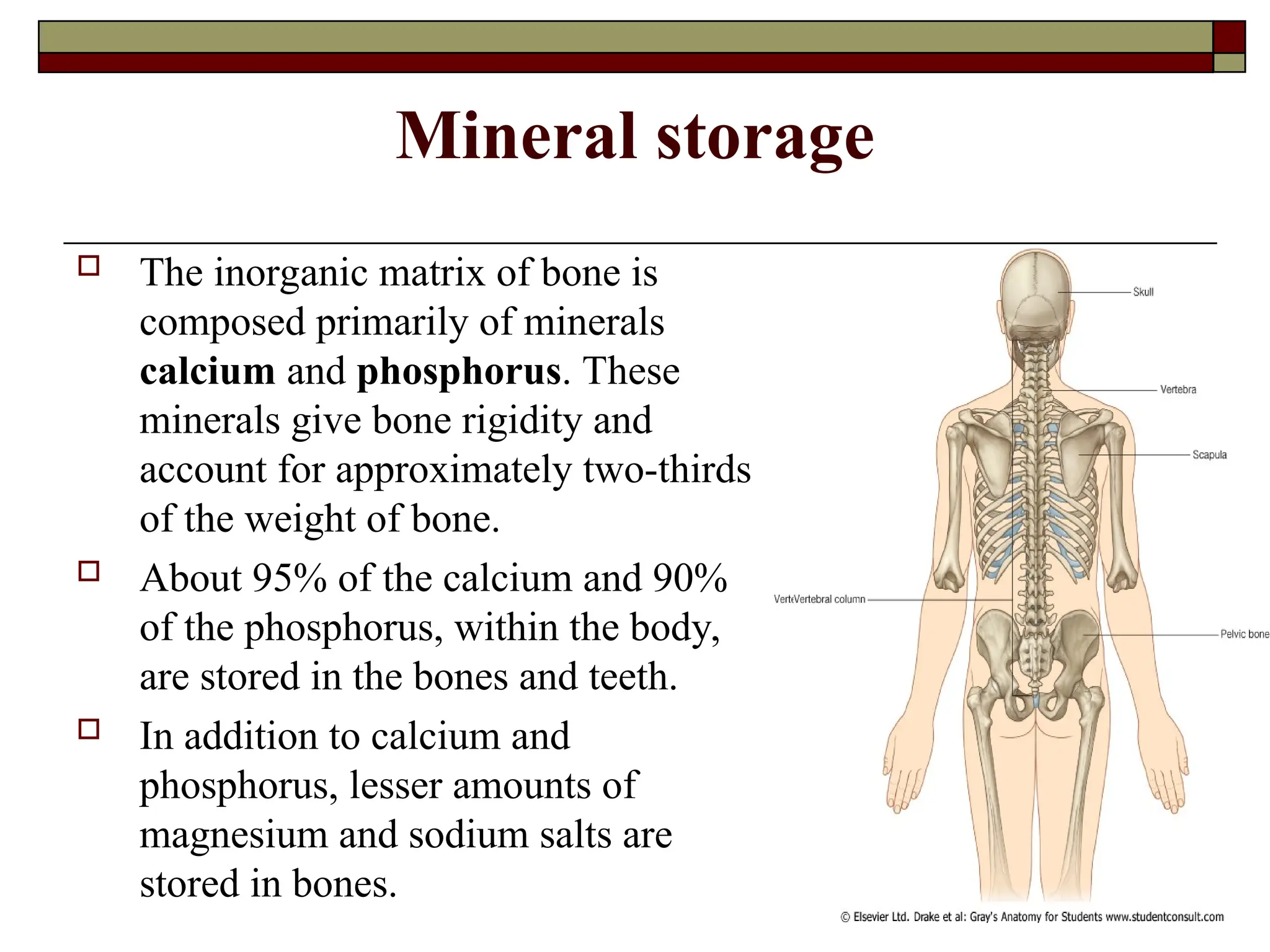
Mineral storage
Theinorganic matrix of bone is
composed primarily of minerals
calcium and phosphorus. These
minerals give bone rigidity and
account for approximately two-thirds
of the weight of bone.
About 95% of the calcium and 90%
of the phosphorus, within the body,
are stored in the bones and teeth.
In addition to calcium and
phosphorus, lesser amounts of
magnesium and sodium salts are
stored in bones.
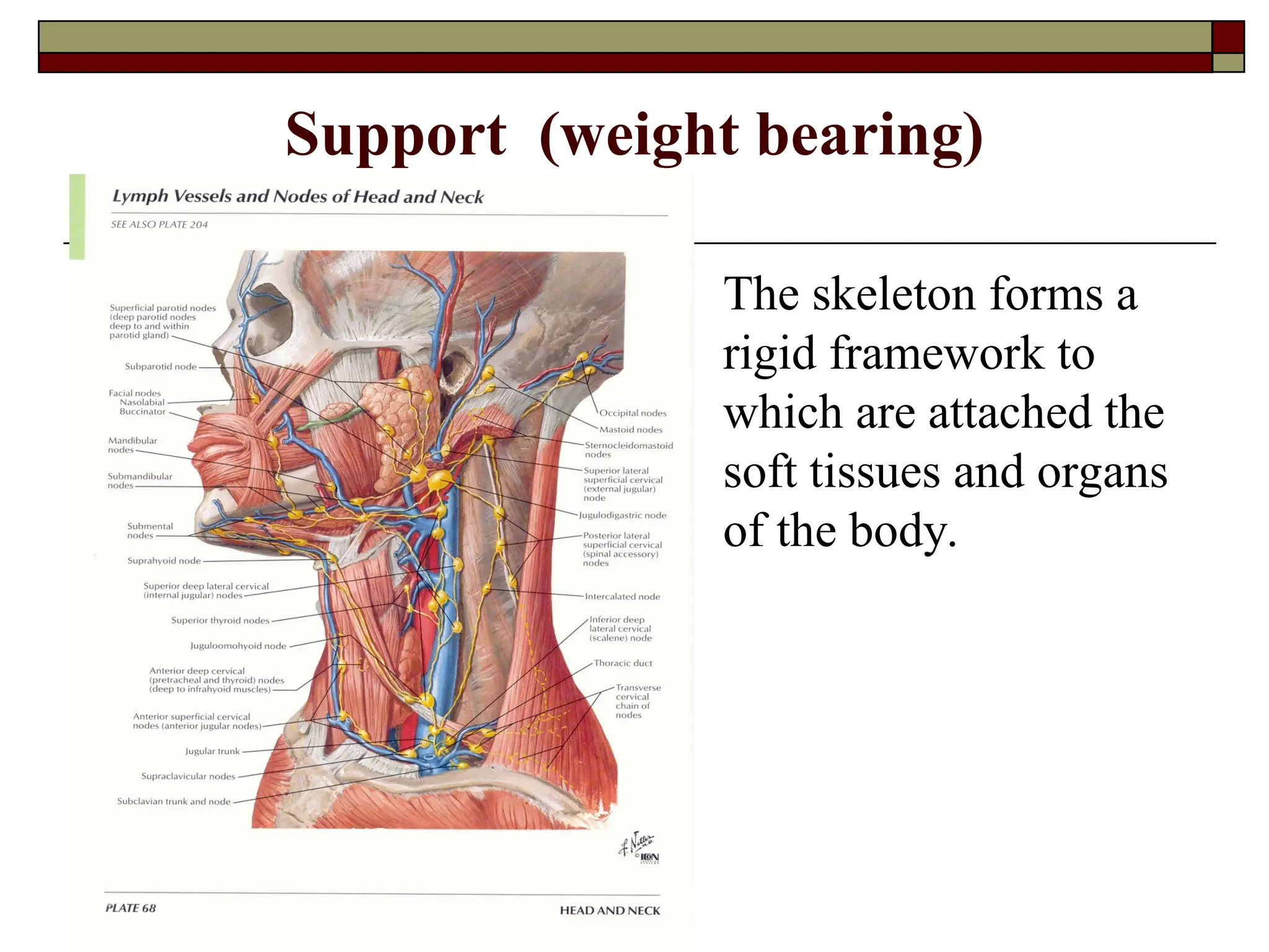
Support (weight bearing)
The skeleton forms a
rigid framework to
which are attached the
soft tissues and organs
of the body.
29.
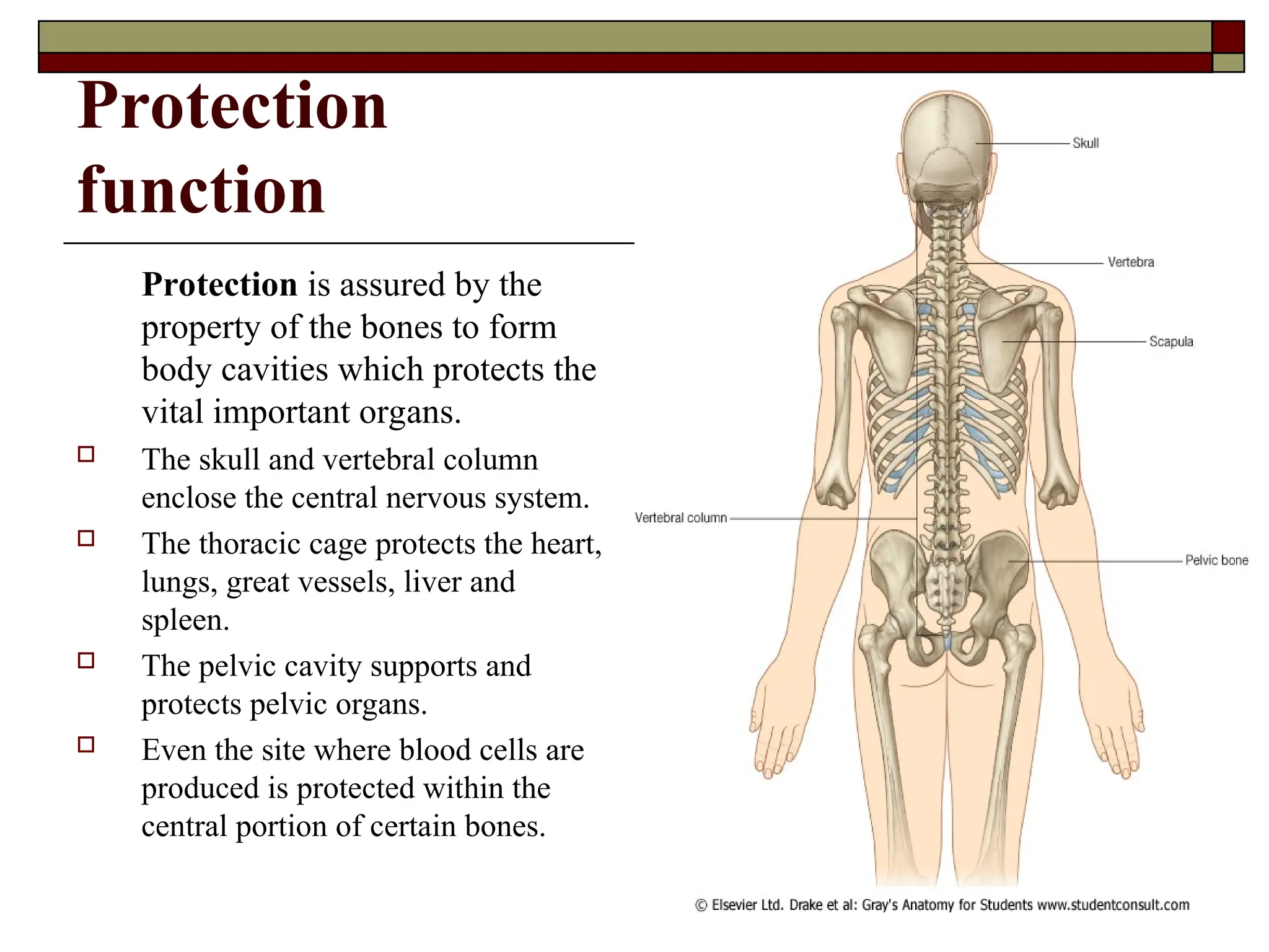
Protection
function
Protection is assuredby the
property of the bones to form
body cavities which protects the
vital important organs.
The skull and vertebral column
enclose the central nervous system.
The thoracic cage protects the heart,
lungs, great vessels, liver and
spleen.
The pelvic cavity supports and
protects pelvic organs.
Even the site where blood cells are
produced is protected within the
central portion of certain bones.
30.
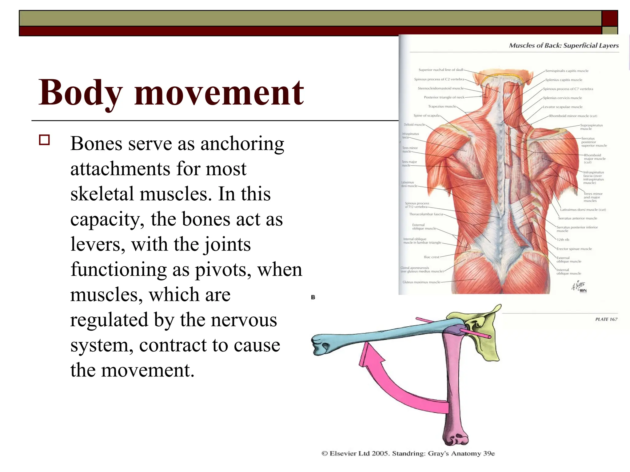
Body movement
Bonesserve as anchoring
attachments for most
skeletal muscles. In this
capacity, the bones act as
levers, with the joints
functioning as pivots, when
muscles, which are
regulated by the nervous
system, contract to cause
the movement.
31.
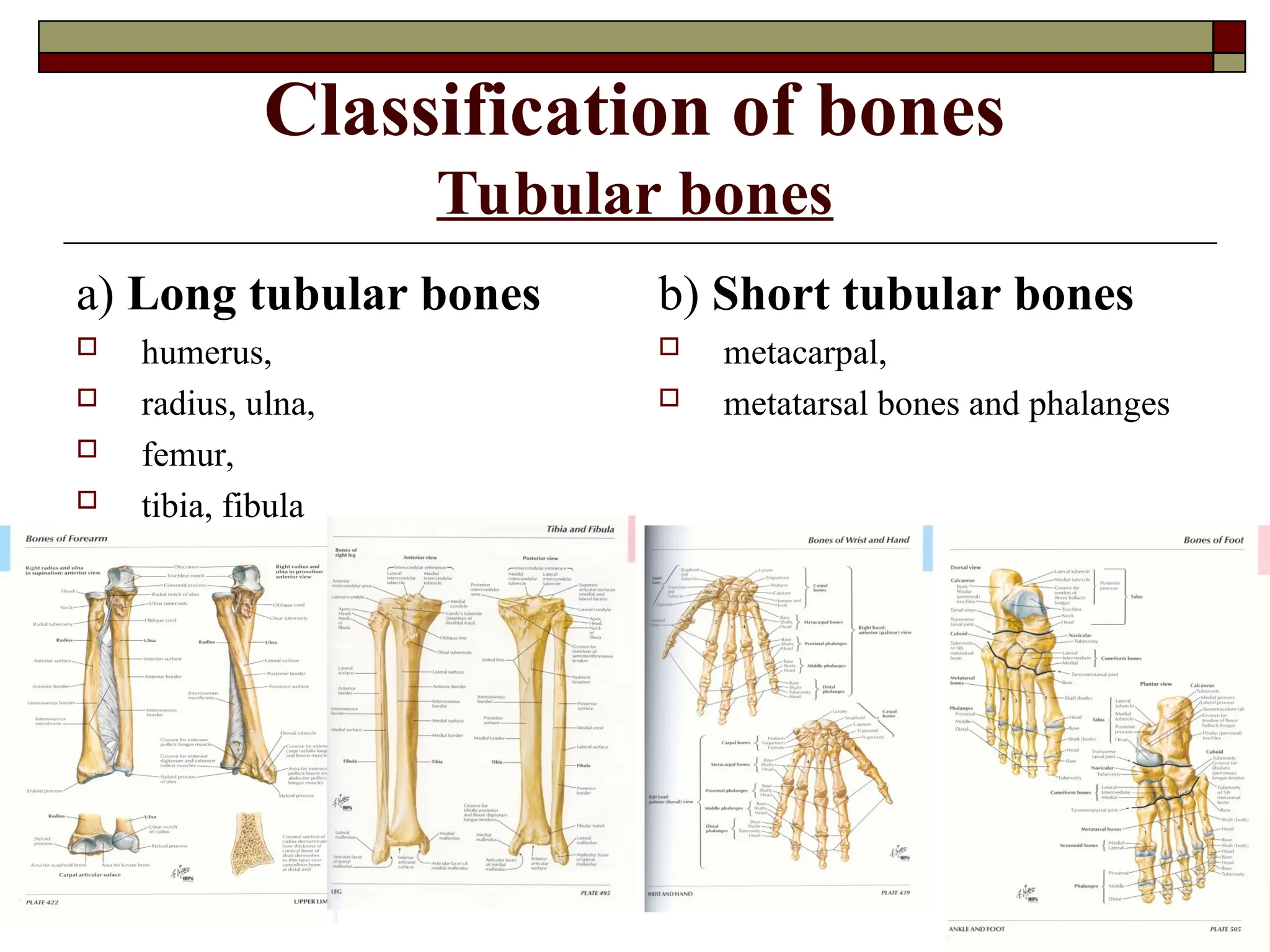
Classification of bones
Tubularbones
a) Long tubular bones
humerus,
radius, ulna,
femur,
tibia, fibula
b) Short tubular bones
metacarpal,
metatarsal bones and phalanges
32.
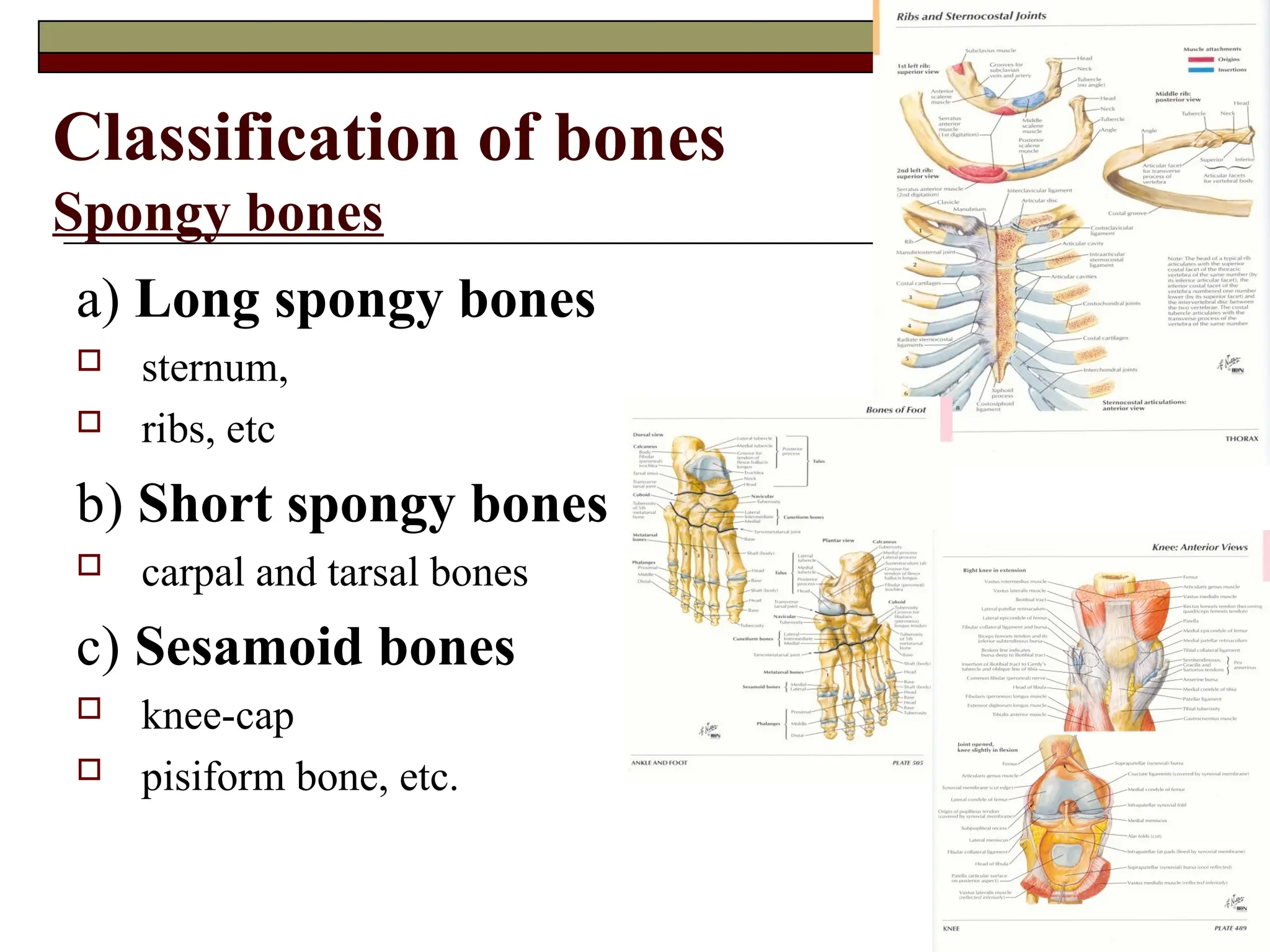
Classification of bones
Spongybones
a) Long spongy bones
sternum,
ribs, etc
b) Short spongy bones
carpal and tarsal bones
c) Sesamoid bones
knee-cap
pisiform bone, etc.
33.
Classification of bones
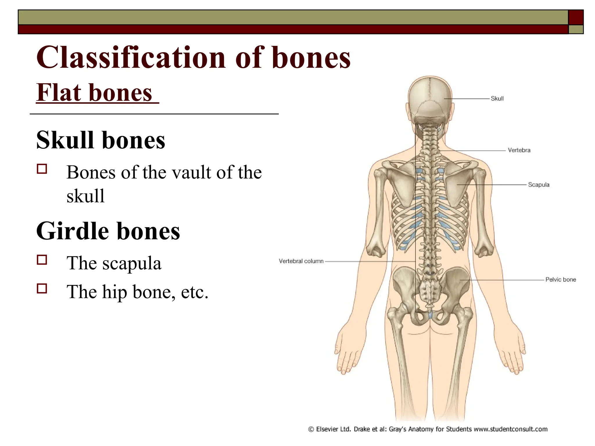
Flatbones
Skull bones
Bones of the vault of the
skull
Girdle bones
The scapula
The hip bone, etc.
34.
Classification of bones
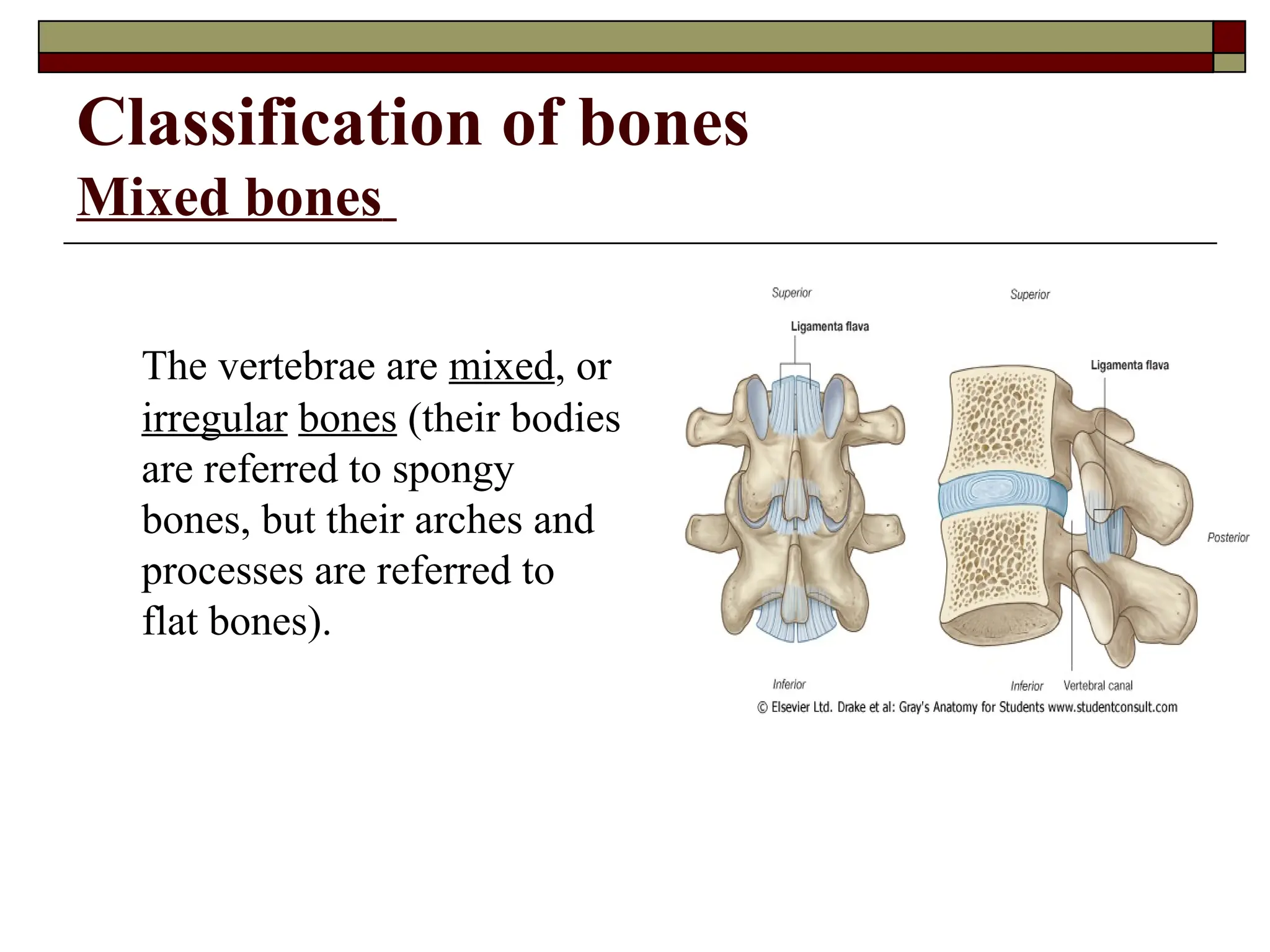
Mixedbones
The vertebrae are mixed, or
irregular bones (their bodies
are referred to spongy
bones, but their arches and
processes are referred to
flat bones).
35.
Classification of bonesdependent on
their development
a) Desmal (tegumentary, or primary bones)
b) Condral (secondary bone)
c) Condro-desmal bone (the vertebrae, the
bones of the base of the skull, the clavicle)
36.
GENERAL NOTIONS CONCERNINGDEVELOPMENT OF BONES
AND THEIR ABNORMALITIES
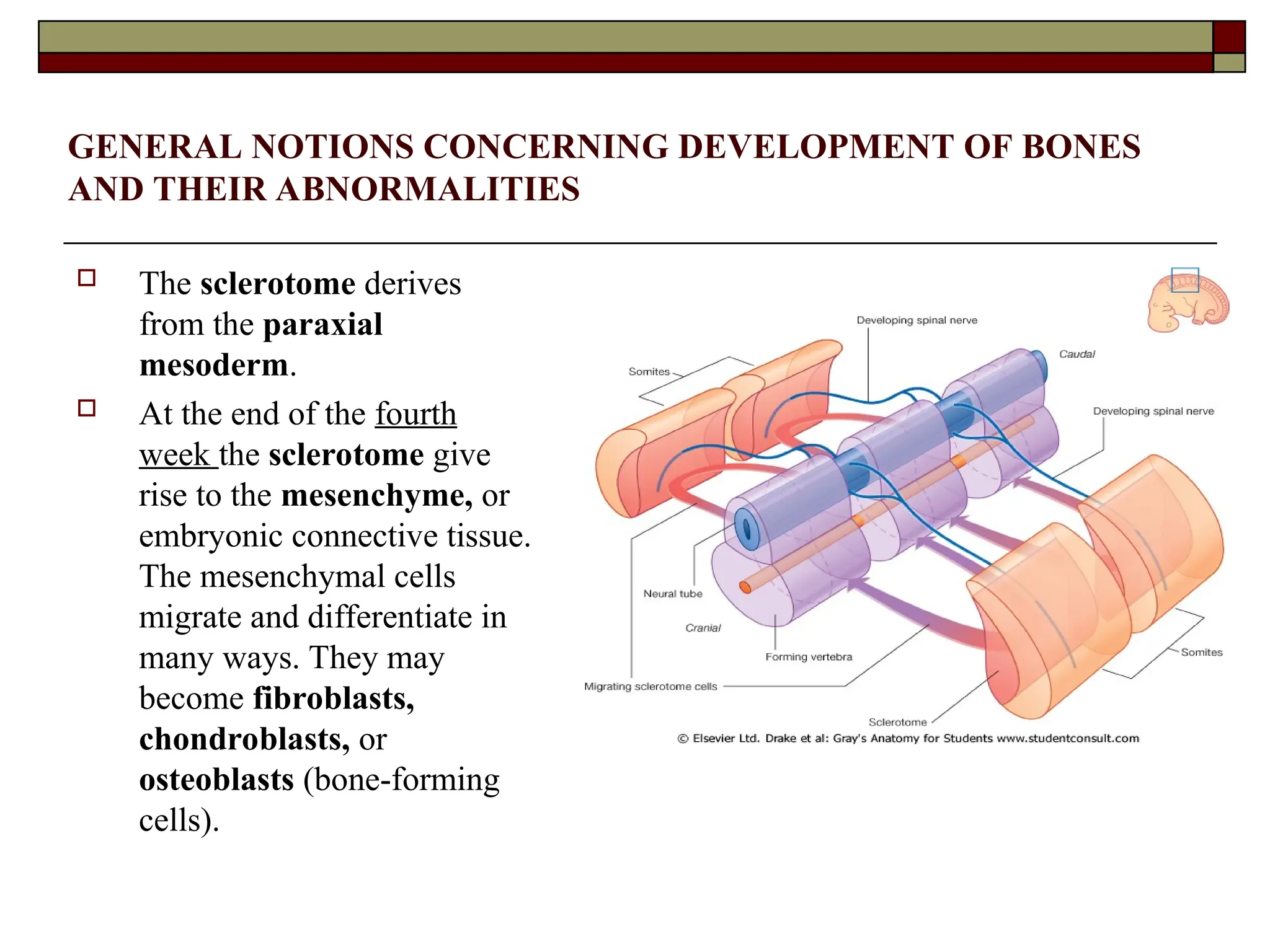
The sclerotome derives
from the paraxial
mesoderm.
At the end of the fourth
week the sclerotome give
rise to the mesenchyme, or
embryonic connective tissue.
The mesenchymal cells
migrate and differentiate in
many ways. They may
become fibroblasts,
chondroblasts, or
osteoblasts (bone-forming
cells).
37.
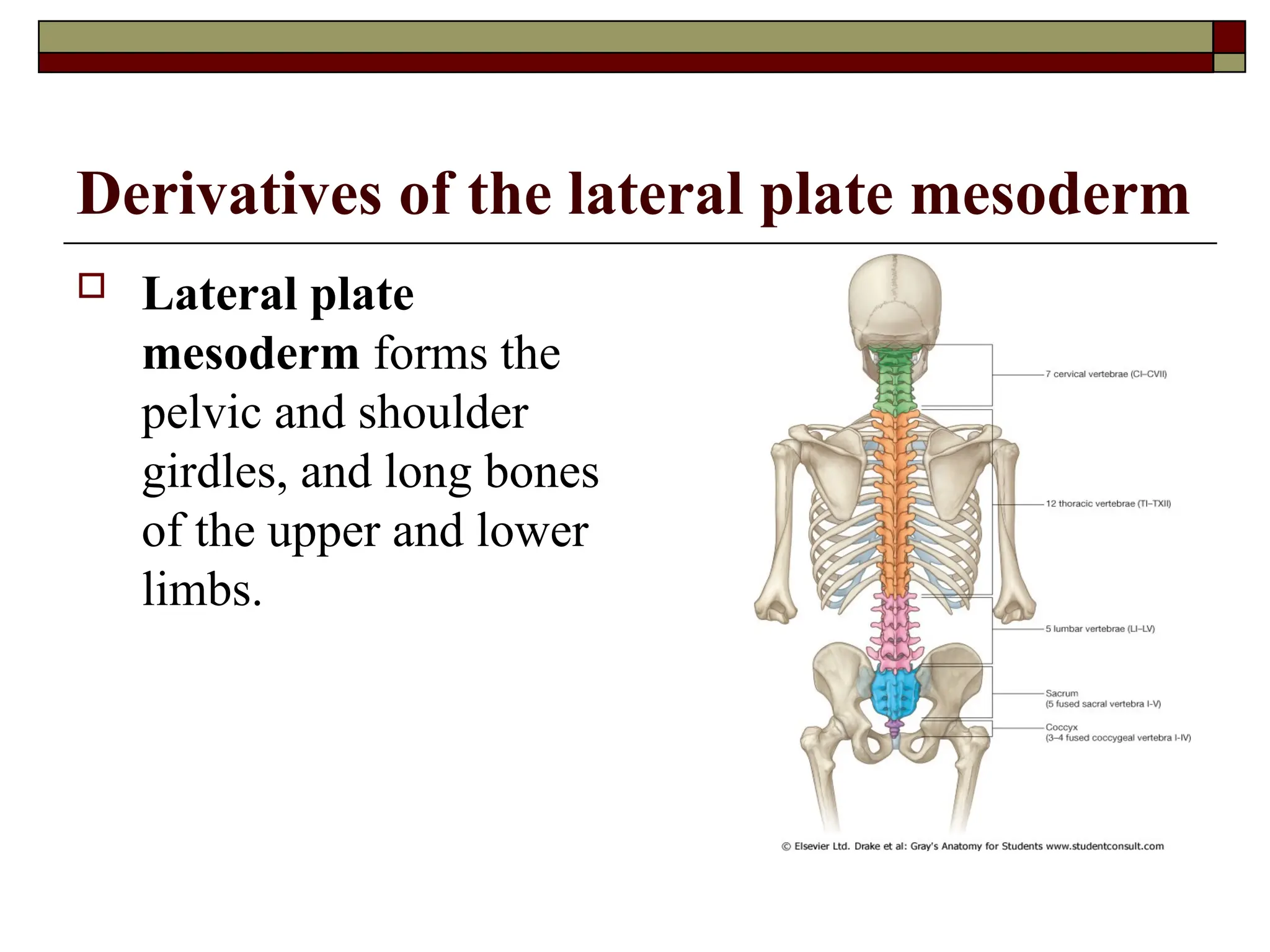
Derivatives of thelateral plate mesoderm
Lateral plate
mesoderm forms the
pelvic and shoulder
girdles, and long bones
of the upper and lower
limbs.
38.
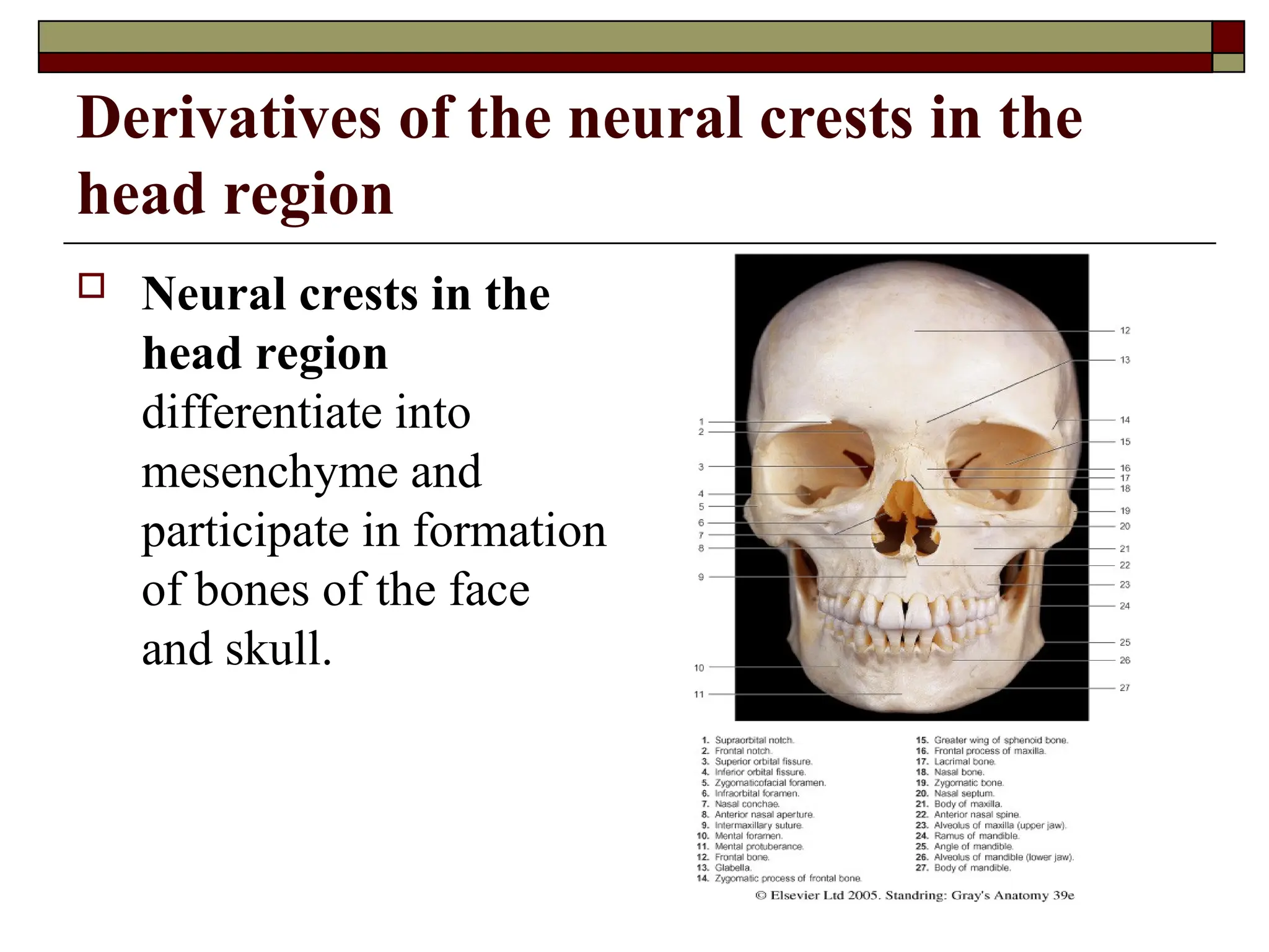
Derivatives of theneural crests in the
head region
Neural crests in the
head region
differentiate into
mesenchyme and
participate in formation
of bones of the face
and skull.
39.
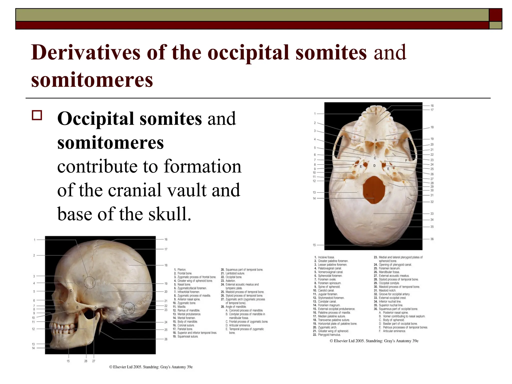
Derivatives of theoccipital somites and
somitomeres
Occipital somites and
somitomeres
contribute to formation
of the cranial vault and
base of the skull.
40.
Stages of developmentof the human
skeleton
Bone formation, or ossification, begins at about the fourth
week of embryonic development, but ossification centers
cannot be readily observed until about the tenth week.
Three stages of development of the human skeleton are
encountered:
Connective-tissue (membranous)
Cartilaginous
Bony
NB: Bones which do not go through the cartilaginous stage of
development are called membrane, or primary bones.
That bones which during their development undergo through all
three stages of development are called secondary bones.
41.
THE LOWS GOVERNINGTHE DEVELOPMENT OF THE
BONES AND THEIR ABNORMALITIES
According to the three developmental stages of the
skeleton bones may develop from connective or
cartilaginous tissue. Four types of ossification
(osteogenesis) are distinguished:
Intramembranous
Perichondral
Periosteal
Encondral, or endochondral
42.
Intramembranous or endesmal
ossification
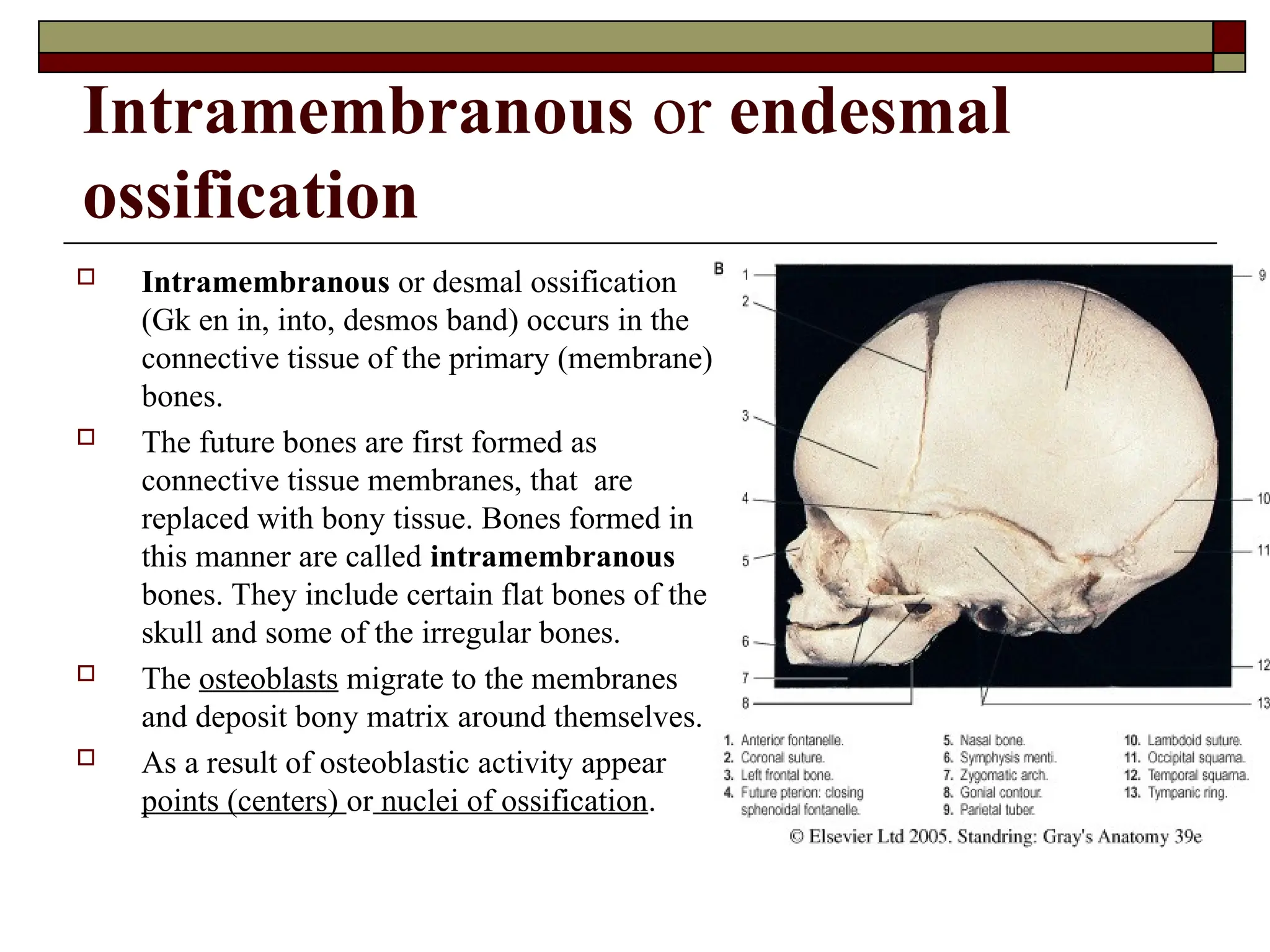
Intramembranous or desmal ossification
(Gk en in, into, desmos band) occurs in the
connective tissue of the primary (membrane)
bones.
The future bones are first formed as
connective tissue membranes, that are
replaced with bony tissue. Bones formed in
this manner are called intramembranous
bones. They include certain flat bones of the
skull and some of the irregular bones.
The osteoblasts migrate to the membranes
and deposit bony matrix around themselves.
As a result of osteoblastic activity appear
points (centers) or nuclei of ossification.
43.
Perichondral ossification(Gk peri around,
chondros cartilage) takes place on the outer
surface of the cartilaginous bone germs with
the participation of the perichondrium. The
perichondral osteoblasts covering the
cartilage replace the cartilaginous tissue
gradually and form a compact bony
substance.
44.
With theconversion of the cartilaginous
model to a bone model, the perichondrium
becomes the periosteum, and further
deposition of bone tissue is accomplished by
the periosteum; this is periosteal
ossification. The perichondral and periosteal
types of ossification are therefore connected
and one follows the other chronologically.
45.
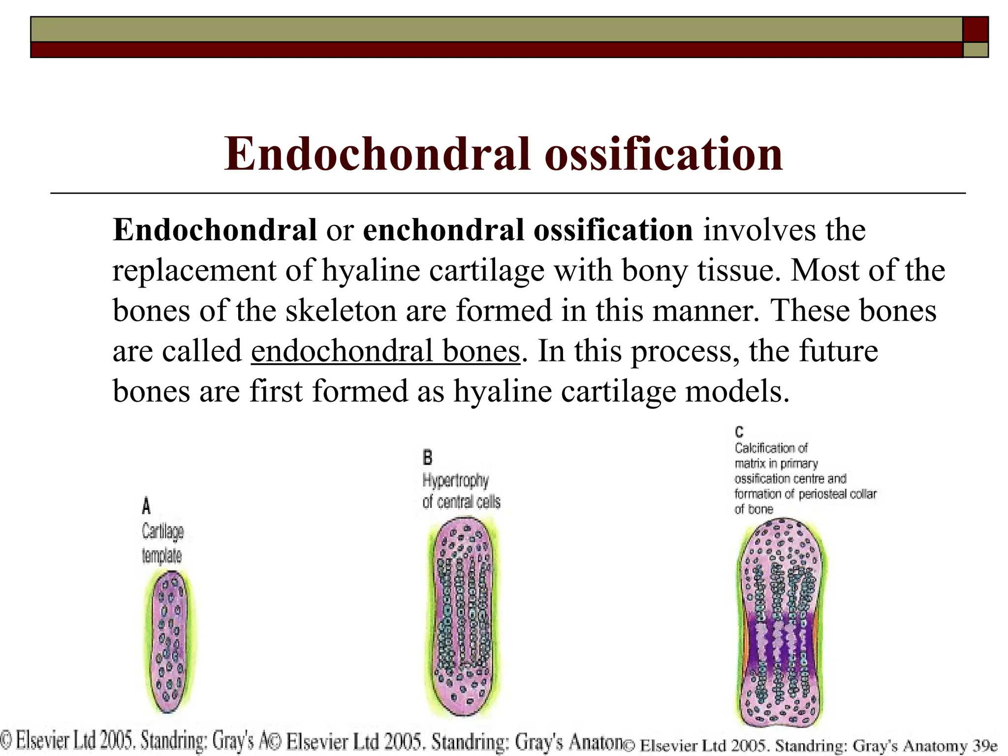
Endochondral ossification
Endochondral orenchondral ossification involves the
replacement of hyaline cartilage with bony tissue. Most of the
bones of the skeleton are formed in this manner. These bones
are called endochondral bones. In this process, the future
bones are first formed as hyaline cartilage models.
46.
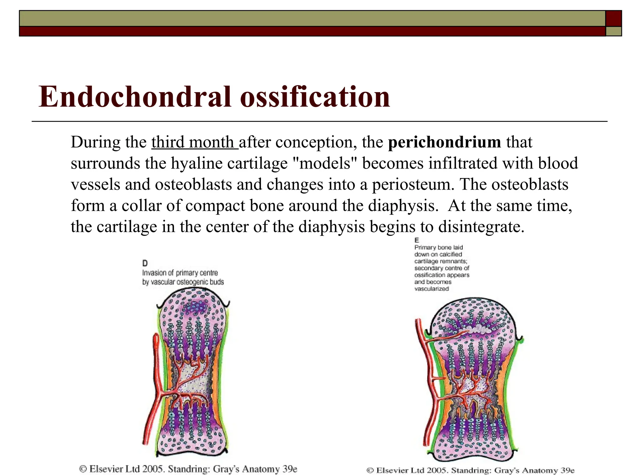
Endochondral ossification
During thethird month after conception, the perichondrium that
surrounds the hyaline cartilage "models" becomes infiltrated with blood
vessels and osteoblasts and changes into a periosteum. The osteoblasts
form a collar of compact bone around the diaphysis. At the same time,
the cartilage in the center of the diaphysis begins to disintegrate.
47.
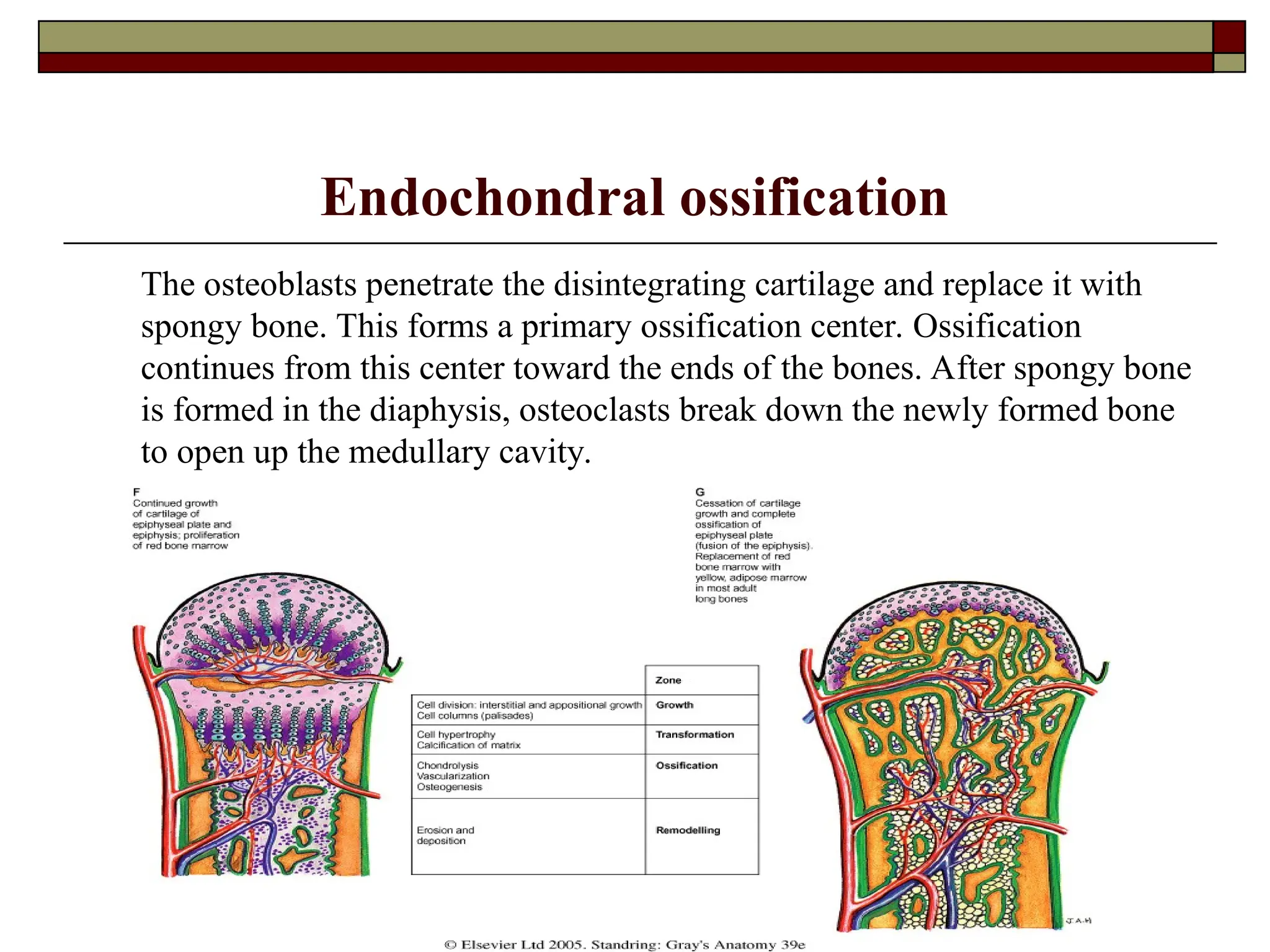
Endochondral ossification
The osteoblastspenetrate the disintegrating cartilage and replace it with
spongy bone. This forms a primary ossification center. Ossification
continues from this center toward the ends of the bones. After spongy bone
is formed in the diaphysis, osteoclasts break down the newly formed bone
to open up the medullary cavity.
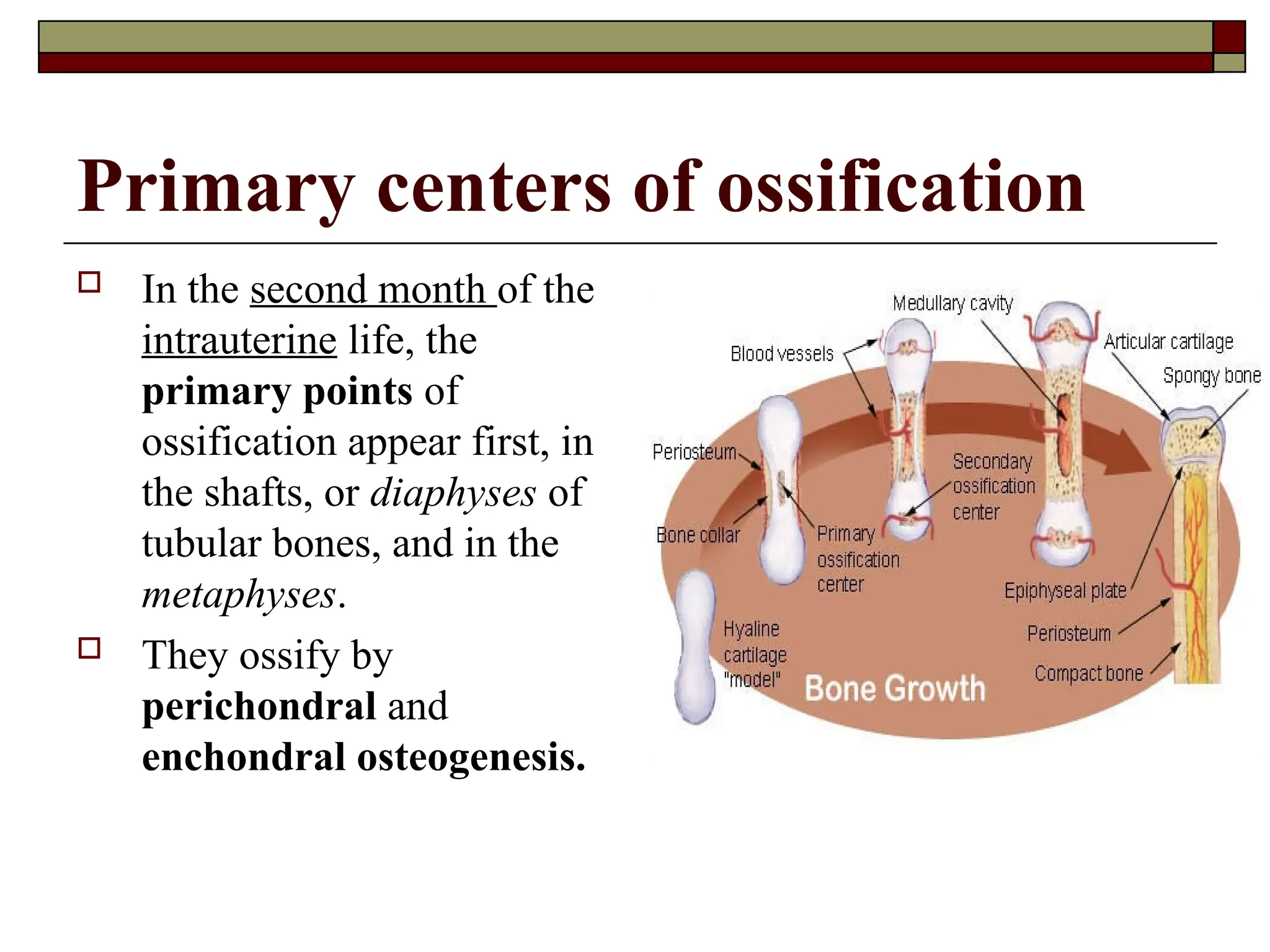
48.
Primary centers ofossification
In the second month of the
intrauterine life, the
primary points of
ossification appear first, in
the shafts, or diaphyses of
tubular bones, and in the
metaphyses.
They ossify by
perichondral and
enchondral osteogenesis.
49.
Secondary and accessory
pointsof ossification
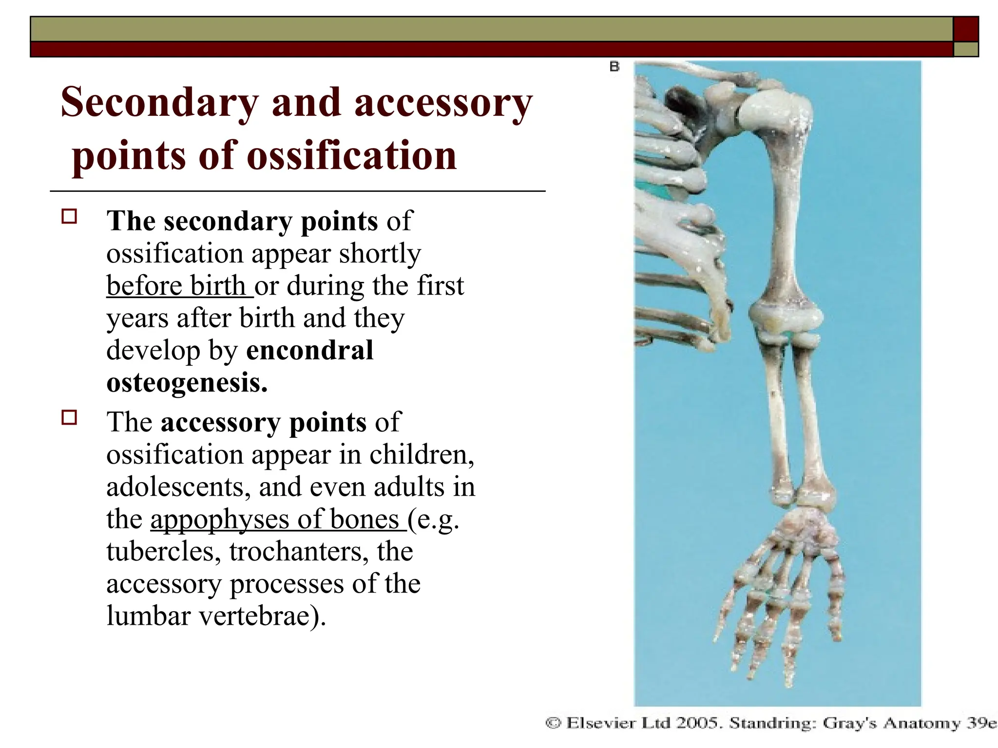
The secondary points of
ossification appear shortly
before birth or during the first
years after birth and they
develop by encondral
osteogenesis.
The accessory points of
ossification appear in children,
adolescents, and even adults in
the appophyses of bones (e.g.
tubercles, trochanters, the
accessory processes of the
lumbar vertebrae).
50.
Growth of bone
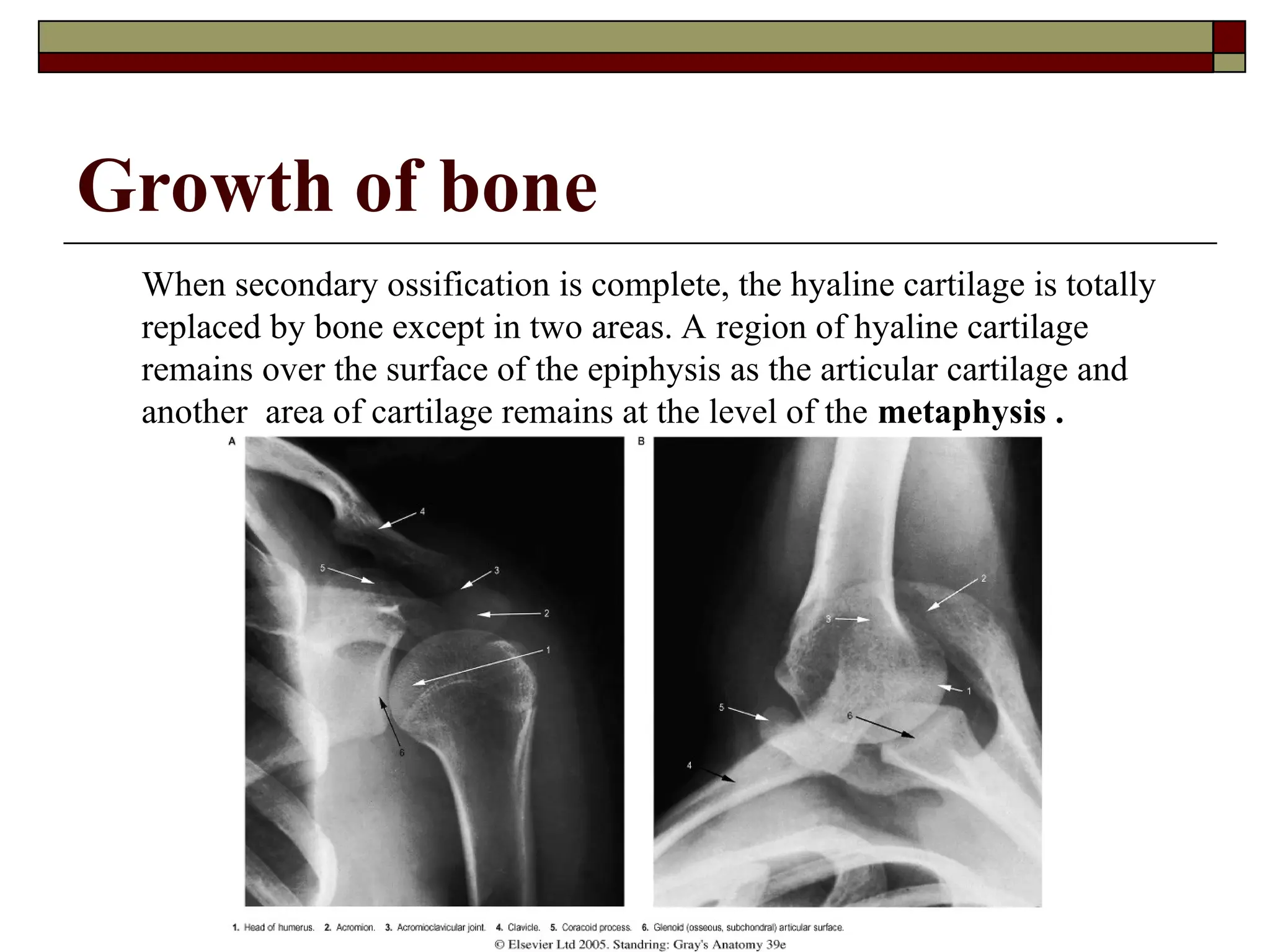
Whensecondary ossification is complete, the hyaline cartilage is totally
replaced by bone except in two areas. A region of hyaline cartilage
remains over the surface of the epiphysis as the articular cartilage and
another area of cartilage remains at the level of the metaphysis .
51.
DEVELOPMENT OF THE
VERTEBRAE
The mesenchyme (sclerotome) gives rise to the skeleton around the
notochord. The vertebral column in its primitive form is made up of upper
and lower cartilaginous arches, which are arranged in a metameric
fashion on the ventral and dorsal aspects of the notochord.
The bodies of the vertebrae grow around the notochord and compress it.
As a result the notochord is replaced by the vertebral bodies and remains
only between the vertebrae as pulpy nucleus (nucleus pulposus) in the
center of the intervertebral discs.
The upper neural arches give rise to the spinous process, to the paired
articular and transverse processes.
The lower ventral arches give rise to the ribs.
After going through the cartilaginous stage, the vertebral column
becomes bony, except the intervertebral discs connecting them.
52.
Abnormality isa deviation from the norm and it can
be of different degrees. Abnormalities of bones are
the result of improper development of bony system.
Different abnormalities of bones are distinguished:
e.g. subdevelopment of bone, absence of bone,
abnormal location of bone, bones can vary in
number (to be more or less that usually), there can
form additional bones, etc.
53.
VARIANTS AND DEVELOPMENTALABNORMALITIES OF THE
VERTEBRAE
Assimilation of the atlas by the cranium, when the first cervical vertebra fusses
with the occipital bone.
Lumbalization when the first sacral vertebra does not fuse with the sacrum and
there are 6 lumbar vertebrae instead of five; or when the last thoracic vertebra is
not joined with a rib and transforms into a lumbar vertebra.
Sacralization when there are 6-7 sacral vertebrae, because the last lumbar
vertebrae fuse with the sacral bone and in this case the number of the lumbar
vertebrae decreases.
Spina bifida – results from a failure of the vertebral arches to fuse. This
abnormality is more commonly for the lumbar and sacral vertebrae.
Intervertebral disc herniation involves the prolapse of the nucleus pulposus
through the defective annulus fibrosus into the vertebral canal.
Spondylolistesis occurs when the pedicles of the vertebral arches fail to fuse with
the vertebral body. Congenital spondylolistesis usually occurs at the level of L5-
S1vertebrae.
Asomia is the absence of the vertebral body.
Hemisomia is the absence of a half of the vertebral body.
54.
DEVELOPMENT OF THESTERNUM AND RIBS
The ribs develop from costal processes that form at
all vertebral levels, but only in the thoracic region
the costal processes grow into ribs.
The sternum or the breastbone develops from two
sternal bars which form in the ventral body wall
independent of the ribs and clavicle. The sternal bars
fuse with each other in a craniocaudal direction to
form the manubrium, the body and the xiphoid
process by week of 8.
55.
VARIANTS AND DEVELOPMENTALABNORMALITIES OF THE
STERNUM AND RIBS
The ribs can vary in number to be more or less than normal
number (12 pairs).
Cervical ribs on one or on both sides, when the VIIth cervical
vertebra joins with a rib. In case of presence of the cervical
ribs, then the VIIth cervical vertebra has appearances of a
thoracic vertebra.
Lumbar ribs in case the Ist lumbar vertebrae joins with a rib.
In rare cases the XIIth rib can be absent from one or from both
sides, and more rarely are cases when the XIth rib is absent.
If there are XIIIth pairs of ribs, then the number of thoracic
vertebrae as well increases.
The anterior extremities of the ribs can fuse to each other, or
on the contrary to bifurcate.
56.
Abnormalities of thesternum
Sternal cleft occurs when the sternal bars do
not fuse completely and the body of the
sternum is split into two halves, it is a rare
abnormality.
Sometimes in the body of the sternum is
present an orifice.
In the xyphoid process can be present an
orifice, or it can be bifurcated.