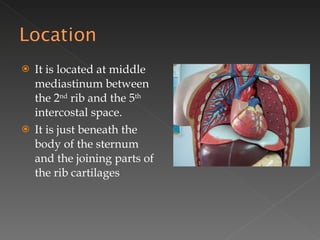
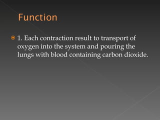
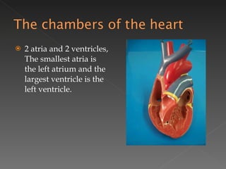
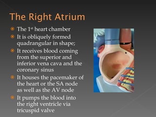
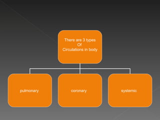
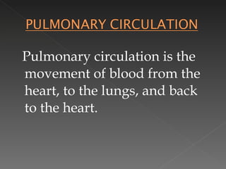
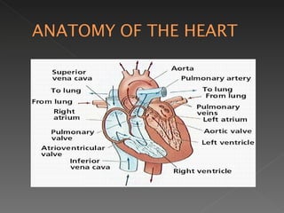
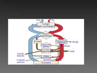
To identify and describe the organs of the circulatory system
Responsible for transporting materials throughout the entire body, these materials include: Blood Oxygen Carbon dioxide Nutrients Water
It is a hallow muscular organ, somewhat conical in shaped wherein the base is located superiorly and the apex is pointing inferiorly It is about the same size of closed fist. It is made-up of cardiac muscle which is involuntary type and striated The weight in male varies from 280-340 gm and in female it is from 230-280 gm
It is located at middle mediastinum between the 2 nd  rib and the 5 th  intercostal space. It is just beneath the body of the sternum and the joining parts of the rib cartilages
1. Each contraction result to transport of oxygen into the system and pouring the lungs with blood containing carbon dioxide.
2 atria and 2 ventricles, The smallest atria is the left atrium and the largest ventricle is the left ventricle.
The 1 st  heart chamber It is obliquely formed quadrangular in shape;  It receives blood coming from the superior and inferior vena cava and the coronary sinus  It houses the pacemaker of the heart or the SA node as well as the AV node It pumps the blood into the right ventricle via tricuspid valve
The 2 nd  heart chamber Triangular in shaped that extends form the right atria to the apex The undersurface is flattened, rests upon the diaphragm
The 3 rd  chamber  and the smallest chamber in the heart. It communicates with pulmunary veins and left ventricle via bicuspid valve
The 4 th  chamber and the biggest of the 4 chambers of the heart It has a thick myocardium and it communicate with aorta by means of its valve
Viceral Pericardium Myocardium Endocardium
Is a fibroserous sac that encloses the heart and the roots of the great vessels. Its function is to restrict excessive movements of the heart as a whole and to serve as a lubricated container in which the different parts of the heart can contract
Pulmonary valve Tricuspid valve
Mitral Valve
Sinuatrial node AKA- Pacemaker It initiates the heart beat and it is located at the walls of the right atrium in the upper part of sulcus terminalis close to SVC Atrioventricular node It is located on the lower part of the atrial septum just above the attachment of the sepyal cusp of tricuspid valve
Atrioventricular bundles – R and L side Is a continuous with the AV node above and purkinje fibers below It descends behind the septal cusp of the tricuspid valve on the membranous part of ventricular septum Purkinje fibers A specialized cardiac muscle fibers that form the conducting system of the heart It is the terminal/ end of the system
There are 3 types Of  Circulations in body pulmonary  coronary  systemic
Pulmonary circulation is the movement of blood from the heart, to the lungs, and back to the heart.
Low O2 blood enters the right atrium via Vena cava. To the right ventricle (via a one way valve) Right ventricle contracts and sends the low O2 blood to lungs through the Pulmonary artery.
In the lungs capillaries, blood exchanges CO2 with O2 O2 rich blood enters into the left atrium via the pulmonary vein Follows by entry to the left ventricle( via a one way valve)
Left ventricle contraction forces the blood in to the Aorta From this point  blood begins its journey throughout the body
While the circulatory system is busy providing oxygen and nourishment to every cell of the body, let's not forget that the heart, which needs nourishment as well as other parts of the body. Coronary circulation refers to the movement of blood through the tissues of the heart.  The heart tissue receives nourishment through the capillaries located in the heart.
Systemic circulation supplies nourishment to all of the tissue located throughout your body, with the exception of the heart and lungs.  In this circulation, the oxygen-rich blood enters the capillaries where the oxygen and nutrients are released. The waste products are collected and the waste-rich blood flows into the veins in order to circulate back to the heart.
 
 
Systemic circulation has two smaller sub circulation within it Renal circulation  Portal circulation
During systemic circulation, blood passes through the kidneys. This phase of systemic circulation is known as renal circulation. During this phase, the kidneys filter much of the waste from the blood.  Blood also passes through the small intestine during systemic circulation. This phase is known as portal circulation. During this phase, the blood from the small intestine collects in the portal vein which passes through the liver. The liver filters sugars from the blood, storing them for later.
The arch of aorta Brachiocephalic Artery Left Common carotid  Artery Left Subclavian Artery
Brachiocephalic Artery Right Subclavian Artery Right Upper  Extremity Right Common  Carotid Artery Right Side of  Head & Neck
Left Common Carotid Artery Left side of  Head & Neck
Left Subclavian Artery Left Upper Extremity
Common Carotid Artery Internal Carotid Artery External  Carotid  Artery Brian  Superior Thyroid Artery (to thyroid gland, neck) Ascending Pharyngeal Artery (Pharynx) Lingual Artery (Tongue) Facial Artery (Face) Occipital Artery (back of the head) Posterior Auricular Artery (back of ear) Superficial Temporal Artery (region of temple) Maxillary Artery (NEXT SLIDE)
Maxillary  Artery Post superior  Alveolar Artery Middle Superior Alveolar Artery Anterior Superior Alveolar Artery Middle Meningeal Artery Inferior Alveolar Artery Upper Molars Upper Bicuspids Upper Canine & Incisors Meninges of the brain All the lower teeth
Right & Left Subclavian Artery Vertebral Artery Int. Thoracic  Artery Thyrocervical trunk Costocervical trunk Brian and spinal cord Anterior thoracic wall including breast Thyroid gland & certain vertebrae Upper thorax & neck
Subclavian Artery Axillary  Artery Brachial Artery Radial Artery Ulnar Artery Superficial Palmar Arch Pollicis Artery Deep Palmar Arch Digital Artery Thumb Digits
Right and Left External Illiac Artery Femoral Artery Popliteal Artery Ant. Tibial Artery Dorsalis  Pedis Artery Hallucis Artery Post. Tibial Artery Lateral Plantar Artery Digital Artery Medial Plantar Artery Toes Big Toe
Big Toe (DORSUM) Hallucis Vein Dorsal Venous Arch Ant. Tibial Vein Popliteal Vein Femoral Vein Ext. Iliac Vein Common Iliac Vein Inferior Vena Cava Long Saphenouse Vein Toes Digital Vein Big Toe (SLOE) Toes Hallucis Vein Digital Vein Medial Plantar Vein Lateral Plantar Vein Post Tibial Vein
Thumb Pollicis Vein Index finger, 3rd 4th, and 5th Digital Vein Dorsal Venous Arch Cephalic Vein Thumb Index 4th & 5th Pollicis Vein Digital Vein Digital Vein Radial Vein Ulnar Vein Brachial Vein Axillary Vein Subclavian Vein Brachiocephalic Vein Superior Vena Cava
Upper Molars   PSA Vein Upper Premolars   MSA Vein  PETRYGOID Upper Anteriors   ASA Vein  PLEXUS OF  TEMPLE All Lower Teeth   IAV Vein  VEINS Superficial Back of head   Ear   Maxillary V  Temporal V  FACE Occipital Vein  Post  Post Facial V  +  Ant Facial V Auricular V BRAIN  Common Facial V  External Jugular V  Internal Jugular V  Lingual V  TONGUE Subclavian V  Brachiocephalic V  Thyroid V  THYROID GLAND Superior Vena Cava  Right Atrium
Inferior Vena  Cava Portal  Vein Hepatic Vein Diaphragm Inf. Phrenic V R adrenal R Suprarenal V Right Kidney R renal V R Ovary / Testicle R Ovarian / Testicular V R Common  Iliac Vein L Common Iliac Vein L Ovary / Testicle L Ovarian / Testicular V Left Kidney L Renal V L adrenal G L Suprarenal V Gastric V Stomach Splenic V Spleen Inf. Mesenteric V Large Intestine Superior Mesenteric V Small Intestine

Circulatory System

  • 1.
  • 2.
    To identify anddescribe the organs of the circulatory system
  • 3.
    Responsible for transportingmaterials throughout the entire body, these materials include: Blood Oxygen Carbon dioxide Nutrients Water
  • 4.
    It is ahallow muscular organ, somewhat conical in shaped wherein the base is located superiorly and the apex is pointing inferiorly It is about the same size of closed fist. It is made-up of cardiac muscle which is involuntary type and striated The weight in male varies from 280-340 gm and in female it is from 230-280 gm
  • 5.
    It is locatedat middle mediastinum between the 2 nd rib and the 5 th intercostal space. It is just beneath the body of the sternum and the joining parts of the rib cartilages
  • 6.
    1. Each contractionresult to transport of oxygen into the system and pouring the lungs with blood containing carbon dioxide.
  • 7.
    2 atria and2 ventricles, The smallest atria is the left atrium and the largest ventricle is the left ventricle.
  • 8.
    The 1 st heart chamber It is obliquely formed quadrangular in shape; It receives blood coming from the superior and inferior vena cava and the coronary sinus It houses the pacemaker of the heart or the SA node as well as the AV node It pumps the blood into the right ventricle via tricuspid valve
  • 9.
    The 2 nd heart chamber Triangular in shaped that extends form the right atria to the apex The undersurface is flattened, rests upon the diaphragm
  • 10.
    The 3 rd chamber and the smallest chamber in the heart. It communicates with pulmunary veins and left ventricle via bicuspid valve
  • 11.
    The 4 th chamber and the biggest of the 4 chambers of the heart It has a thick myocardium and it communicate with aorta by means of its valve
  • 12.
  • 13.
    Is a fibroseroussac that encloses the heart and the roots of the great vessels. Its function is to restrict excessive movements of the heart as a whole and to serve as a lubricated container in which the different parts of the heart can contract
  • 14.
  • 15.
  • 16.
    Sinuatrial node AKA-Pacemaker It initiates the heart beat and it is located at the walls of the right atrium in the upper part of sulcus terminalis close to SVC Atrioventricular node It is located on the lower part of the atrial septum just above the attachment of the sepyal cusp of tricuspid valve
  • 17.
    Atrioventricular bundles –R and L side Is a continuous with the AV node above and purkinje fibers below It descends behind the septal cusp of the tricuspid valve on the membranous part of ventricular septum Purkinje fibers A specialized cardiac muscle fibers that form the conducting system of the heart It is the terminal/ end of the system
  • 18.
    There are 3types Of Circulations in body pulmonary coronary systemic
  • 19.
    Pulmonary circulation isthe movement of blood from the heart, to the lungs, and back to the heart.
  • 20.
    Low O2 bloodenters the right atrium via Vena cava. To the right ventricle (via a one way valve) Right ventricle contracts and sends the low O2 blood to lungs through the Pulmonary artery.
  • 21.
    In the lungscapillaries, blood exchanges CO2 with O2 O2 rich blood enters into the left atrium via the pulmonary vein Follows by entry to the left ventricle( via a one way valve)
  • 22.
    Left ventricle contractionforces the blood in to the Aorta From this point blood begins its journey throughout the body
  • 23.
    While the circulatorysystem is busy providing oxygen and nourishment to every cell of the body, let's not forget that the heart, which needs nourishment as well as other parts of the body. Coronary circulation refers to the movement of blood through the tissues of the heart. The heart tissue receives nourishment through the capillaries located in the heart.
  • 24.
    Systemic circulation suppliesnourishment to all of the tissue located throughout your body, with the exception of the heart and lungs. In this circulation, the oxygen-rich blood enters the capillaries where the oxygen and nutrients are released. The waste products are collected and the waste-rich blood flows into the veins in order to circulate back to the heart.
  • 25.
  • 26.
  • 27.
    Systemic circulation hastwo smaller sub circulation within it Renal circulation Portal circulation
  • 28.
    During systemic circulation,blood passes through the kidneys. This phase of systemic circulation is known as renal circulation. During this phase, the kidneys filter much of the waste from the blood. Blood also passes through the small intestine during systemic circulation. This phase is known as portal circulation. During this phase, the blood from the small intestine collects in the portal vein which passes through the liver. The liver filters sugars from the blood, storing them for later.
  • 29.
    The arch ofaorta Brachiocephalic Artery Left Common carotid Artery Left Subclavian Artery
  • 30.
    Brachiocephalic Artery RightSubclavian Artery Right Upper Extremity Right Common Carotid Artery Right Side of Head & Neck
  • 31.
    Left Common CarotidArtery Left side of Head & Neck
  • 32.
    Left Subclavian ArteryLeft Upper Extremity
  • 33.
    Common Carotid ArteryInternal Carotid Artery External Carotid Artery Brian Superior Thyroid Artery (to thyroid gland, neck) Ascending Pharyngeal Artery (Pharynx) Lingual Artery (Tongue) Facial Artery (Face) Occipital Artery (back of the head) Posterior Auricular Artery (back of ear) Superficial Temporal Artery (region of temple) Maxillary Artery (NEXT SLIDE)
  • 34.
    Maxillary ArteryPost superior Alveolar Artery Middle Superior Alveolar Artery Anterior Superior Alveolar Artery Middle Meningeal Artery Inferior Alveolar Artery Upper Molars Upper Bicuspids Upper Canine & Incisors Meninges of the brain All the lower teeth
  • 35.
    Right & LeftSubclavian Artery Vertebral Artery Int. Thoracic Artery Thyrocervical trunk Costocervical trunk Brian and spinal cord Anterior thoracic wall including breast Thyroid gland & certain vertebrae Upper thorax & neck
  • 36.
    Subclavian Artery Axillary Artery Brachial Artery Radial Artery Ulnar Artery Superficial Palmar Arch Pollicis Artery Deep Palmar Arch Digital Artery Thumb Digits
  • 37.
    Right and LeftExternal Illiac Artery Femoral Artery Popliteal Artery Ant. Tibial Artery Dorsalis Pedis Artery Hallucis Artery Post. Tibial Artery Lateral Plantar Artery Digital Artery Medial Plantar Artery Toes Big Toe
  • 38.
    Big Toe (DORSUM)Hallucis Vein Dorsal Venous Arch Ant. Tibial Vein Popliteal Vein Femoral Vein Ext. Iliac Vein Common Iliac Vein Inferior Vena Cava Long Saphenouse Vein Toes Digital Vein Big Toe (SLOE) Toes Hallucis Vein Digital Vein Medial Plantar Vein Lateral Plantar Vein Post Tibial Vein
  • 39.
    Thumb Pollicis VeinIndex finger, 3rd 4th, and 5th Digital Vein Dorsal Venous Arch Cephalic Vein Thumb Index 4th & 5th Pollicis Vein Digital Vein Digital Vein Radial Vein Ulnar Vein Brachial Vein Axillary Vein Subclavian Vein Brachiocephalic Vein Superior Vena Cava
  • 40.
    Upper Molars PSA Vein Upper Premolars MSA Vein PETRYGOID Upper Anteriors ASA Vein PLEXUS OF TEMPLE All Lower Teeth IAV Vein VEINS Superficial Back of head Ear Maxillary V Temporal V FACE Occipital Vein Post Post Facial V + Ant Facial V Auricular V BRAIN Common Facial V External Jugular V Internal Jugular V Lingual V TONGUE Subclavian V Brachiocephalic V Thyroid V THYROID GLAND Superior Vena Cava Right Atrium
  • 41.
    Inferior Vena Cava Portal Vein Hepatic Vein Diaphragm Inf. Phrenic V R adrenal R Suprarenal V Right Kidney R renal V R Ovary / Testicle R Ovarian / Testicular V R Common Iliac Vein L Common Iliac Vein L Ovary / Testicle L Ovarian / Testicular V Left Kidney L Renal V L adrenal G L Suprarenal V Gastric V Stomach Splenic V Spleen Inf. Mesenteric V Large Intestine Superior Mesenteric V Small Intestine