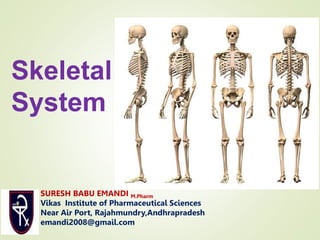
SlideShare a Scribd company logo
1 of 78
Skeletal
System
SURESH BABU EMANDI M.Pharm
Vikas Institute of Pharmaceutical Sciences
Near Air Port, Rajahmundry,Andhrapradesh
emandi2008@gmail.com
Although bones are
often thought to be
static or permanent
they are highly
vascular living
structures that are
continuously being
remodelled
Types of bones
Bones are classified as
Long bones
Short bones
Irregular bones
Flat bones
Sesamoid bones.
Long bones. These consist of a shaft and two extremities.
As the name suggests the length is much greater than the
width. Examples include the femur, tibia and fibula.
Short, irregular, flat and sesamoid bones. These have
no shafts or extremities and are diverse in shape and size.
Examples include:
• short bones — carpals (wrist)
• irregular bones—vertebrae and some skull bones
• flat bones — sternum, ribs and most skull bones
• sesamoid bones — patella (knee cap).
Bone is a strong and durable type of
connective tissue.
It consists of:
• water (25%)
• organic constituents including osteoid
(the carbon containing part of the
matrix) and bone cells (25%)
• inorganic constituents, mainly calcium
phosphate (50%).
⦁ The bones primarily give support to the appendages
and protect the fragile organs of the body such as
the brain, spinal cord, heart, and lungs.
⦁ They also store calcium and phoshorus in the hard
bone matrix, which can be released when needed
elsewhere.
⦁ The bumps, ridges, and grooves on the surface of
bones provide attachment sites for the skeletal
muscles.
⦁ Additionally, many bones contain a soft tissue called
marrow that produces new blood cells and stores fat
⦁ Bone is one of the hardest parts of the body
⦁ Very lightweight
⦁ Can resist tension
◦ Calcium salts make it hard
◦ Collagen fibers make it strong and flexible
⚫ Support
⚫ Internal framework of body
⚫ Ex: Legs support body trunk,
rib cage supports chest wall
⚫ Protection
⚫ Protects soft body organs (skull
protects brain, ribs protect
heart and lungs, etc)
⚫ Movement*
⚫ Muscles use bones as levers to
move body
⚫ NOT a function skeletal system
can do on it’s own
⚫ Storage
⚫ Fat storage
⚫ In yellow marrow
⚫ Mineral storage
⚫ Calcium, phosphorus, and
others.
⚫ Blood cell formation
⚫ Hematopoeisis
⚫ Occurs in bone marrow of long
bones
⦁ The adult human skeleton is a framework of 206
bones and is anatomically divided into two parts,
◦ The axial skeleton
◦ The appendicular skeleton
⦁ Two types of bone
◦ Compact bone
🞄 Homogeneous appearance
🞄 Dense, looks smooth
◦ Spongy bone
🞄 Small needle-like pieces
🞄 Lots of open spaces
🞄 (Looks like a sponge, hence the name)
⦁ In embryos…
◦ Skeleton made of hyaline cartilage
◦ Gets replaced by bone with age
⦁ Cartilage remains in a few places…
◦ Bridge of nose
◦ Parts of ribs
◦ Joints
⚫ Epiphyseal plates allow for growth of long bone
during childhood
⚫New cartilage is continuously formed
⚫Older cartilage becomes ossified
⚫Cartilage is broken down
⚫Bone replaces cartilage
⚫ Bones remodeled, lengthened until growth stops
⚫Bones change shape somewhat
⚫Bones grow in width
⦁ Osteocyte
◦ Mature bone cell
⦁ Osteoblast
◦ “Bone builders”
⦁ Osteoclast
◦ “Bone destroyers”
◦ Break down bone matrix to remodel and/or release
calcium
⚫ Bone remodeling is done by osteoblasts and osteoclasts
⚫ Essential for bones to keep normal proportions and
strength
⚫ Bones get larger/thicker when…
⚫Large muscles are attached to them
⚫Need more bone to attach the large muscle to
⚫ Bones get smaller/weaker when…
⚫Physical inactivity
⚫Non-use of musclesatrophy
⚫Small muscles don’t need large bones to attach to
⚫ Bones remodeled to become smaller
⚫ Long Bones
⚫Longer than wide
⚫Contain shaft with head at
each end
⚫Mostly compact bone
⚫Limbs (not ankle or wrist
bones)
⚫ Short Bones
⚫Cube-shaped
⚫Mostly spongy bone
⚫Wrist and ankle bones
⚫Patella (kneecap)
⚫ Flat Bones
⚫Thin, flat, usually curved
⚫Two layers of compact
bone with spongy bone in
between
⚫Skull, ribs, breastbone
⚫ Irregular Bones
⚫Don’t fit into the other
categories
⚫Vertebrae, hip bones
⚫ Diaphysis
⚫Shaft
⚫Made of compact bone
⚫ Epiphyses
⚫Ends of long bone
⚫Made of compact
bone surrounding
spongy bone
⚫Covered with articular
cartilage
⚫Smooth, glassy, decreases
friction at joints
⦁ Epiphyseal Line
◦ Line of bone tissue that
looks different from rest
of bone
◦ Remnant of epiphyseal
plate
🞄 Hyaline cartilage
🞄 Causes long bones to grow
lengthwise
⦁ Periosteum
◦ Outer covering of shaft
◦ Like “skin” on chicken
bone
⦁ Sharpey’s fibers
◦ Attach periosteum to
bone
⦁ Arteries
◦ Supply nutrients to bone
cells
⦁ Medullary Cavity
◦ In shaft
◦ Contains yellow marrow
(adipose) in adults
◦ Contains red marrow in
infants (for blood cell
formation)
⦁ Osteon (Haversian system)
◦ A “unit” of bone
⦁ Central (Haversian) canal
◦ Opening in center of osteon
◦ Contains blood vessels
⦁ Perforating (Volkman’s) canal
◦ Perpendicular to central canal
◦ Contains blood vessels and nerves
⚫ Lacunae
⚫Cavities that contain
osteocytes (bone cells)
⚫Arranged in concentric
rings (like tree rings)
⚫ Lamellae
⚫Rings around the central
canal
⚫Where the lacunae
can be found
⦁ Canaliculi
◦ Tiny canals
◦ Radiate from central
canal to lacunae
◦ Transport nutrients to
bone cells
⦁ The human skeleton can be divided into two
◦ The axial skeleton
◦ The appendicular skeleton.
⦁ The axial skeleton is formed by the vertebral
column, the rib cage, the skull and other associated
bones.
⦁ The appendicular skeleton, which is attached to the
axial skeleton, is formed by the shoulder girdle,
the pelvic girdle and the bones of the upper and
lower limbs.
⦁ The core of the skeleton is referred to as the axial
skeleton. It consists of the following 80 bones.
⦁ The upright posture of humans is maintained by the
axial skeleton, which transmits the weight from the
head, the trunk, and the upper extremities down to
the lower extremities at the hip joints.
◦ Skull: 22 bones
◦ Hyoid: 1 bone
◦ Vertebrae: 32 -34 bones
◦ Ribs: 24 bones
◦ Sternum: 1 bone
⦁ The skull consists of 22 bones and 7 associated
bones. All the larger skull bones are shown in this
exploded view. They fall into two main groups.
◦ One group (including the frontal, parietal, and temporal
bones) surrounds the brain and is fused together to form the
cranium.
◦ The remainder of the bones form the face.
⦁ Sutures
◦ Interlocking, immovable joints holding most skullbones
together
🞄 Mandible-only freely movable bone in skull
⦁ This highly flexible structure, also called the
vertebral column, supports the head and body.
⦁ It also protects the delicate tissues of the spinal
cord.
⦁ It is made up of 32-34 bones called vertebrae,
separated by intervertebral discs, which act as shock
absorbers.
⦁ The bones of the spine are kept in place and
supported by attached ligaments and muscles.
⦁ Hollow portions of bone around nasal cavity
◦ Named for bones they are found in
◦ Lighten skull
◦ Amplify sounds when speaking/ give resonance
⦁ Large in comparison
to body length of
infant
⦁ Contains fontanelles
◦ Soft fibrous membranes
connecting cranial bones
🞄“Soft Spots”
🞄 Usually convert to bone
around 24 months after
birth
⦁ Vertebrae named for
their location
◦ 7 cervical vertebrae
🞄 C1 = Atlas
🞄 C2 = Axis
◦ 12 thoracic vertebrae
◦ 5 lumbar vertebrae
◦ Sacrum
🞄 5 fused vertebrae
◦ Coccyx
🞄 4 fused vertebrae
⦁ Vertebrae separated by
fibrocartilage discs
◦ Cushions/absorbs shock
⦁ Forms a cage to protect
organs
⦁ Three parts
◦ Sternum
🞄 Flat bone
🞄 3 fused bones
🞄 Manubrium
🞄 Body
🞄 Xyphoid process
🞄 Attached to first 7 pair of
ribs
◦ Rib Cage
🞄 12 pairs
🞄 True Ribs= 7 pair
🞄 False Ribs = 5 pair
🞄 Floating Ribs = 2 pair
◦ Thoracic vertebrae
The appendicular skeleton is
divided into six major regions:
⦁ Pectoral girdles (4 bones) –
◦ Left and right clavicle (2)
◦ Scapula (2).
⦁ Arms and forearms (6 bones) –
◦ Left and right humerus (2) (arm)
◦ Ulna (2)
◦ Radius (2) (forearm).
⦁ Hands (54 bones) –
◦ Left and right carpals (16) (wrist)
◦ Metacarpals(10)
◦ Proximal phalanges (10),
◦ Intermediate phalanges (8)
◦ Distal phalanges (10)
⦁ Pelvis (2 bones) –
◦ Left and right hip bone (2).
⦁ Thighs and legs (8 bones) –
◦ Left and right femur (2) (thigh)
◦ Patella (2) (knee)
◦ Tibia (2)
◦ Fibula (2) (leg).
⦁ Feet and ankles (52 bones)
⦁ Left and right tarsals (14) (ankle)
⦁ Metatarsals (10)
⦁ Proximal phalanges (10)
⦁ Intermediate phalanges (8)
⦁ Distal phalanges (10).
⦁ Pectoral girdles (4 bones)
◦ Left and right clavicle (2)
◦ Scapula (2).
⦁ Arms and forearms (6 bones) –
◦ Left and right humerus (2) (arm)
◦ Ulna (2)
◦ Radius (2) (forearm).
⦁ Hands (54 bones) –
◦ Left and right carpals (16) (wrist)
◦ Metacarpals (10)
◦ Proximal phalanges (10),
◦ Intermediate phalanges (8)
◦ Distal phalanges (10)
⦁ Three pairs of fused
bones
◦ Ilium
◦ Ischium
◦ Pubic bone
⦁ Carries total weight of
upper body
⦁ Protects
◦ Reproductive organs
◦ Bladder
◦ Part of large intestine
⦁ Female inlet larger/more
circular
⦁ Female pelvis is shallower,
bones lighter/thinner
⦁ Female hip bones flare
wider
⦁ Female sacrum is
shorter/less curved
⦁ Female pubic arch is more
rounded/angle is greater
◦ Left and right femur
(2) (thigh)
◦ Patella (2) (knee)
◦ Tibia (2)
◦ Fibula (2) (leg).
⦁ Left and
right tarsals (14)
(ankle)
⦁ Metatarsals (10)
⦁ Proximal
phalanges (10)
⦁ Intermediate
phalanges (8)
⦁ Distal phalanges (10).
A joint is the site at which any two or
more bones articulate
or come together. Some joints have no
movement
(fibrous), some only slight movement
(cartilaginous) and
some are freely movable (synovial).
⦁ Articulations of bones
⦁ Functions
◦ Hold bones together
◦ Mobility
⦁ Classified by
structure/function
⦁ Fibrous joints or Immovable
🞄 EX: sutures of skull
⦁ Cartilaginous joints OR slightly movable
EX: Pubic symphysis and Vertebrae
Synovial joints
Freely movable
Fibrous /immovable/fixed Joints
 A fibrous joint is a fixed joint where collagenous
fibrous connective tissue connects two bones.
 Fibrous joints (synarthroses) are usually immovable and
have no joint cavity.
 They are subdivided further into sutures, gomphoses,
and syndesmoses.
 Sutures are immobile joints found only in the cranium.
Fibrous/immovable/fixed joints
Cartilaginous or slightly movable joints
 There is a pad of fibrocartilage between the ends of
the bones that form the joint which allows for very
slight movement where the pad of cartilage is
compressed.
 Examples include the symphysis pubis and the joints
between the vertebral bodies.
Cartilaginous or
Slightly movable joints
Synovial or freely movable joints
Synovial joints have characteristic features that
enable a wide range of movements.
They are classified according to the range of
movement possible or to the shape of the
articulating parts of the bones involved
Classification of synovial joints
Ball and socket
Hinge joints
Gliding joints
Pivot joints.
Condyloid
Saddle joints
Ball and socket.
The head or ball of one bone articulates with a
socket of another and the shape of the bones
allows for a wide range of movement.
Those possible are flexion, extension, adduction,
abduction, rotation and circumduction.
Examples are the shoulder and hip.
Hinge joints.
 These allow the movements of flexion
 and extension only. They are the elbow, knee, ankle, the
 joints between the atlas and the occipital bone, and the
 interphalangeal joints of the fingers and toes.
Gliding joints
 . The articular surfaces glide over each
 other, e.g. sternoclavicular joints, acromioclavicular joints
 and joints between the carpal bones and those between
 the tarsal bones.
Pivot joints.
Movement is round one axis (rotation),
e.g. proximal and distal radioulnar joints and
the joint between the atlas and the odontoid
process of the axis
Condyloid and saddle joints
 Movements take place round two axes, permitting
flexion, extension, abduction, adduction and
circumduction, e.g. the wrist,
temporomandibular,metacarpophalangeal and
metatarsophalangeal joints.
Characteristics of a synovial joint
 All synovial joints have certain characteristics in common.
 Articular or hyaline cartilage
 The parts of the bones which are in contact are always covered with
hyaline cartilage.
 It provides a smooth articular surface and is strong enough to absorb
compression forces and bear the weight of the body.
 The cartilage lining, which is up to 7mm thick in young
people, becomes thinner and less compressible with age.
 This leads to increasing stress on other structures in the
joint.
 Cartilage has no blood supply and receives its
nourishmentfrom synovial fluid.
Capsule or capsular ligament
 The joint is surrounded and enclosed by a sleeve of
fibrous tissue which holds the bones together.
 It is sufficiently loose to allow freedom of movement
but strong enough to protect it from injury.
Synovial membrane
This is composed of epithelial cells and is
found: lining the capsule covering those parts
of the bones within the joint not covered by
articular cartilage
covering all intra-capsular structures that do
not bear weight.
Synovial fluid
This is a thick sticky fluid, of egg-white
consistency, secreted by synovial membranes
into the synovial cavity.
Provides nutrients for the structures within
the joint cavity.
Synovial fluid
 contains phagocytes, which remove microbes and cellular
debris.
 Acts as a lubricant.
 Maintains joint stability.
 Prevents the ends of the bones from being separated, as
does a little water between two glass surfaces.
 Little sacs of synovial fluid or bursae are present in some joints, e.g.
the knee.
 They act as cushions to prevent friction between a bone and a
ligament or tendon, or skin where a bone in a joint is near the
surface.
Other intra-capsular structures
Some joints have structures within the capsule,
but outside the synovial membrane, which assist
in maintenance of stability, e.g. fat pads and
menisci in the knee joint.
When these structures do not bear weight they
are covered by synovial membrane.
Extracapsular structures
Ligaments that blend with the capsule provide
additional stability at most joints.
Muscles or their tendons also provide stability and
stretch across the joints they move.
When the muscle contracts it shortens, pulling one
bone towards the other
⦁ Articulating bones
separated by joint cavity
◦ Articular cartilage covers
bone ends
◦ Joint surfaces enclosed by
fibrous capsule
◦ Cavity contains synovial
fluid
◦ Ligaments help reinforce
the joint
⦁ Associated Joint
Structures
◦ Bursae
🞄Flat fibrous sacs
🞄 Lined with synovial
membrane
🞄Contains synovial fluid
🞄 Common where ligaments,
muscles, skin, tendons rub
together
◦ Tendon sheath
🞄 Elongated bursa wrapping
around a tendon that deals
with lots of friction
🞄 Like a bun around hot dog
⦁ A break in a bone
⦁ Types of Fractures
◦ Simple (closed) fracture
🞄 Bone breaks cleanly, does not penetrate skin
◦ Compound (open) Fracture
🞄 When bone ends penetrate through skin
⦁ Treated by reduction and immobilization
◦ Realignment of bone ends
⚫ Hematoma forms
⚫Blood vessels rupture
when bone breaks
⚫Blood filled swelling
⚫Cells without nutrients die
⚫ Fibrocartilage callus
forms
⚫Made of connective tissue
⚫Splints the break
⚫Phagocytes dispose of dead
tissue
⚫ Bony callus forms
⚫Osteoblasts/clasts move in,
create callus of spongy
bone
⚫ Remodeling
⚫Osteoblasts/clasts remodel
in response to stress on the
bone
⚫Helps strengthen bone
⚫Creates a permanent “patch”
at the site
⚫Often, a healed fracture can
be stronger than the rest of
the bone
THANK YOU

More Related Content

Similar to SKELETAL SYSYTEM

Aaaaaa a skeletal system
Aaaaaa a skeletal systemAaaaaa a skeletal system
Aaaaaa a skeletal systemBobbyPabores1
 
Skeletal System 1
Skeletal System 1Skeletal System 1
Skeletal System 1Marc Potter
 
Anatomy & Physiology Lecture Notes - Skeletal system
 Anatomy & Physiology Lecture Notes - Skeletal system Anatomy & Physiology Lecture Notes - Skeletal system
Anatomy & Physiology Lecture Notes - Skeletal systemAmmedicine Medicine
 
Skeletalsystem 111027103336-phpapp01
Skeletalsystem 111027103336-phpapp01Skeletalsystem 111027103336-phpapp01
Skeletalsystem 111027103336-phpapp01BobbyPabores1
 
THE SKELETAL SYSTEM ppt.pptx
THE SKELETAL SYSTEM ppt.pptxTHE SKELETAL SYSTEM ppt.pptx
THE SKELETAL SYSTEM ppt.pptxAbhishek Borkar15
 
Lecture 1 (introduction to bone)
Lecture 1 (introduction to bone)Lecture 1 (introduction to bone)
Lecture 1 (introduction to bone)Ayub Abdi
 
THE SKELETAL SYSTEM Bachelors zoology botany chemistry
THE SKELETAL SYSTEM Bachelors zoology botany chemistryTHE SKELETAL SYSTEM Bachelors zoology botany chemistry
THE SKELETAL SYSTEM Bachelors zoology botany chemistryabhiasido310101
 
THE SKELETAL SYSTEM.pdf
THE SKELETAL SYSTEM.pdfTHE SKELETAL SYSTEM.pdf
THE SKELETAL SYSTEM.pdfUmaymaMatoobe
 
skeletal-system-220930065657-a14b470a (1).pdf
skeletal-system-220930065657-a14b470a (1).pdfskeletal-system-220930065657-a14b470a (1).pdf
skeletal-system-220930065657-a14b470a (1).pdfVrindaGupta60
 
Skeletal System
Skeletal SystemSkeletal System
Skeletal SystemRahul SIR
 

Similar to SKELETAL SYSYTEM (20)

Aaaaaa a skeletal system
Aaaaaa a skeletal systemAaaaaa a skeletal system
Aaaaaa a skeletal system
 
Skeletal System 1
Skeletal System 1Skeletal System 1
Skeletal System 1
 
Anatomy & Physiology Lecture Notes - Skeletal system
 Anatomy & Physiology Lecture Notes - Skeletal system Anatomy & Physiology Lecture Notes - Skeletal system
Anatomy & Physiology Lecture Notes - Skeletal system
 
Skeletal system
Skeletal systemSkeletal system
Skeletal system
 
Skeletalsystem 111027103336-phpapp01
Skeletalsystem 111027103336-phpapp01Skeletalsystem 111027103336-phpapp01
Skeletalsystem 111027103336-phpapp01
 
THE SKELETAL SYSTEM ppt.pptx
THE SKELETAL SYSTEM ppt.pptxTHE SKELETAL SYSTEM ppt.pptx
THE SKELETAL SYSTEM ppt.pptx
 
Lecture 1 (introduction to bone)
Lecture 1 (introduction to bone)Lecture 1 (introduction to bone)
Lecture 1 (introduction to bone)
 
Skeletal system
Skeletal systemSkeletal system
Skeletal system
 
Skeletal system
Skeletal systemSkeletal system
Skeletal system
 
Skeletonbonel2
Skeletonbonel2Skeletonbonel2
Skeletonbonel2
 
THE SKELETAL SYSTEM Bachelors zoology botany chemistry
THE SKELETAL SYSTEM Bachelors zoology botany chemistryTHE SKELETAL SYSTEM Bachelors zoology botany chemistry
THE SKELETAL SYSTEM Bachelors zoology botany chemistry
 
Chapter 7&8
Chapter 7&8Chapter 7&8
Chapter 7&8
 
THE SKELETAL SYSTEM.pdf
THE SKELETAL SYSTEM.pdfTHE SKELETAL SYSTEM.pdf
THE SKELETAL SYSTEM.pdf
 
THE SKELETAL SYSTEM.pdf
THE SKELETAL SYSTEM.pdfTHE SKELETAL SYSTEM.pdf
THE SKELETAL SYSTEM.pdf
 
Skeletal System
Skeletal SystemSkeletal System
Skeletal System
 
Unit 4 the skeletal system
Unit 4 the skeletal systemUnit 4 the skeletal system
Unit 4 the skeletal system
 
skeletal-system.ppt
skeletal-system.pptskeletal-system.ppt
skeletal-system.ppt
 
skeletal-system-220930065657-a14b470a (1).pdf
skeletal-system-220930065657-a14b470a (1).pdfskeletal-system-220930065657-a14b470a (1).pdf
skeletal-system-220930065657-a14b470a (1).pdf
 
GENERAL OSTEOLOGY.pptx
GENERAL OSTEOLOGY.pptxGENERAL OSTEOLOGY.pptx
GENERAL OSTEOLOGY.pptx
 
Skeletal System
Skeletal SystemSkeletal System
Skeletal System
 

More from SURESH BABU EMANDI DEPARTMENT OF PHARMACOGNOSY Vikas Institute of Pharmaceutical scienes

More from SURESH BABU EMANDI DEPARTMENT OF PHARMACOGNOSY Vikas Institute of Pharmaceutical scienes (20)

B;LOOD COMPOSITION AND FUNCTIOS AND DIFFERENT FORMED ELEMENTS
B;LOOD COMPOSITION AND FUNCTIOS AND DIFFERENT FORMED ELEMENTSB;LOOD COMPOSITION AND FUNCTIOS AND DIFFERENT FORMED ELEMENTS
B;LOOD COMPOSITION AND FUNCTIOS AND DIFFERENT FORMED ELEMENTS
 
CRANIAL NERVES.
CRANIAL NERVES.CRANIAL NERVES.
CRANIAL NERVES.
 
TISSUES
TISSUESTISSUES
TISSUES
 
NERVOUS SYSTEM
NERVOUS SYSTEMNERVOUS SYSTEM
NERVOUS SYSTEM
 
DIGESTIVE SYSTEM
DIGESTIVE SYSTEM DIGESTIVE SYSTEM
DIGESTIVE SYSTEM
 
GENRAL PRINCIPLES OF POISONING.pptx
GENRAL PRINCIPLES OF POISONING.pptxGENRAL PRINCIPLES OF POISONING.pptx
GENRAL PRINCIPLES OF POISONING.pptx
 
GENERAL PRINCIPLES OF POISONING.pptx
GENERAL PRINCIPLES OF POISONING.pptxGENERAL PRINCIPLES OF POISONING.pptx
GENERAL PRINCIPLES OF POISONING.pptx
 
DIGESTIVE SYSTEM
DIGESTIVE SYSTEM DIGESTIVE SYSTEM
DIGESTIVE SYSTEM
 
Cardiovascular system
Cardiovascular systemCardiovascular system
Cardiovascular system
 
URINARY SYSTEM
URINARY SYSTEM URINARY SYSTEM
URINARY SYSTEM
 
ENDOCRINE SYSTEM
ENDOCRINE SYSTEM ENDOCRINE SYSTEM
ENDOCRINE SYSTEM
 
DIGESTIVE SYSTEM
DIGESTIVE SYSTEM DIGESTIVE SYSTEM
DIGESTIVE SYSTEM
 
NERVOUS SYSTEM 2022
NERVOUS SYSTEM 2022NERVOUS SYSTEM 2022
NERVOUS SYSTEM 2022
 
HISTORY SCOPE DEVELOPMENT OF PHARMACOGNOSY.pptx
HISTORY SCOPE DEVELOPMENT OF PHARMACOGNOSY.pptxHISTORY SCOPE DEVELOPMENT OF PHARMACOGNOSY.pptx
HISTORY SCOPE DEVELOPMENT OF PHARMACOGNOSY.pptx
 
SOURSE OF CRUDE DRUGS.
SOURSE OF CRUDE DRUGS.SOURSE OF CRUDE DRUGS.
SOURSE OF CRUDE DRUGS.
 
PLANT HORMONES
PLANT HORMONESPLANT HORMONES
PLANT HORMONES
 
EVALUATION OF CRUDE DRUGS 2022.pptx
EVALUATION OF CRUDE DRUGS 2022.pptxEVALUATION OF CRUDE DRUGS 2022.pptx
EVALUATION OF CRUDE DRUGS 2022.pptx
 
URINARY SYSTEM
URINARY SYSTEMURINARY SYSTEM
URINARY SYSTEM
 
Reticuloendothelial System
Reticuloendothelial SystemReticuloendothelial System
Reticuloendothelial System
 
AUTONOMIC NERVOUS SYSTEM
AUTONOMIC NERVOUS SYSTEMAUTONOMIC NERVOUS SYSTEM
AUTONOMIC NERVOUS SYSTEM
 

Recently uploaded

Q4-W6-Restating Informational Text Grade 3
Q4-W6-Restating Informational Text Grade 3Q4-W6-Restating Informational Text Grade 3
Q4-W6-Restating Informational Text Grade 3JemimahLaneBuaron
 
Z Score,T Score, Percential Rank and Box Plot Graph
Z Score,T Score, Percential Rank and Box Plot GraphZ Score,T Score, Percential Rank and Box Plot Graph
Z Score,T Score, Percential Rank and Box Plot GraphThiyagu K
 
18-04-UA_REPORT_MEDIALITERAСY_INDEX-DM_23-1-final-eng.pdf
18-04-UA_REPORT_MEDIALITERAСY_INDEX-DM_23-1-final-eng.pdf18-04-UA_REPORT_MEDIALITERAСY_INDEX-DM_23-1-final-eng.pdf
18-04-UA_REPORT_MEDIALITERAСY_INDEX-DM_23-1-final-eng.pdfssuser54595a
 
Interactive Powerpoint_How to Master effective communication
Interactive Powerpoint_How to Master effective communicationInteractive Powerpoint_How to Master effective communication
Interactive Powerpoint_How to Master effective communicationnomboosow
 
The basics of sentences session 2pptx copy.pptx
The basics of sentences session 2pptx copy.pptxThe basics of sentences session 2pptx copy.pptx
The basics of sentences session 2pptx copy.pptxheathfieldcps1
 
Paris 2024 Olympic Geographies - an activity
Paris 2024 Olympic Geographies - an activityParis 2024 Olympic Geographies - an activity
Paris 2024 Olympic Geographies - an activityGeoBlogs
 
“Oh GOSH! Reflecting on Hackteria's Collaborative Practices in a Global Do-It...
“Oh GOSH! Reflecting on Hackteria's Collaborative Practices in a Global Do-It...“Oh GOSH! Reflecting on Hackteria's Collaborative Practices in a Global Do-It...
“Oh GOSH! Reflecting on Hackteria's Collaborative Practices in a Global Do-It...Marc Dusseiller Dusjagr
 
Grant Readiness 101 TechSoup and Remy Consulting
Grant Readiness 101 TechSoup and Remy ConsultingGrant Readiness 101 TechSoup and Remy Consulting
Grant Readiness 101 TechSoup and Remy ConsultingTechSoup
 
1029-Danh muc Sach Giao Khoa khoi 6.pdf
1029-Danh muc Sach Giao Khoa khoi  6.pdf1029-Danh muc Sach Giao Khoa khoi  6.pdf
1029-Danh muc Sach Giao Khoa khoi 6.pdfQucHHunhnh
 
Student login on Anyboli platform.helpin
Student login on Anyboli platform.helpinStudent login on Anyboli platform.helpin
Student login on Anyboli platform.helpinRaunakKeshri1
 
mini mental status format.docx
mini    mental       status     format.docxmini    mental       status     format.docx
mini mental status format.docxPoojaSen20
 
Sanyam Choudhary Chemistry practical.pdf
Sanyam Choudhary Chemistry practical.pdfSanyam Choudhary Chemistry practical.pdf
Sanyam Choudhary Chemistry practical.pdfsanyamsingh5019
 
Presentation by Andreas Schleicher Tackling the School Absenteeism Crisis 30 ...
Presentation by Andreas Schleicher Tackling the School Absenteeism Crisis 30 ...Presentation by Andreas Schleicher Tackling the School Absenteeism Crisis 30 ...
Presentation by Andreas Schleicher Tackling the School Absenteeism Crisis 30 ...EduSkills OECD
 
Call Girls in Dwarka Mor Delhi Contact Us 9654467111
Call Girls in Dwarka Mor Delhi Contact Us 9654467111Call Girls in Dwarka Mor Delhi Contact Us 9654467111
Call Girls in Dwarka Mor Delhi Contact Us 9654467111Sapana Sha
 
Kisan Call Centre - To harness potential of ICT in Agriculture by answer farm...
Kisan Call Centre - To harness potential of ICT in Agriculture by answer farm...Kisan Call Centre - To harness potential of ICT in Agriculture by answer farm...
Kisan Call Centre - To harness potential of ICT in Agriculture by answer farm...Krashi Coaching
 
Beyond the EU: DORA and NIS 2 Directive's Global Impact
Beyond the EU: DORA and NIS 2 Directive's Global ImpactBeyond the EU: DORA and NIS 2 Directive's Global Impact
Beyond the EU: DORA and NIS 2 Directive's Global ImpactPECB
 
URLs and Routing in the Odoo 17 Website App
URLs and Routing in the Odoo 17 Website AppURLs and Routing in the Odoo 17 Website App
URLs and Routing in the Odoo 17 Website AppCeline George
 

Recently uploaded (20)

Q4-W6-Restating Informational Text Grade 3
Q4-W6-Restating Informational Text Grade 3Q4-W6-Restating Informational Text Grade 3
Q4-W6-Restating Informational Text Grade 3
 
Z Score,T Score, Percential Rank and Box Plot Graph
Z Score,T Score, Percential Rank and Box Plot GraphZ Score,T Score, Percential Rank and Box Plot Graph
Z Score,T Score, Percential Rank and Box Plot Graph
 
18-04-UA_REPORT_MEDIALITERAСY_INDEX-DM_23-1-final-eng.pdf
18-04-UA_REPORT_MEDIALITERAСY_INDEX-DM_23-1-final-eng.pdf18-04-UA_REPORT_MEDIALITERAСY_INDEX-DM_23-1-final-eng.pdf
18-04-UA_REPORT_MEDIALITERAСY_INDEX-DM_23-1-final-eng.pdf
 
Interactive Powerpoint_How to Master effective communication
Interactive Powerpoint_How to Master effective communicationInteractive Powerpoint_How to Master effective communication
Interactive Powerpoint_How to Master effective communication
 
Código Creativo y Arte de Software | Unidad 1
Código Creativo y Arte de Software | Unidad 1Código Creativo y Arte de Software | Unidad 1
Código Creativo y Arte de Software | Unidad 1
 
Staff of Color (SOC) Retention Efforts DDSD
Staff of Color (SOC) Retention Efforts DDSDStaff of Color (SOC) Retention Efforts DDSD
Staff of Color (SOC) Retention Efforts DDSD
 
The basics of sentences session 2pptx copy.pptx
The basics of sentences session 2pptx copy.pptxThe basics of sentences session 2pptx copy.pptx
The basics of sentences session 2pptx copy.pptx
 
Paris 2024 Olympic Geographies - an activity
Paris 2024 Olympic Geographies - an activityParis 2024 Olympic Geographies - an activity
Paris 2024 Olympic Geographies - an activity
 
“Oh GOSH! Reflecting on Hackteria's Collaborative Practices in a Global Do-It...
“Oh GOSH! Reflecting on Hackteria's Collaborative Practices in a Global Do-It...“Oh GOSH! Reflecting on Hackteria's Collaborative Practices in a Global Do-It...
“Oh GOSH! Reflecting on Hackteria's Collaborative Practices in a Global Do-It...
 
Mattingly "AI & Prompt Design: Structured Data, Assistants, & RAG"
Mattingly "AI & Prompt Design: Structured Data, Assistants, & RAG"Mattingly "AI & Prompt Design: Structured Data, Assistants, & RAG"
Mattingly "AI & Prompt Design: Structured Data, Assistants, & RAG"
 
Grant Readiness 101 TechSoup and Remy Consulting
Grant Readiness 101 TechSoup and Remy ConsultingGrant Readiness 101 TechSoup and Remy Consulting
Grant Readiness 101 TechSoup and Remy Consulting
 
1029-Danh muc Sach Giao Khoa khoi 6.pdf
1029-Danh muc Sach Giao Khoa khoi  6.pdf1029-Danh muc Sach Giao Khoa khoi  6.pdf
1029-Danh muc Sach Giao Khoa khoi 6.pdf
 
Student login on Anyboli platform.helpin
Student login on Anyboli platform.helpinStudent login on Anyboli platform.helpin
Student login on Anyboli platform.helpin
 
mini mental status format.docx
mini    mental       status     format.docxmini    mental       status     format.docx
mini mental status format.docx
 
Sanyam Choudhary Chemistry practical.pdf
Sanyam Choudhary Chemistry practical.pdfSanyam Choudhary Chemistry practical.pdf
Sanyam Choudhary Chemistry practical.pdf
 
Presentation by Andreas Schleicher Tackling the School Absenteeism Crisis 30 ...
Presentation by Andreas Schleicher Tackling the School Absenteeism Crisis 30 ...Presentation by Andreas Schleicher Tackling the School Absenteeism Crisis 30 ...
Presentation by Andreas Schleicher Tackling the School Absenteeism Crisis 30 ...
 
Call Girls in Dwarka Mor Delhi Contact Us 9654467111
Call Girls in Dwarka Mor Delhi Contact Us 9654467111Call Girls in Dwarka Mor Delhi Contact Us 9654467111
Call Girls in Dwarka Mor Delhi Contact Us 9654467111
 
Kisan Call Centre - To harness potential of ICT in Agriculture by answer farm...
Kisan Call Centre - To harness potential of ICT in Agriculture by answer farm...Kisan Call Centre - To harness potential of ICT in Agriculture by answer farm...
Kisan Call Centre - To harness potential of ICT in Agriculture by answer farm...
 
Beyond the EU: DORA and NIS 2 Directive's Global Impact
Beyond the EU: DORA and NIS 2 Directive's Global ImpactBeyond the EU: DORA and NIS 2 Directive's Global Impact
Beyond the EU: DORA and NIS 2 Directive's Global Impact
 
URLs and Routing in the Odoo 17 Website App
URLs and Routing in the Odoo 17 Website AppURLs and Routing in the Odoo 17 Website App
URLs and Routing in the Odoo 17 Website App
 

SKELETAL SYSYTEM

  • 1. Skeletal System SURESH BABU EMANDI M.Pharm Vikas Institute of Pharmaceutical Sciences Near Air Port, Rajahmundry,Andhrapradesh emandi2008@gmail.com
  • 2. Although bones are often thought to be static or permanent they are highly vascular living structures that are continuously being remodelled
  • 3. Types of bones Bones are classified as Long bones Short bones Irregular bones Flat bones Sesamoid bones.
  • 4. Long bones. These consist of a shaft and two extremities. As the name suggests the length is much greater than the width. Examples include the femur, tibia and fibula. Short, irregular, flat and sesamoid bones. These have no shafts or extremities and are diverse in shape and size. Examples include: • short bones — carpals (wrist) • irregular bones—vertebrae and some skull bones • flat bones — sternum, ribs and most skull bones • sesamoid bones — patella (knee cap).
  • 5. Bone is a strong and durable type of connective tissue. It consists of: • water (25%) • organic constituents including osteoid (the carbon containing part of the matrix) and bone cells (25%) • inorganic constituents, mainly calcium phosphate (50%).
  • 6. ⦁ The bones primarily give support to the appendages and protect the fragile organs of the body such as the brain, spinal cord, heart, and lungs. ⦁ They also store calcium and phoshorus in the hard bone matrix, which can be released when needed elsewhere. ⦁ The bumps, ridges, and grooves on the surface of bones provide attachment sites for the skeletal muscles. ⦁ Additionally, many bones contain a soft tissue called marrow that produces new blood cells and stores fat
  • 7. ⦁ Bone is one of the hardest parts of the body ⦁ Very lightweight ⦁ Can resist tension ◦ Calcium salts make it hard ◦ Collagen fibers make it strong and flexible
  • 8. ⚫ Support ⚫ Internal framework of body ⚫ Ex: Legs support body trunk, rib cage supports chest wall ⚫ Protection ⚫ Protects soft body organs (skull protects brain, ribs protect heart and lungs, etc) ⚫ Movement* ⚫ Muscles use bones as levers to move body ⚫ NOT a function skeletal system can do on it’s own ⚫ Storage ⚫ Fat storage ⚫ In yellow marrow ⚫ Mineral storage ⚫ Calcium, phosphorus, and others. ⚫ Blood cell formation ⚫ Hematopoeisis ⚫ Occurs in bone marrow of long bones
  • 9. ⦁ The adult human skeleton is a framework of 206 bones and is anatomically divided into two parts, ◦ The axial skeleton ◦ The appendicular skeleton ⦁ Two types of bone ◦ Compact bone 🞄 Homogeneous appearance 🞄 Dense, looks smooth ◦ Spongy bone 🞄 Small needle-like pieces 🞄 Lots of open spaces 🞄 (Looks like a sponge, hence the name)
  • 10. ⦁ In embryos… ◦ Skeleton made of hyaline cartilage ◦ Gets replaced by bone with age ⦁ Cartilage remains in a few places… ◦ Bridge of nose ◦ Parts of ribs ◦ Joints
  • 11. ⚫ Epiphyseal plates allow for growth of long bone during childhood ⚫New cartilage is continuously formed ⚫Older cartilage becomes ossified ⚫Cartilage is broken down ⚫Bone replaces cartilage ⚫ Bones remodeled, lengthened until growth stops ⚫Bones change shape somewhat ⚫Bones grow in width
  • 12.
  • 13.
  • 14. ⦁ Osteocyte ◦ Mature bone cell ⦁ Osteoblast ◦ “Bone builders” ⦁ Osteoclast ◦ “Bone destroyers” ◦ Break down bone matrix to remodel and/or release calcium
  • 15. ⚫ Bone remodeling is done by osteoblasts and osteoclasts ⚫ Essential for bones to keep normal proportions and strength ⚫ Bones get larger/thicker when… ⚫Large muscles are attached to them ⚫Need more bone to attach the large muscle to ⚫ Bones get smaller/weaker when… ⚫Physical inactivity ⚫Non-use of musclesatrophy ⚫Small muscles don’t need large bones to attach to ⚫ Bones remodeled to become smaller
  • 16. ⚫ Long Bones ⚫Longer than wide ⚫Contain shaft with head at each end ⚫Mostly compact bone ⚫Limbs (not ankle or wrist bones) ⚫ Short Bones ⚫Cube-shaped ⚫Mostly spongy bone ⚫Wrist and ankle bones ⚫Patella (kneecap) ⚫ Flat Bones ⚫Thin, flat, usually curved ⚫Two layers of compact bone with spongy bone in between ⚫Skull, ribs, breastbone ⚫ Irregular Bones ⚫Don’t fit into the other categories ⚫Vertebrae, hip bones
  • 17. ⚫ Diaphysis ⚫Shaft ⚫Made of compact bone ⚫ Epiphyses ⚫Ends of long bone ⚫Made of compact bone surrounding spongy bone ⚫Covered with articular cartilage ⚫Smooth, glassy, decreases friction at joints
  • 18. ⦁ Epiphyseal Line ◦ Line of bone tissue that looks different from rest of bone ◦ Remnant of epiphyseal plate 🞄 Hyaline cartilage 🞄 Causes long bones to grow lengthwise
  • 19. ⦁ Periosteum ◦ Outer covering of shaft ◦ Like “skin” on chicken bone ⦁ Sharpey’s fibers ◦ Attach periosteum to bone ⦁ Arteries ◦ Supply nutrients to bone cells
  • 20. ⦁ Medullary Cavity ◦ In shaft ◦ Contains yellow marrow (adipose) in adults ◦ Contains red marrow in infants (for blood cell formation)
  • 21. ⦁ Osteon (Haversian system) ◦ A “unit” of bone ⦁ Central (Haversian) canal ◦ Opening in center of osteon ◦ Contains blood vessels ⦁ Perforating (Volkman’s) canal ◦ Perpendicular to central canal ◦ Contains blood vessels and nerves
  • 22.
  • 23. ⚫ Lacunae ⚫Cavities that contain osteocytes (bone cells) ⚫Arranged in concentric rings (like tree rings) ⚫ Lamellae ⚫Rings around the central canal ⚫Where the lacunae can be found
  • 24. ⦁ Canaliculi ◦ Tiny canals ◦ Radiate from central canal to lacunae ◦ Transport nutrients to bone cells
  • 25. ⦁ The human skeleton can be divided into two ◦ The axial skeleton ◦ The appendicular skeleton. ⦁ The axial skeleton is formed by the vertebral column, the rib cage, the skull and other associated bones. ⦁ The appendicular skeleton, which is attached to the axial skeleton, is formed by the shoulder girdle, the pelvic girdle and the bones of the upper and lower limbs.
  • 26.
  • 27. ⦁ The core of the skeleton is referred to as the axial skeleton. It consists of the following 80 bones. ⦁ The upright posture of humans is maintained by the axial skeleton, which transmits the weight from the head, the trunk, and the upper extremities down to the lower extremities at the hip joints. ◦ Skull: 22 bones ◦ Hyoid: 1 bone ◦ Vertebrae: 32 -34 bones ◦ Ribs: 24 bones ◦ Sternum: 1 bone
  • 28.
  • 29. ⦁ The skull consists of 22 bones and 7 associated bones. All the larger skull bones are shown in this exploded view. They fall into two main groups. ◦ One group (including the frontal, parietal, and temporal bones) surrounds the brain and is fused together to form the cranium. ◦ The remainder of the bones form the face. ⦁ Sutures ◦ Interlocking, immovable joints holding most skullbones together 🞄 Mandible-only freely movable bone in skull
  • 30.
  • 31. ⦁ This highly flexible structure, also called the vertebral column, supports the head and body. ⦁ It also protects the delicate tissues of the spinal cord. ⦁ It is made up of 32-34 bones called vertebrae, separated by intervertebral discs, which act as shock absorbers. ⦁ The bones of the spine are kept in place and supported by attached ligaments and muscles.
  • 32. ⦁ Hollow portions of bone around nasal cavity ◦ Named for bones they are found in ◦ Lighten skull ◦ Amplify sounds when speaking/ give resonance
  • 33. ⦁ Large in comparison to body length of infant ⦁ Contains fontanelles ◦ Soft fibrous membranes connecting cranial bones 🞄“Soft Spots” 🞄 Usually convert to bone around 24 months after birth
  • 34. ⦁ Vertebrae named for their location ◦ 7 cervical vertebrae 🞄 C1 = Atlas 🞄 C2 = Axis ◦ 12 thoracic vertebrae ◦ 5 lumbar vertebrae ◦ Sacrum 🞄 5 fused vertebrae ◦ Coccyx 🞄 4 fused vertebrae ⦁ Vertebrae separated by fibrocartilage discs ◦ Cushions/absorbs shock
  • 35.
  • 36. ⦁ Forms a cage to protect organs ⦁ Three parts ◦ Sternum 🞄 Flat bone 🞄 3 fused bones 🞄 Manubrium 🞄 Body 🞄 Xyphoid process 🞄 Attached to first 7 pair of ribs ◦ Rib Cage 🞄 12 pairs 🞄 True Ribs= 7 pair 🞄 False Ribs = 5 pair 🞄 Floating Ribs = 2 pair ◦ Thoracic vertebrae
  • 37. The appendicular skeleton is divided into six major regions: ⦁ Pectoral girdles (4 bones) – ◦ Left and right clavicle (2) ◦ Scapula (2). ⦁ Arms and forearms (6 bones) – ◦ Left and right humerus (2) (arm) ◦ Ulna (2) ◦ Radius (2) (forearm). ⦁ Hands (54 bones) – ◦ Left and right carpals (16) (wrist) ◦ Metacarpals(10) ◦ Proximal phalanges (10), ◦ Intermediate phalanges (8) ◦ Distal phalanges (10) ⦁ Pelvis (2 bones) – ◦ Left and right hip bone (2). ⦁ Thighs and legs (8 bones) – ◦ Left and right femur (2) (thigh) ◦ Patella (2) (knee) ◦ Tibia (2) ◦ Fibula (2) (leg). ⦁ Feet and ankles (52 bones) ⦁ Left and right tarsals (14) (ankle) ⦁ Metatarsals (10) ⦁ Proximal phalanges (10) ⦁ Intermediate phalanges (8) ⦁ Distal phalanges (10).
  • 38.
  • 39. ⦁ Pectoral girdles (4 bones) ◦ Left and right clavicle (2) ◦ Scapula (2).
  • 40. ⦁ Arms and forearms (6 bones) – ◦ Left and right humerus (2) (arm) ◦ Ulna (2) ◦ Radius (2) (forearm).
  • 41. ⦁ Hands (54 bones) – ◦ Left and right carpals (16) (wrist) ◦ Metacarpals (10) ◦ Proximal phalanges (10), ◦ Intermediate phalanges (8) ◦ Distal phalanges (10)
  • 42. ⦁ Three pairs of fused bones ◦ Ilium ◦ Ischium ◦ Pubic bone ⦁ Carries total weight of upper body ⦁ Protects ◦ Reproductive organs ◦ Bladder ◦ Part of large intestine
  • 43. ⦁ Female inlet larger/more circular ⦁ Female pelvis is shallower, bones lighter/thinner ⦁ Female hip bones flare wider ⦁ Female sacrum is shorter/less curved ⦁ Female pubic arch is more rounded/angle is greater
  • 44. ◦ Left and right femur (2) (thigh) ◦ Patella (2) (knee) ◦ Tibia (2) ◦ Fibula (2) (leg).
  • 45. ⦁ Left and right tarsals (14) (ankle) ⦁ Metatarsals (10) ⦁ Proximal phalanges (10) ⦁ Intermediate phalanges (8) ⦁ Distal phalanges (10).
  • 46.
  • 47. A joint is the site at which any two or more bones articulate or come together. Some joints have no movement (fibrous), some only slight movement (cartilaginous) and some are freely movable (synovial).
  • 48. ⦁ Articulations of bones ⦁ Functions ◦ Hold bones together ◦ Mobility ⦁ Classified by structure/function
  • 49. ⦁ Fibrous joints or Immovable 🞄 EX: sutures of skull ⦁ Cartilaginous joints OR slightly movable EX: Pubic symphysis and Vertebrae Synovial joints Freely movable
  • 50. Fibrous /immovable/fixed Joints  A fibrous joint is a fixed joint where collagenous fibrous connective tissue connects two bones.  Fibrous joints (synarthroses) are usually immovable and have no joint cavity.  They are subdivided further into sutures, gomphoses, and syndesmoses.  Sutures are immobile joints found only in the cranium.
  • 52. Cartilaginous or slightly movable joints  There is a pad of fibrocartilage between the ends of the bones that form the joint which allows for very slight movement where the pad of cartilage is compressed.  Examples include the symphysis pubis and the joints between the vertebral bodies.
  • 54. Synovial or freely movable joints Synovial joints have characteristic features that enable a wide range of movements. They are classified according to the range of movement possible or to the shape of the articulating parts of the bones involved
  • 55. Classification of synovial joints Ball and socket Hinge joints Gliding joints Pivot joints. Condyloid Saddle joints
  • 56. Ball and socket. The head or ball of one bone articulates with a socket of another and the shape of the bones allows for a wide range of movement. Those possible are flexion, extension, adduction, abduction, rotation and circumduction. Examples are the shoulder and hip.
  • 57. Hinge joints.  These allow the movements of flexion  and extension only. They are the elbow, knee, ankle, the  joints between the atlas and the occipital bone, and the  interphalangeal joints of the fingers and toes.
  • 58. Gliding joints  . The articular surfaces glide over each  other, e.g. sternoclavicular joints, acromioclavicular joints  and joints between the carpal bones and those between  the tarsal bones.
  • 59. Pivot joints. Movement is round one axis (rotation), e.g. proximal and distal radioulnar joints and the joint between the atlas and the odontoid process of the axis
  • 60. Condyloid and saddle joints  Movements take place round two axes, permitting flexion, extension, abduction, adduction and circumduction, e.g. the wrist, temporomandibular,metacarpophalangeal and metatarsophalangeal joints.
  • 61. Characteristics of a synovial joint  All synovial joints have certain characteristics in common.  Articular or hyaline cartilage  The parts of the bones which are in contact are always covered with hyaline cartilage.  It provides a smooth articular surface and is strong enough to absorb compression forces and bear the weight of the body.
  • 62.  The cartilage lining, which is up to 7mm thick in young people, becomes thinner and less compressible with age.  This leads to increasing stress on other structures in the joint.  Cartilage has no blood supply and receives its nourishmentfrom synovial fluid.
  • 63. Capsule or capsular ligament  The joint is surrounded and enclosed by a sleeve of fibrous tissue which holds the bones together.  It is sufficiently loose to allow freedom of movement but strong enough to protect it from injury.
  • 64. Synovial membrane This is composed of epithelial cells and is found: lining the capsule covering those parts of the bones within the joint not covered by articular cartilage covering all intra-capsular structures that do not bear weight.
  • 65. Synovial fluid This is a thick sticky fluid, of egg-white consistency, secreted by synovial membranes into the synovial cavity. Provides nutrients for the structures within the joint cavity.
  • 66. Synovial fluid  contains phagocytes, which remove microbes and cellular debris.  Acts as a lubricant.  Maintains joint stability.  Prevents the ends of the bones from being separated, as does a little water between two glass surfaces.
  • 67.  Little sacs of synovial fluid or bursae are present in some joints, e.g. the knee.  They act as cushions to prevent friction between a bone and a ligament or tendon, or skin where a bone in a joint is near the surface.
  • 68. Other intra-capsular structures Some joints have structures within the capsule, but outside the synovial membrane, which assist in maintenance of stability, e.g. fat pads and menisci in the knee joint. When these structures do not bear weight they are covered by synovial membrane.
  • 69. Extracapsular structures Ligaments that blend with the capsule provide additional stability at most joints. Muscles or their tendons also provide stability and stretch across the joints they move. When the muscle contracts it shortens, pulling one bone towards the other
  • 70. ⦁ Articulating bones separated by joint cavity ◦ Articular cartilage covers bone ends ◦ Joint surfaces enclosed by fibrous capsule ◦ Cavity contains synovial fluid ◦ Ligaments help reinforce the joint ⦁ Associated Joint Structures ◦ Bursae 🞄Flat fibrous sacs 🞄 Lined with synovial membrane 🞄Contains synovial fluid 🞄 Common where ligaments, muscles, skin, tendons rub together ◦ Tendon sheath 🞄 Elongated bursa wrapping around a tendon that deals with lots of friction 🞄 Like a bun around hot dog
  • 71.
  • 72.
  • 73.
  • 74. ⦁ A break in a bone ⦁ Types of Fractures ◦ Simple (closed) fracture 🞄 Bone breaks cleanly, does not penetrate skin ◦ Compound (open) Fracture 🞄 When bone ends penetrate through skin ⦁ Treated by reduction and immobilization ◦ Realignment of bone ends
  • 75.
  • 76. ⚫ Hematoma forms ⚫Blood vessels rupture when bone breaks ⚫Blood filled swelling ⚫Cells without nutrients die ⚫ Fibrocartilage callus forms ⚫Made of connective tissue ⚫Splints the break ⚫Phagocytes dispose of dead tissue ⚫ Bony callus forms ⚫Osteoblasts/clasts move in, create callus of spongy bone ⚫ Remodeling ⚫Osteoblasts/clasts remodel in response to stress on the bone ⚫Helps strengthen bone ⚫Creates a permanent “patch” at the site ⚫Often, a healed fracture can be stronger than the rest of the bone
  • 77.