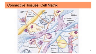
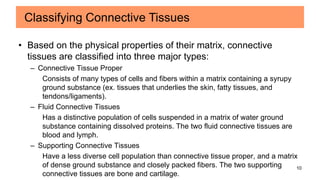
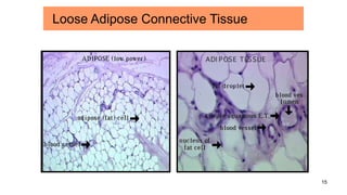
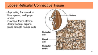
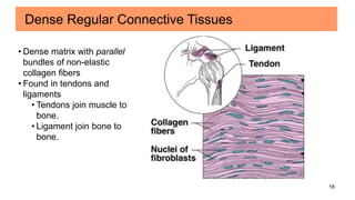
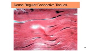
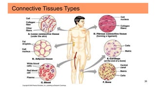
1
Unit 3
Cells, Tissues & Membranes
SECTION 2: CONNECTIVE TISSUES
Anatomy & Physiology
NeQuelle DeFord
2
• Most abundant and diverse tissues of the body.
• Grouped because of similar developmental origin.
• Distributed throughout the body but never exposed to the
outside environment.
• Many are highly vascular and innervated.
• Have three basic components:
1. Specialized Cell(s)
– Matrix
2. Solid, extracellular protein fibers
3. Fluid, extracellular ground substance – amorphous, clear viscous
material that fills the space between cells (slows movement of pathogen)
Connective Tissues: Characteristics
3
• Mechanical Support and Protection
– Establishment of a bony structural framework for the body that
protects delicate organs.
• Metabolic Functions
– Medium for the exchange of nutrients and waste products
• Energy Store and Thermal Regulation
• Defense
– Barrier
– Engulf invading microorganisms
– Antibody production
Connective Tissues: Functions
4
• Fibroblasts/cytes
–most abundant cells in connective tissue proper
–produce/maintains connective tissue fibers and ground
substance
• Macrophages
–scattered throughout the matrix
–phagocytize (engulf) damaged cells or pathogens
–release chemicals the mobilize the immune system
Cells of Connective Tissue
5
• Adipoblasts/cytes
–fat cells
–permanent residents
–number of fat cells vary
• Mast Cells
–small, mobile cells often found near blood vessels
–cytoplasm is packed with vesicles filled with chemicals that
are released to begin the body’s defensive activities after
an injury or infection
Cells of Connective Tissue
6
• Chondroblasts/cytes
–produce/maintain cartilage
• Osteobasts/cytes
–produce/maintain bone
• Hematopoietic stem cells
–produce/maintain blood
• Plasma Cells
–produce antibodies
Cells of Connective Tissue
7
1. Collagen Fibers
• long, straight, unbranched
• strong, but flexible
• most common
• Example: Achilles tendon
2. Elastic Fibers
• branched and wavy
• return to their original length after stretching
• contain the protein elastin
• Example: skin
3 Basic Types of Connective Tissue Fibers
8
3. Reticular Fibers
• made of same protein subunit as collagen fibers but
arranged differently
• thinner than collagen fibers
• form branching, interwoven framework in various
organs
• least common
3 Basic Types of Connective Tissue Fibers
9
Connective Tissues: Cell Matrix
10
• Based on the physical properties of their matrix, connective
tissues are classified into three major types:
– Connective Tissue Proper
Consists of many types of cells and fibers within a matrix containing a syrupy
ground substance (ex. tissues that underlies the skin, fatty tissues, and
tendons/ligaments).
– Fluid Connective Tissues
Has a distinctive population of cells suspended in a matrix of water ground
substance containing dissolved proteins. The two fluid connective tissues are
blood and lymph.
– Supporting Connective Tissues
Have a less diverse cell population than connective tissue proper, and a matrix
of dense ground substance and closely packed fibers. The two supporting
connective tissues are bone and cartilage.
Classifying Connective Tissues
11
• Loose Connective Tissue
–“Packing materials” of the body
–More ground substance, fewer fibers
–Example: areolar tissue, adipose tissue, reticular tissue
• Dense Connective Tissue
– Tough, strong, and durable
– More fibers, less ground substance
– Example: dense regular (tendons/ligaments), dense
irregular
Connective Tissue Proper
12
Fibroblasts
Elastic fibers
Collagen fibers
Location: subcutaneous layer of skin
Function: wraps and cushions organs, provides strength, elasticity, and support
Loose Areolor Connective Tissue
Gel-like ground, with both
elastic and non-elastic fibers
running through the ground in
many different directions.
13
Loose Areolar Connective Tissues
14
Blood vessel
•Contains fat pads and
insulates the body;
supports and protects
•Stores energy
•Each cell contains a large
fat droplet that swells when
fat is stored; shrinks when
fat is used
•Very little matrix
Loose Adipose Connective Tissue
15
Loose Adipose Connective Tissue
16
• Supporting framework of
liver, spleen, and lymph
nodes
• Function: forms stroma
(framework) of organs,
binds smooth muscle cells
Loose Reticular Connective Tissue
17
Loose Reticular Connective Tissue
18
Dense Regular Connective Tissues
• Dense matrix with parallel
bundles of non-elastic
collagen fibers
• Found in tendons and
ligaments
• Tendons join muscle to
bone.
• Ligament join bone to
bone.
19
Dense Regular Connective Tissues
20
Dense Irregular Connective Tissues
• Irregularly arranged collagen fibers
• Able to withstand tension exerted in many directions
• Located in skin and fibrous joint capsules.
21
Dense Irregular Connective Tissues
22
Dense Elastic Connective Tissues
• Found in aorta
• Stretches and recoils to
propel blood.
23
Dense Elastic Connective Tissues
aorta
24
• Bone
–Strong (hard calcium compounds)
–Resists shattering (flexible collagen fibers)
–Bone cells (osteocytes) arranged around central canals
within matrix.
Supporting Connective Tissue
25
• Cartilage
– Hyaline Cartilage
• Stiff, flexible support
• Reduces friction between bones
• Found in synovial joints, rib tips, sternum, and trachea
– Elastic Cartilage
• Supportive but bends easily
• Found in external ear and epiglottis
– Fibrocartilage
• Limits movement
• Prevents bone-to-bone contact
• Found between pubic bones and intervertebral discs
Supporting Connective Tissue
26
Bone (Osseous Tissue)
• Hard, calcified matrix containing many collagen fibers that forms the skeleton
• Very well vascularized and innervated
• Supports, protects, and provides levers for muscles to act on
27
Hyaline Cartilage
• Most abundant in the body
• Firm matrix; provides firm
support
• Composes entire fetal
skeleton
• Nose tip is hyaline cartilage
• It covers long ends on bones
and provides compression on
joints, preventing bones from
rubbing against one another.
• Holds trachea open
28
Larynx
Hyaline Cartilage
29
Elastic Cartilage
• Similar to hyaline cartilage but with
more elastic fibers
• Provides shape and structure with
flexibility
• Supports ear
30
Elastic Cartilage
31
Fibrocartilage
• Thick collagen fibers
• Can absorb shock; it is
compressible and has tension
resistance
• Found where heavy amounts
of pressure are required, such
as in inter-vertebral joints and
knee joints
32
Fibrocartilage
33
• Blood and Lymph
–Watery matrix of dissolved proteins
–Carry specific cell types (formed elements)
• Formed elements of blood
–Erythrocytes (Red Blood Cells or RBC): transport
–Leucocytes (White Blood Cells or WBC): defense
–Thrombocytes (platelets): protection
Fluid Connective Tissues
34
Blood (Vascular Tissue)
• Liquid matrix of nonliving fluid
called blood plasma
•90% water, 10% plasma
proteins, electrolytes,
hormones, oxygen, glucose,
etc
• Transports gases, nutrients,
wastes and other substances
• About ½ cells and ½ fluid
35
Blood
• Formed elements:
(a) Erythrocytes (RBCs) – 48 billion (females) to 54 billion (males) cells/ml
of blood in humans. Mammals are anucleated while the rest of the
vertebrates have nuclei. RBC’s contain hemoglobin to allow for oxygen to be
carried to the tissues.
(b, c, & d) Leukocytes (WBCs) – about 7.5 million cells/ml of blood
Thrombocytes (Platelets) – blood clotting
36
Connective Tissues Types

Connective Tissue PPT

  • 1.
    1 Unit 3 Cells, Tissues& Membranes SECTION 2: CONNECTIVE TISSUES Anatomy & Physiology NeQuelle DeFord
  • 2.
    2 • Most abundantand diverse tissues of the body. • Grouped because of similar developmental origin. • Distributed throughout the body but never exposed to the outside environment. • Many are highly vascular and innervated. • Have three basic components: 1. Specialized Cell(s) – Matrix 2. Solid, extracellular protein fibers 3. Fluid, extracellular ground substance – amorphous, clear viscous material that fills the space between cells (slows movement of pathogen) Connective Tissues: Characteristics
  • 3.
    3 • Mechanical Supportand Protection – Establishment of a bony structural framework for the body that protects delicate organs. • Metabolic Functions – Medium for the exchange of nutrients and waste products • Energy Store and Thermal Regulation • Defense – Barrier – Engulf invading microorganisms – Antibody production Connective Tissues: Functions
  • 4.
    4 • Fibroblasts/cytes –most abundantcells in connective tissue proper –produce/maintains connective tissue fibers and ground substance • Macrophages –scattered throughout the matrix –phagocytize (engulf) damaged cells or pathogens –release chemicals the mobilize the immune system Cells of Connective Tissue
  • 5.
    5 • Adipoblasts/cytes –fat cells –permanentresidents –number of fat cells vary • Mast Cells –small, mobile cells often found near blood vessels –cytoplasm is packed with vesicles filled with chemicals that are released to begin the body’s defensive activities after an injury or infection Cells of Connective Tissue
  • 6.
    6 • Chondroblasts/cytes –produce/maintain cartilage •Osteobasts/cytes –produce/maintain bone • Hematopoietic stem cells –produce/maintain blood • Plasma Cells –produce antibodies Cells of Connective Tissue
  • 7.
    7 1. Collagen Fibers •long, straight, unbranched • strong, but flexible • most common • Example: Achilles tendon 2. Elastic Fibers • branched and wavy • return to their original length after stretching • contain the protein elastin • Example: skin 3 Basic Types of Connective Tissue Fibers
  • 8.
    8 3. Reticular Fibers •made of same protein subunit as collagen fibers but arranged differently • thinner than collagen fibers • form branching, interwoven framework in various organs • least common 3 Basic Types of Connective Tissue Fibers
  • 9.
  • 10.
    10 • Based onthe physical properties of their matrix, connective tissues are classified into three major types: – Connective Tissue Proper Consists of many types of cells and fibers within a matrix containing a syrupy ground substance (ex. tissues that underlies the skin, fatty tissues, and tendons/ligaments). – Fluid Connective Tissues Has a distinctive population of cells suspended in a matrix of water ground substance containing dissolved proteins. The two fluid connective tissues are blood and lymph. – Supporting Connective Tissues Have a less diverse cell population than connective tissue proper, and a matrix of dense ground substance and closely packed fibers. The two supporting connective tissues are bone and cartilage. Classifying Connective Tissues
  • 11.
    11 • Loose ConnectiveTissue –“Packing materials” of the body –More ground substance, fewer fibers –Example: areolar tissue, adipose tissue, reticular tissue • Dense Connective Tissue – Tough, strong, and durable – More fibers, less ground substance – Example: dense regular (tendons/ligaments), dense irregular Connective Tissue Proper
  • 12.
    12 Fibroblasts Elastic fibers Collagen fibers Location:subcutaneous layer of skin Function: wraps and cushions organs, provides strength, elasticity, and support Loose Areolor Connective Tissue Gel-like ground, with both elastic and non-elastic fibers running through the ground in many different directions.
  • 13.
  • 14.
    14 Blood vessel •Contains fatpads and insulates the body; supports and protects •Stores energy •Each cell contains a large fat droplet that swells when fat is stored; shrinks when fat is used •Very little matrix Loose Adipose Connective Tissue
  • 15.
  • 16.
    16 • Supporting frameworkof liver, spleen, and lymph nodes • Function: forms stroma (framework) of organs, binds smooth muscle cells Loose Reticular Connective Tissue
  • 17.
  • 18.
    18 Dense Regular ConnectiveTissues • Dense matrix with parallel bundles of non-elastic collagen fibers • Found in tendons and ligaments • Tendons join muscle to bone. • Ligament join bone to bone.
  • 19.
  • 20.
    20 Dense Irregular ConnectiveTissues • Irregularly arranged collagen fibers • Able to withstand tension exerted in many directions • Located in skin and fibrous joint capsules.
  • 21.
  • 22.
    22 Dense Elastic ConnectiveTissues • Found in aorta • Stretches and recoils to propel blood.
  • 23.
  • 24.
    24 • Bone –Strong (hardcalcium compounds) –Resists shattering (flexible collagen fibers) –Bone cells (osteocytes) arranged around central canals within matrix. Supporting Connective Tissue
  • 25.
    25 • Cartilage – HyalineCartilage • Stiff, flexible support • Reduces friction between bones • Found in synovial joints, rib tips, sternum, and trachea – Elastic Cartilage • Supportive but bends easily • Found in external ear and epiglottis – Fibrocartilage • Limits movement • Prevents bone-to-bone contact • Found between pubic bones and intervertebral discs Supporting Connective Tissue
  • 26.
    26 Bone (Osseous Tissue) •Hard, calcified matrix containing many collagen fibers that forms the skeleton • Very well vascularized and innervated • Supports, protects, and provides levers for muscles to act on
  • 27.
    27 Hyaline Cartilage • Mostabundant in the body • Firm matrix; provides firm support • Composes entire fetal skeleton • Nose tip is hyaline cartilage • It covers long ends on bones and provides compression on joints, preventing bones from rubbing against one another. • Holds trachea open
  • 28.
  • 29.
    29 Elastic Cartilage • Similarto hyaline cartilage but with more elastic fibers • Provides shape and structure with flexibility • Supports ear
  • 30.
  • 31.
    31 Fibrocartilage • Thick collagenfibers • Can absorb shock; it is compressible and has tension resistance • Found where heavy amounts of pressure are required, such as in inter-vertebral joints and knee joints
  • 32.
  • 33.
    33 • Blood andLymph –Watery matrix of dissolved proteins –Carry specific cell types (formed elements) • Formed elements of blood –Erythrocytes (Red Blood Cells or RBC): transport –Leucocytes (White Blood Cells or WBC): defense –Thrombocytes (platelets): protection Fluid Connective Tissues
  • 34.
    34 Blood (Vascular Tissue) •Liquid matrix of nonliving fluid called blood plasma •90% water, 10% plasma proteins, electrolytes, hormones, oxygen, glucose, etc • Transports gases, nutrients, wastes and other substances • About ½ cells and ½ fluid
  • 35.
    35 Blood • Formed elements: (a)Erythrocytes (RBCs) – 48 billion (females) to 54 billion (males) cells/ml of blood in humans. Mammals are anucleated while the rest of the vertebrates have nuclei. RBC’s contain hemoglobin to allow for oxygen to be carried to the tissues. (b, c, & d) Leukocytes (WBCs) – about 7.5 million cells/ml of blood Thrombocytes (Platelets) – blood clotting
  • 36.