Download as PDF, PPTX


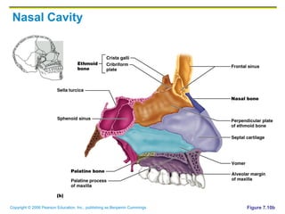


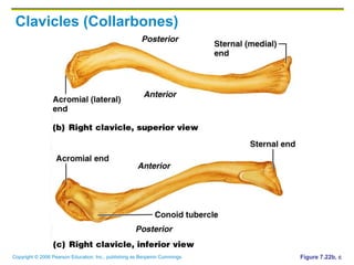


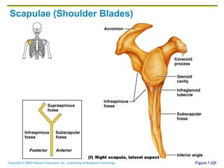


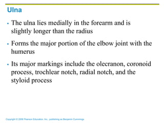


The document provides information about the skeletal system, including: 1. It describes the two divisions of the skeleton - the axial skeleton which includes the skull, vertebral column, and thorax, and the appendicular skeleton which includes the limbs and girdles. 2. It discusses the main functions of bones which are support, protection, movement, storage, and blood cell formation. 3. It provides details about the types of bones, classifications of bones, bones of the axial skeleton including the skull, vertebrae, and thoracic cage.


![Human Reproduction [ Reproductive System ] Notes @irfanullah_mehar Irfanullah...](https://cdn.slidesharecdn.com/ss_thumbnails/humanreproductionreproductivesystemnotesirfanullahmeharirfanullahmeharjanantantra-260111172350-56e85778-thumbnail.jpg?width=640&height=640&fit=bounds)