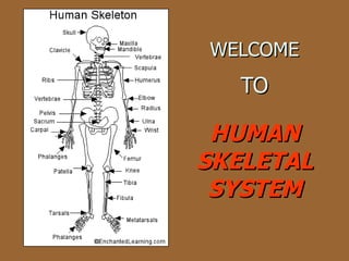
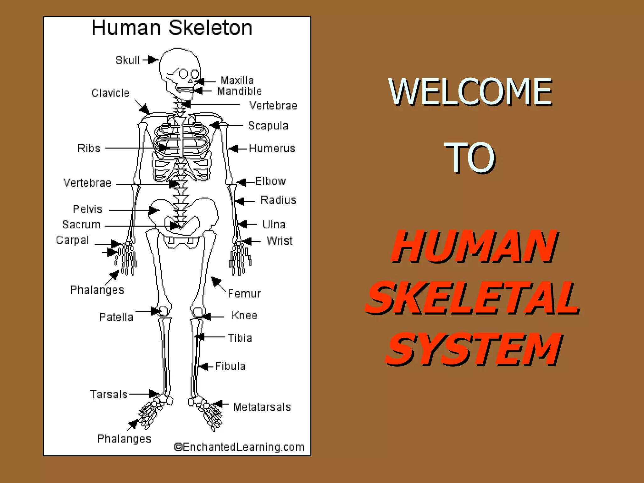
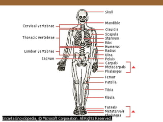
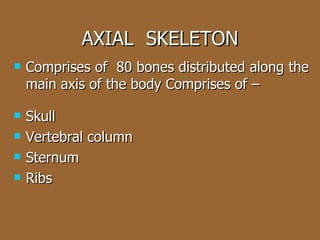
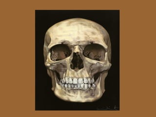
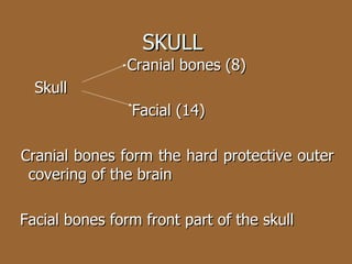
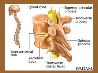
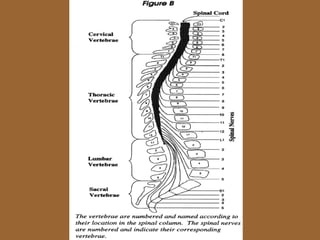
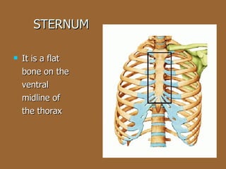
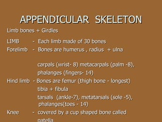
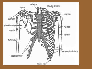
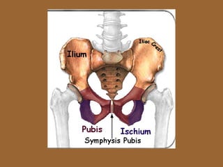
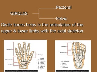
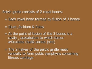
WELCOME   TO HUMAN SKELETAL SYSTEM
PRESENTED BY MR.ATUL MANKAR P.G.T. (BIOLOGY) Kendriya Vidyalaya DIPHU
OBJECTIVES To impart knowledge about human  skeletal system To make the viewers realize their body  framework
CONTENTS INTRODUCTION DIVISIONS OF SKELETON FUNCTIONS
The human skeletal system consists  of frame work of      Cartilages    Bones  (206)  (few) INTRODUCTION
Bones & Cartilages are specialized connective  tissues BONES  CARTILAGE Has hard matrix  Has slightly pliable  matrix Presence of Ca salts  Presence of chondroitin  salts
 
  Human Skeleton   Grouped into 2 main divisions Axial skeleton  Appendicular skeleton HUMAN  SKELETON
AXIAL  SKELETON Comprises of  80 bones distributed along the main axis of the body Comprises of – Skull Vertebral column  Sternum  Ribs
 
SKULL  Cranial bones (8) Skull  Facial (14) Cranial bones form the hard protective outer covering of the brain Facial bones form front part of the skull
Hyoid It is a single U shaped bone present at the base of buccal cavity  Also included in the skull
Our each middle ear contain 3 tiny bones- Mallaeus ,Incus & Stapes Called ear ossicles
Human skull is  dicondylic  as it articulates with the superior region of vertebral column with the help of 2 occipital condyles.
VERTEBRAL COLUMN Formed of 26 serially arranged units called vertebrae It is dorsally placed Extends from base of the skull Constitutes main framework of the trunk The spinal cord passes through the central hollow neural canal of the vertebrae
 
 
1 st  vertebra - Atlas It articulates with the occipital condyles 7 cervical vertebrae - make up the neck part 12 thoracic vertebrae 5 lumbar vertebrae 1 fused sacral vertebrae 1 fused coccygeal vertebrae
Function of Vertebral Column Protects the spinal cord Supports the head Serves as point of attachment for the ribs & musculature of the back
STERNUM It is a flat bone on the ventral midline of the thorax
 
RIBS 12 pairs of ribs Each rib is a thin flat bone connected dorsally to the vertebral column & ventrally to the sternum Has 2 articulation surface on it’s dorsal end (dicephalic) 1 st  7pairs of ribs -true ribs. attached dorsally to the  vertebral column & ventrally to the  sternum with the help of hyaline cartilage 8 th  ,9 th & 10 th  pairs of ribs called false ribs. Do not articulate directly with the sternum but join the 7 th  rib with the  help of hyaline cartilage (vertebrochondral rib) 11 th  & 12 th  pairs of ribs called floating ribs. They are not connected ventrally RIB CAGE  ---  THORACIC VERTEBRAE + RIBS+ STERNUM
 
APPENDICULAR  SKELETON Limb bones + Girdles LIMB  -  Each limb made of 30 bones Forelimb  -  Bones are humerus , radius  + ulna  carpals (wrist- 8) metacarpals (palm -8),  phalanges (fingers- 14) Hind limb  - Bones are femur (thigh bone - longest) tibia + fibula tarsals  (ankle-7), metatarsals (sole -5),   phalanges(toes - 14) Knee  -  covered by a cup shaped bone called  patella
 
 
  Pectoral GIRDLES        Pelvic Girdle bones helps in the articulation of the  upper & lower limbs with the axial skeleton
Each half of pectoral girdle consists of a CLAVICLE & a SCAPULA Scapula-large triangular flat bone Situated in the dorsal part of the thorax between the 2 nd  & 7 th  ribs Dorsal side has a slightly elevated ridge called spine which extends as a flat expanded  acromian process   It articulates with the clavicle Below acromian a depression called glenoid cavity which atriculates with head of humerus to form shoulder joint (ball & socket joint) Clavicle  -  called collar bone   -  Long slender bone with 2 curvatures
Pelvic girdle consists of 2 coxal bones: = Each coxal bone formed by fusion of 3 bones = Ilium ,Ischium & Pubis = At the point of fusion of the 3 bones is a  cavity , acetabulum to which femur  articulates (ball& socket joint) = The 2 halves of the pelvic girdle meet  ventrally to form pubic symphysis containing  fibrous cartilage

Skeletal System

  • 1.
    WELCOME TO HUMAN SKELETAL SYSTEM
  • 2.
    PRESENTED BY MR.ATULMANKAR P.G.T. (BIOLOGY) Kendriya Vidyalaya DIPHU
  • 3.
    OBJECTIVES To impartknowledge about human skeletal system To make the viewers realize their body framework
  • 4.
    CONTENTS INTRODUCTION DIVISIONSOF SKELETON FUNCTIONS
  • 5.
    The human skeletalsystem consists of frame work of Cartilages Bones (206) (few) INTRODUCTION
  • 6.
    Bones & Cartilagesare specialized connective tissues BONES CARTILAGE Has hard matrix Has slightly pliable matrix Presence of Ca salts Presence of chondroitin salts
  • 7.
  • 8.
    HumanSkeleton Grouped into 2 main divisions Axial skeleton Appendicular skeleton HUMAN SKELETON
  • 9.
    AXIAL SKELETONComprises of 80 bones distributed along the main axis of the body Comprises of – Skull Vertebral column Sternum Ribs
  • 10.
  • 11.
    SKULL Cranialbones (8) Skull Facial (14) Cranial bones form the hard protective outer covering of the brain Facial bones form front part of the skull
  • 12.
    Hyoid It isa single U shaped bone present at the base of buccal cavity Also included in the skull
  • 13.
    Our each middleear contain 3 tiny bones- Mallaeus ,Incus & Stapes Called ear ossicles
  • 14.
    Human skull is dicondylic as it articulates with the superior region of vertebral column with the help of 2 occipital condyles.
  • 15.
    VERTEBRAL COLUMN Formedof 26 serially arranged units called vertebrae It is dorsally placed Extends from base of the skull Constitutes main framework of the trunk The spinal cord passes through the central hollow neural canal of the vertebrae
  • 16.
  • 17.
  • 18.
    1 st vertebra - Atlas It articulates with the occipital condyles 7 cervical vertebrae - make up the neck part 12 thoracic vertebrae 5 lumbar vertebrae 1 fused sacral vertebrae 1 fused coccygeal vertebrae
  • 19.
    Function of VertebralColumn Protects the spinal cord Supports the head Serves as point of attachment for the ribs & musculature of the back
  • 20.
    STERNUM It isa flat bone on the ventral midline of the thorax
  • 21.
  • 22.
    RIBS 12 pairsof ribs Each rib is a thin flat bone connected dorsally to the vertebral column & ventrally to the sternum Has 2 articulation surface on it’s dorsal end (dicephalic) 1 st 7pairs of ribs -true ribs. attached dorsally to the vertebral column & ventrally to the sternum with the help of hyaline cartilage 8 th ,9 th & 10 th pairs of ribs called false ribs. Do not articulate directly with the sternum but join the 7 th rib with the help of hyaline cartilage (vertebrochondral rib) 11 th & 12 th pairs of ribs called floating ribs. They are not connected ventrally RIB CAGE --- THORACIC VERTEBRAE + RIBS+ STERNUM
  • 23.
  • 24.
    APPENDICULAR SKELETONLimb bones + Girdles LIMB - Each limb made of 30 bones Forelimb - Bones are humerus , radius + ulna carpals (wrist- 8) metacarpals (palm -8), phalanges (fingers- 14) Hind limb - Bones are femur (thigh bone - longest) tibia + fibula tarsals (ankle-7), metatarsals (sole -5), phalanges(toes - 14) Knee - covered by a cup shaped bone called patella
  • 25.
  • 26.
  • 27.
    PectoralGIRDLES Pelvic Girdle bones helps in the articulation of the upper & lower limbs with the axial skeleton
  • 28.
    Each half ofpectoral girdle consists of a CLAVICLE & a SCAPULA Scapula-large triangular flat bone Situated in the dorsal part of the thorax between the 2 nd & 7 th ribs Dorsal side has a slightly elevated ridge called spine which extends as a flat expanded acromian process It articulates with the clavicle Below acromian a depression called glenoid cavity which atriculates with head of humerus to form shoulder joint (ball & socket joint) Clavicle - called collar bone - Long slender bone with 2 curvatures
  • 29.
    Pelvic girdle consistsof 2 coxal bones: = Each coxal bone formed by fusion of 3 bones = Ilium ,Ischium & Pubis = At the point of fusion of the 3 bones is a cavity , acetabulum to which femur articulates (ball& socket joint) = The 2 halves of the pelvic girdle meet ventrally to form pubic symphysis containing fibrous cartilage