Downloaded 106 times


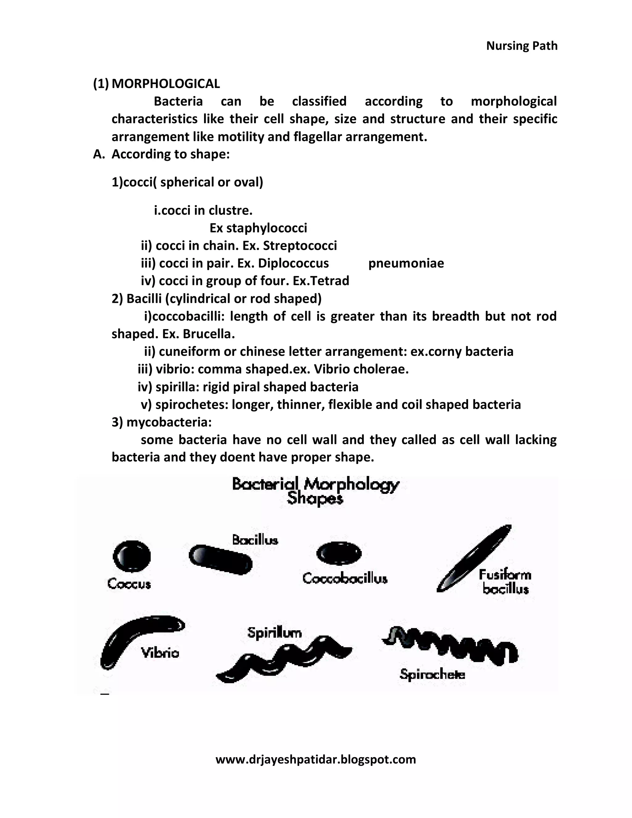


The document extensively covers characteristics, classification, and anatomy of bacteria, detailing their unicellular nature, reproduction through binary fission, and fundamental structures like the cell wall and flagella. It also discusses various classification systems based on morphology, biochemical reactions, staining responses, and pathogenicity, as well as important bacterial features such as spores and capsules. Key differences between gram-positive and gram-negative bacteria are highlighted, particularly in relation to their cell wall structures and resilience to environmental stress.