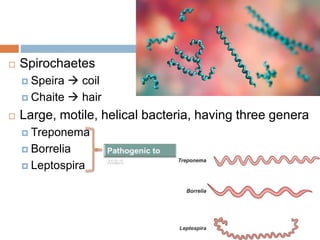
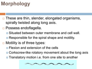
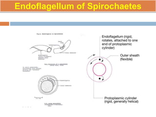
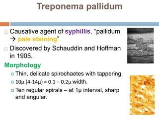
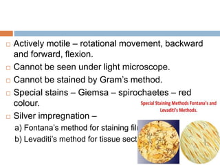
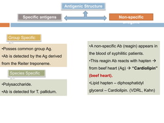
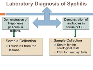
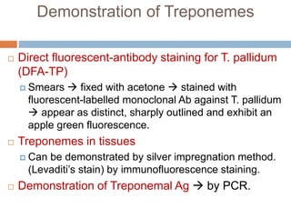
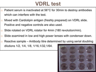
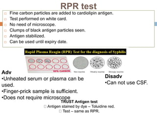
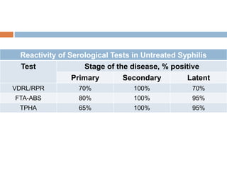
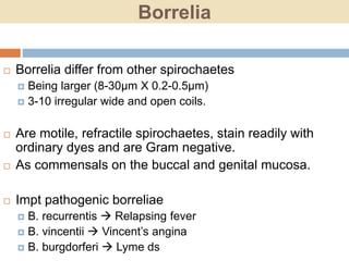
SPIROCHAETES
By Dr. Rakesh Prasad Sah
Associate Professor, Microbiology
 Spirochaetes
 Speira  coil
 Chaite  hair
 Large, motile, helical bacteria, having three genera
 Treponema
 Borrelia
 Leptospira
Morphology
 These are thin, slender, elongated organisms,
spirally twisted along long axis.
 Possess endoflagella.
 Situated between outer membrane and cell wall.
 Responsible for the spiral shape and motility.
 Motility is of three types:
 Flexion and extension of the cells
 Corkscrew-like rotatory movement about the long axis
 Translatory motion i.e. from one site to another
Endoflagellum of Spirochaetes
 Gram negative type cell wall composed of an outer
membrane, a peptidoglycan layer and a
cytoplasmic membrane.
 Can be seen by Dark ground microscopy, silver
impregnation method and immunoflurescence.
Diseases
Genus Species Disease
Treponema T. Pallidum Syphilis
T. Pertenue Yaws
T. Carateum Pinta
T. Endemicum Endemic syphilis
Borrelia B. Recurrentis Relapsing fever
B. Vincentii Vincent’s angina
B. Burgdorferi Lyme disease
Leptospira L. Interrogans Leptospirosis
L. Biflexa Saprophytes
Treponema
 Treponemes
 Trepos  turn
 Nema  thread
 Are slender spirochaetes with fine spirals
having pointed ends.
 Some of them are commensals in mouth and
genitalia while others are pathogenic
 T.pallidum – syphilis
 T.pertenue – yaws (In Africa, Asia and USA)
 T.carateum – pinta
Treponema pallidum
 Causative agent of syphillis. “pallidum
 pale staining”
 Discovered by Schauddin and Hoffman
in 1905.
Morphology
 Thin, delicate spirochaetes with tappering.
 10μ (4-14μ) × 0.1 – 0.2μ width.
 Ten regular spirals – at 1μ interval, sharp
and angular.
 Actively motile – rotational movement, backward
and forward, flexion.
 Cannot be seen under light microscope.
 Cannot be stained by Gram’s method.
 Special stains – Giemsa – spirochaetes – red
colour.
 Silver impregnation –
a) Fontana’s method for staining films.
b) Levaditi’s method for tissue sections.
Cultivation
 Pathogenic treponemes do not grow in artificial
culture media but are maintained by subcuture in
susceptible animals.
 can be maintained for years by testicular passage
in rabbits.
 Nicholl’s strain – virulent strains
 Non pathogenic strain ( Reiter strain) can be
cultivated in
cultures. (Thioglycollate medium).
Antigenic Structure
Non-specific
antigens
Specific antigens
Group Specific
Ag
Species Specific
Ag
•Posses common group Ag.
•Ab is detected by the Ag derived
from the Reiter treponeme.
•Polysaccharide.
•Ab is detected for T. pallidum.
•A non-specific Ab (reagin) appears in
the blood of syphilitic patients.
•This reagin Ab reacts with hapten 
from beef heart (Ag)  “Cardiolipin”
(beef heart).
•Lipid hapten – diphosphatidyl
glycerol – Cardiolipin. (VDRL, Kahn)
Pathogenesis
 Treponema pallidum causes syphilis.
 Acquired by:
 1. Sexual contact.
 2. Congenital infection from infected mother.
 3. Iatrogenic.
 Infective dose is small – 60 treponemes can infect
50% contacts.
 Slow multiplication – generation time 30-33 hrs.
 Incubation period – about 1 month (10-90 days).
 Three clinical stages of the disease in an untreated
case – Primary, Secondary and Tertiary
Primary Syphilis
 A papule  genital area 
ulcerates  forming a
classical chancre of primary
syphillis, called hard
chancre.
 Painless, relatively
avascular, indurated,
covered by a thick exudates
 rich in spirochaetes.
 Regional lymph nodes are
swollen, discrete, non-tender
Primary Syphilis
 Entry site  lymph & bloodstream, even prior to appearance of
chancre, hence patient may be infectious during the late
incubation period.
 Chancre heals within 10-40 days even without treatment,
leaving scar.
 Persistent or multiple chancres  seen in HIV or
immunocompromised patients.
Secondary Syphilis
 After primary lesion heals the patient remains asymptomatic for
2-6 months  secondary syphilis sets in.
 Sec lesions  widespread multiplication of the treponemes and
dissemination through the blood.
 Characterised by macular rash on mucous membranes and
skin which contain numerous treponemes.
 Is most infectious during secondary stage.
Secondary Syphilis
 Rash  coalesce in
intertrigionous area especially in
the perianal region, producing
wart-like condylomata.
 May be retinitis, meningitis,
periostitis and arthritis.
 Lesions  spontaneous healing
in some instances  as long as
4-5yrs.
 After sec lesion disappears 
30% of cases remain dormant
(latent) for many years without
any clinical symptoms but with
positive serology.
 In many cases, natural cure
Tertiary Syphilis or late stage
 Contain few treponemes.
 May be CVS lesions including aneurysms,
chronic granulomata (gummata) and
meningovascular manifestation.
 In few cases, neurosyphilis or general
paralysis develop. These are known as late
tertiary or quaternary syphilis.
Congenital Syphilis
 Can cross placental barrier occurs from primary and
secondary infection of the mother
 Usually develop after the fourth month of gestation.
 Antenatal screening and treatment of positive cases
during pregnancy may prevent congenital syphillis.
 Untreated syphilitic pregnant woman may lead to
abortions and stillbirths.
Syphilis acquired non-venereally
 doctors or nurses contact with patient’s lesion 
examination.
 Chancre is extra-genital  usually on fingers than
venereal syphilis.
 Rarely when syphilis is transmitted by blood
transfusion, the primary chancre does not develop.
Laboratory Diagnosis of Syphilis
 Sample Collection
 Exudates from the
lesions
Demonstration of
Treponema
pallidum in
lesions.
Demonstration of
antibodies in
serum or CSF.
 Sample Collection
 Serum for the
serological tests
 CSF for neurosyphillis.
Demonstration of Treponemes
 Method of sample collection – Clean with saline.
Scrape.
 Apply pressure at base of lesion.
 Serous exudate collected in capillary tubes. Sealed
and transported to laboratory.
 Wet films and smears are prepared.
 Wet Films – Examined by Dark ground
microscope.
Demonstration of Treponemes
 Direct fluorescent-antibody staining for T. pallidum
(DFA-TP)
 Smears  fixed with acetone  stained with
fluorescent-labelled monoclonal Ab against T. pallidum
 appear as distinct, sharply outlined and exhibit an
apple green fluorescence.
 Treponemes in tissues
 Can be demonstrated by silver impregnation method.
(Levaditi’s stain) by immunofluorescence staining.
 Demonstration of Treponemal Ag  by PCR.
Serological Tests
 Non-treponemal Tests
 In the standard test for syphillis (STS)  reagin Abs 
detected by cardiolipin Ag (extracted from beef heart
tissue, in which lecithin and cholesterol are added).
 It includes, VDRL, RPR, kahn test and Wassermann
reaction.
Non-specific Ab
(reagin Ab)
Secific anti-treponemal
Ab
Non-treponemal tests
(cardiolipin or lipoidal
Ag)
Treponemal tests
(treponemes are used
as the Ag)
Flocculation tests CFT
VDRL test
 Patient serum is inactivated at 56°C for 30min to destroy antibodies
which can interfere with the test.
 Mixed with Cardiolipin antigen (freshly prepared) on VDRL slide.
Positive and negative controls are also used.
 Slide rotated on VDRL rotator for 4min (180 revolution/min).
 Slide examined in low and high power lenses with condenser down.
 Reactive sample – Antibody titre determined by using serial doubling
dilutions 1/2, 1/4, 1/8, 1/16,1/32,1/64.
RPR test
 Fine carbon particles are added to cardiolipin antigen.
 Test performed on white card.
 No need of microscope.
 Clumps of black antigen particles seen.
 Antigen stabilized.
 Can be used until expiry date.
Adv
•Unheated serum or plasma can be
used.
•Finger-prick sample is sufficient.
•Does not require microscope
Disadv
•Can not use CSF.
TRUST Antigen test
Antigen stained by dye – Toluidine red.
Test – same as RPR.
Biological false positive
reactions
 STS can give positive
reactions in many other
diseases.
 Acute biological false
positive reactions
(lasting few
weeks/months)
 Leprosy. (lepromatous
leprosy)
 Malaria.
 Relapsing fever.
 Infectious mononucleosis.
 Hepatitis
Chronic Biological false
positive
(lasting > 6 months)
•Systemic lupus
erythematosus.
•Other collagen diseases like
•rheumatoid arthritis,
•polyarteritis nodosa.
•Samples giving positive
reactions with VDRL, RPR
should
be retested with more specific
tests like FTA-Abs or
TPHA.
Treponemal Tests
 Group specific test- using cultivate Reiter’s strain –
RPCF.
 Specific tests – using pathogenic Treponemes
(Nicolle’s strain).
 Using live T. pallidum
 TPI (Treponema pallidum immobilisation test)
 Using Killed T. pallidum
 TPA (Treponema pallidum agglutionation test)
 TPIA (Treponema pallidum immune adherence test)
 Fluorescent treponemal antibody (FTA) test
 Using an extract of T. pallidum
 Treponema pallidum haemagglutination assay (TPHA)
 Enzyme immunoassay (EIA)
 Using live T. pallidum
 TPI (Treponema pallidum immobilisation test)  serum +
actively motile Nichol’s strain of T. pallidum  incubated 
Abs +nt  treponemes are immobilised (non-motile) 
dark ground illumination.
 Using Killed T. pallidum
 TPA (Treponema pallidum agglutination test)  T. pallidum
inactivated by formalin mixed with serum  examined
under dark ground microscopy found agglutinated in the
+ce of Abs.
 TPIA (Treponema pallidum immune adherence test) 
suspension of inactivated T. pallidum  mixed with serum,
complement and fresh heparinized whole blood from
normal individual  incubated  treponemes will be found
to adhere to the RBCs.
 FTA (Fluorescent treponemal antibody test)
 Is an indirect immunofluorescence test
 Smears of killed T. pallidum (Nicholo’s strain) fixed on
slide  patient’s serum  bind  fluorescein labelled
antihuman immunoglobulin (conjugate)  incubate
washing off conjugate  examined under fluroescent
microscope.
 FTA-ABS (Fluorescent trepnemal antibody-
absorption test)
 Patient’s serum  absorbed with an extract of non-
pathogenic treponemes (Reiter treponemes) to remove
reagin and the group-reactive Ab.
 Method is same as FTA.
 High sensitive and Specific.
 Can be performed on CSF.
Using extract of T. pallidum
 TPHA (Treponema pallidum haemagglutination
assay)
 Sheep RBCs sensitized with an extract of T. pallidum
 mixed with patient’s serum  Clump formation.
 Like FTA-ABS test, serum is preabsorbed with an
extract of Reiter treponemes to remove group-reactive
Abs.
 Used to detect localised production of Abs in CSF.
 less sensitive than FTA-ABS in primary syphilis
(65%), is sensitive as FTA-ABS in other stages (II ary
and late).
 Standard confirmatory test.
Reactivity of Serological Tests in Untreated Syphilis
Test Stage of the disease, % positive
Primary Secondary Latent
VDRL/RPR 70% 100% 70%
FTA-ABS 80% 100% 95%
TPHA 65% 100% 95%
NONVENEREAL TREPONEMATOSES
Occur d/t poor standard of hygiene
1) YAWS – T. pertenue , in children ulcerating
papule in arms / legs spread - direct contact /
vector – flies II ry / III ry stages seen , I ry lesion
extra genital
2) PINTA - T. carateum , all ages non - ulcerating
extra genital papule spread – direct contact / insect
II ry lesions - hyperpigmentation / hypo CNS , CVS
- involvement rare
3)Endemic syphilis – T.endemicum EX- Bejel / Siti
 seen in children– poor personal hygiene spread –
direct contact / sharing contaminated utensils
primary chancre not seen – seen on the nipples of
the mother infected by her children manifestations-
IIry Syphilis + progresses – IIIry lesions –
gummatous lesions- skin / bones
Thank You
Borrelia
 Borrelia differ from other spirochaetes
 Being larger (8-30µm X 0.2-0.5µm)
 3-10 irregular wide and open coils.
 Are motile, refractile spirochaetes, stain readily with
ordinary dyes and are Gram negative.
 As commensals on the buccal and genital mucosa.
 Impt pathogenic borreliae
 B. recurrentis  Relapsing fever
 B. vincentii  Vincent’s angina
 B. burgdorferi  Lyme ds
Borrelia recurrentis
Louse borne
Tick borne
Epidemic
typhus
Endemic
typhus
B.
recurrentis
Morphology
 Various strains of Borrelia  morphologically indistinguishable
 exhibited some Agenic differences
 Measure 8-20 x 0.2-0.5 µm.
 Motile, Gram negative, possess 5-8 irregular spirals at intervals
of 2µm with pointed ends.
 Stains with Giemsa or Wright stains.
Culture
 Microaerophilic
 Opt tempr 28-300C.
 Grown on fluid media containing blood, serum or tissue, CAM of
chick embryo and in peritoneal cavity of mouse.
Pathogenesis
 MOT
 Body louse (Pediculus humanus corporis) in B.
recurrentis
 Tick (Ornithodorus) in B. duttoni
 I.P.  2-10 days
 Recurrent febrile episodes
 Febrile episodes of sudden onset.
 Lasting for 3-5 days intervening with afebrile periods of 4-14
days.
 Relapse can occur up to 10 days
 Subsequent episodes are shorter
 Hemorrhages
 Petechiae (a small red or purple spot caused by
bleeding into the skin.)
 Epistaxis and blood-tinged sputum are more likely in
epidemic RF
 Neurologic features
 Meningitis
 Seizure
 Focal deficits (problem with nerve, spinal cord, or
brain function)
 Paraplegia (inability to voluntarily move the lower
parts of the body)
 Pyschosis (10-30%)
Lab Diagnosis
 Sample Blood collected during febrile period
 Dark ground Microscopy
 Geimsa or Leishman stain
 Direct fluorescent Ab test (speciation by mono
Clonal Ab)
 Culture  Blood Culture
 Serology  ELISA
 Molecular Methods  RT PCR
Borrelia Vincentii (Treponema
vincentii)
 Motile spirocheate
 5-20µm long and 0.2-0.6µm wide with 3-8 coils of
variable size.
 Easily stained with
 dilute carbol fuchsin
 methyl violet and
 Gram negative.
 Is a normal pathogen of mouth commensal but a potential
pathogen.
 Under predisposing conditions such as
 Malnutrition
 Viral infections
Ulcerative
Gingivostomatitis
or
Oropharyngitis
Vincent’s Angina
Vincent’s Angina B. Vincentii
Associated with
fusiform bacilli
(Fusobacterium
fusiforme)
fusospirochetosis
Laboratory Diagnosis
 Demonstration of Spirocheates and fusiform bacilli in the
stained smears of exudates from the lesions.
 Cultivation  difficult  can be done in enriched media
anaerobically.
Borrelia burgdorferi
 First reported in 1975 in Lyme, USA.
 Causes Lyme disease.
 Morphology
 4-30µm x 0.2-0.25µm.
 Flexible, helical and Gram negative
 Reservoir
 Rodents
 Deers
Culture
 Microaerophilic
 Fastidious bacterium
 Special media  BSK (Barbour-Stoenner-Kelly) medium 
2 weeks at 33-370C.
Pathogenesis
 MOT tick bite (Ixodes ricinus complex)
 Incubation period  3-30 days.
 Clinical Manifestations
 Stage 1: Early localized infection
 Stage 2: Early disseminated infection
 Stage 3: Late persistent infection (Lyme arthritis)
Stage 1: Early localized infection
 An annular maculopapular lesion  develops at the site of tick
bite  called erythema migrans  commonly involving thigh
and groin.
Stage 2: Early disseminated infection
 Spreads hematogenously  many sites resulting in 
secondary annular skin lesions, arthralgia, malaise and
neurological abnormalities
Stage 3: Late persistent infection (Lyme arthritis)
 About 60% of the patients  develops arthritis of large joints
(e.g. knees), lasting for months  refractory to the treatment.
Laboratory Diagnosis
 Isolation
 Skin lesions
 CSF
 Blood
 Serology
 ELISA
 IF
Spirochete (Treponema and Borrelia)

Spirochete (Treponema and Borrelia)

  • 1.
    SPIROCHAETES By Dr. RakeshPrasad Sah Associate Professor, Microbiology
  • 2.
     Spirochaetes  Speira coil  Chaite  hair  Large, motile, helical bacteria, having three genera  Treponema  Borrelia  Leptospira
  • 3.
    Morphology  These arethin, slender, elongated organisms, spirally twisted along long axis.  Possess endoflagella.  Situated between outer membrane and cell wall.  Responsible for the spiral shape and motility.  Motility is of three types:  Flexion and extension of the cells  Corkscrew-like rotatory movement about the long axis  Translatory motion i.e. from one site to another
  • 4.
  • 5.
     Gram negativetype cell wall composed of an outer membrane, a peptidoglycan layer and a cytoplasmic membrane.  Can be seen by Dark ground microscopy, silver impregnation method and immunoflurescence.
  • 6.
    Diseases Genus Species Disease TreponemaT. Pallidum Syphilis T. Pertenue Yaws T. Carateum Pinta T. Endemicum Endemic syphilis Borrelia B. Recurrentis Relapsing fever B. Vincentii Vincent’s angina B. Burgdorferi Lyme disease Leptospira L. Interrogans Leptospirosis L. Biflexa Saprophytes
  • 8.
    Treponema  Treponemes  Trepos turn  Nema  thread  Are slender spirochaetes with fine spirals having pointed ends.  Some of them are commensals in mouth and genitalia while others are pathogenic  T.pallidum – syphilis  T.pertenue – yaws (In Africa, Asia and USA)  T.carateum – pinta
  • 9.
    Treponema pallidum  Causativeagent of syphillis. “pallidum  pale staining”  Discovered by Schauddin and Hoffman in 1905. Morphology  Thin, delicate spirochaetes with tappering.  10μ (4-14μ) × 0.1 – 0.2μ width.  Ten regular spirals – at 1μ interval, sharp and angular.
  • 10.
     Actively motile– rotational movement, backward and forward, flexion.  Cannot be seen under light microscope.  Cannot be stained by Gram’s method.  Special stains – Giemsa – spirochaetes – red colour.  Silver impregnation – a) Fontana’s method for staining films. b) Levaditi’s method for tissue sections.
  • 11.
    Cultivation  Pathogenic treponemesdo not grow in artificial culture media but are maintained by subcuture in susceptible animals.  can be maintained for years by testicular passage in rabbits.  Nicholl’s strain – virulent strains  Non pathogenic strain ( Reiter strain) can be cultivated in cultures. (Thioglycollate medium).
  • 12.
    Antigenic Structure Non-specific antigens Specific antigens GroupSpecific Ag Species Specific Ag •Posses common group Ag. •Ab is detected by the Ag derived from the Reiter treponeme. •Polysaccharide. •Ab is detected for T. pallidum. •A non-specific Ab (reagin) appears in the blood of syphilitic patients. •This reagin Ab reacts with hapten  from beef heart (Ag)  “Cardiolipin” (beef heart). •Lipid hapten – diphosphatidyl glycerol – Cardiolipin. (VDRL, Kahn)
  • 13.
    Pathogenesis  Treponema pallidumcauses syphilis.  Acquired by:  1. Sexual contact.  2. Congenital infection from infected mother.  3. Iatrogenic.  Infective dose is small – 60 treponemes can infect 50% contacts.  Slow multiplication – generation time 30-33 hrs.  Incubation period – about 1 month (10-90 days).  Three clinical stages of the disease in an untreated case – Primary, Secondary and Tertiary
  • 14.
    Primary Syphilis  Apapule  genital area  ulcerates  forming a classical chancre of primary syphillis, called hard chancre.  Painless, relatively avascular, indurated, covered by a thick exudates  rich in spirochaetes.  Regional lymph nodes are swollen, discrete, non-tender
  • 15.
    Primary Syphilis  Entrysite  lymph & bloodstream, even prior to appearance of chancre, hence patient may be infectious during the late incubation period.  Chancre heals within 10-40 days even without treatment, leaving scar.  Persistent or multiple chancres  seen in HIV or immunocompromised patients.
  • 16.
    Secondary Syphilis  Afterprimary lesion heals the patient remains asymptomatic for 2-6 months  secondary syphilis sets in.  Sec lesions  widespread multiplication of the treponemes and dissemination through the blood.  Characterised by macular rash on mucous membranes and skin which contain numerous treponemes.  Is most infectious during secondary stage.
  • 17.
    Secondary Syphilis  Rash coalesce in intertrigionous area especially in the perianal region, producing wart-like condylomata.  May be retinitis, meningitis, periostitis and arthritis.  Lesions  spontaneous healing in some instances  as long as 4-5yrs.  After sec lesion disappears  30% of cases remain dormant (latent) for many years without any clinical symptoms but with positive serology.  In many cases, natural cure
  • 18.
    Tertiary Syphilis orlate stage  Contain few treponemes.  May be CVS lesions including aneurysms, chronic granulomata (gummata) and meningovascular manifestation.  In few cases, neurosyphilis or general paralysis develop. These are known as late tertiary or quaternary syphilis.
  • 19.
    Congenital Syphilis  Cancross placental barrier occurs from primary and secondary infection of the mother  Usually develop after the fourth month of gestation.  Antenatal screening and treatment of positive cases during pregnancy may prevent congenital syphillis.  Untreated syphilitic pregnant woman may lead to abortions and stillbirths.
  • 20.
    Syphilis acquired non-venereally doctors or nurses contact with patient’s lesion  examination.  Chancre is extra-genital  usually on fingers than venereal syphilis.  Rarely when syphilis is transmitted by blood transfusion, the primary chancre does not develop.
  • 21.
    Laboratory Diagnosis ofSyphilis  Sample Collection  Exudates from the lesions Demonstration of Treponema pallidum in lesions. Demonstration of antibodies in serum or CSF.  Sample Collection  Serum for the serological tests  CSF for neurosyphillis.
  • 22.
    Demonstration of Treponemes Method of sample collection – Clean with saline. Scrape.  Apply pressure at base of lesion.  Serous exudate collected in capillary tubes. Sealed and transported to laboratory.  Wet films and smears are prepared.  Wet Films – Examined by Dark ground microscope.
  • 24.
    Demonstration of Treponemes Direct fluorescent-antibody staining for T. pallidum (DFA-TP)  Smears  fixed with acetone  stained with fluorescent-labelled monoclonal Ab against T. pallidum  appear as distinct, sharply outlined and exhibit an apple green fluorescence.  Treponemes in tissues  Can be demonstrated by silver impregnation method. (Levaditi’s stain) by immunofluorescence staining.  Demonstration of Treponemal Ag  by PCR.
  • 25.
    Serological Tests  Non-treponemalTests  In the standard test for syphillis (STS)  reagin Abs  detected by cardiolipin Ag (extracted from beef heart tissue, in which lecithin and cholesterol are added).  It includes, VDRL, RPR, kahn test and Wassermann reaction. Non-specific Ab (reagin Ab) Secific anti-treponemal Ab Non-treponemal tests (cardiolipin or lipoidal Ag) Treponemal tests (treponemes are used as the Ag) Flocculation tests CFT
  • 26.
    VDRL test  Patientserum is inactivated at 56°C for 30min to destroy antibodies which can interfere with the test.  Mixed with Cardiolipin antigen (freshly prepared) on VDRL slide. Positive and negative controls are also used.  Slide rotated on VDRL rotator for 4min (180 revolution/min).  Slide examined in low and high power lenses with condenser down.  Reactive sample – Antibody titre determined by using serial doubling dilutions 1/2, 1/4, 1/8, 1/16,1/32,1/64.
  • 27.
    RPR test  Finecarbon particles are added to cardiolipin antigen.  Test performed on white card.  No need of microscope.  Clumps of black antigen particles seen.  Antigen stabilized.  Can be used until expiry date. Adv •Unheated serum or plasma can be used. •Finger-prick sample is sufficient. •Does not require microscope Disadv •Can not use CSF. TRUST Antigen test Antigen stained by dye – Toluidine red. Test – same as RPR.
  • 28.
    Biological false positive reactions STS can give positive reactions in many other diseases.  Acute biological false positive reactions (lasting few weeks/months)  Leprosy. (lepromatous leprosy)  Malaria.  Relapsing fever.  Infectious mononucleosis.  Hepatitis Chronic Biological false positive (lasting > 6 months) •Systemic lupus erythematosus. •Other collagen diseases like •rheumatoid arthritis, •polyarteritis nodosa. •Samples giving positive reactions with VDRL, RPR should be retested with more specific tests like FTA-Abs or TPHA.
  • 29.
    Treponemal Tests  Groupspecific test- using cultivate Reiter’s strain – RPCF.  Specific tests – using pathogenic Treponemes (Nicolle’s strain).  Using live T. pallidum  TPI (Treponema pallidum immobilisation test)  Using Killed T. pallidum  TPA (Treponema pallidum agglutionation test)  TPIA (Treponema pallidum immune adherence test)  Fluorescent treponemal antibody (FTA) test  Using an extract of T. pallidum  Treponema pallidum haemagglutination assay (TPHA)  Enzyme immunoassay (EIA)
  • 30.
     Using liveT. pallidum  TPI (Treponema pallidum immobilisation test)  serum + actively motile Nichol’s strain of T. pallidum  incubated  Abs +nt  treponemes are immobilised (non-motile)  dark ground illumination.  Using Killed T. pallidum  TPA (Treponema pallidum agglutination test)  T. pallidum inactivated by formalin mixed with serum  examined under dark ground microscopy found agglutinated in the +ce of Abs.  TPIA (Treponema pallidum immune adherence test)  suspension of inactivated T. pallidum  mixed with serum, complement and fresh heparinized whole blood from normal individual  incubated  treponemes will be found to adhere to the RBCs.
  • 31.
     FTA (Fluorescenttreponemal antibody test)  Is an indirect immunofluorescence test  Smears of killed T. pallidum (Nicholo’s strain) fixed on slide  patient’s serum  bind  fluorescein labelled antihuman immunoglobulin (conjugate)  incubate washing off conjugate  examined under fluroescent microscope.  FTA-ABS (Fluorescent trepnemal antibody- absorption test)  Patient’s serum  absorbed with an extract of non- pathogenic treponemes (Reiter treponemes) to remove reagin and the group-reactive Ab.  Method is same as FTA.  High sensitive and Specific.  Can be performed on CSF.
  • 32.
    Using extract ofT. pallidum  TPHA (Treponema pallidum haemagglutination assay)  Sheep RBCs sensitized with an extract of T. pallidum  mixed with patient’s serum  Clump formation.  Like FTA-ABS test, serum is preabsorbed with an extract of Reiter treponemes to remove group-reactive Abs.  Used to detect localised production of Abs in CSF.  less sensitive than FTA-ABS in primary syphilis (65%), is sensitive as FTA-ABS in other stages (II ary and late).  Standard confirmatory test.
  • 33.
    Reactivity of SerologicalTests in Untreated Syphilis Test Stage of the disease, % positive Primary Secondary Latent VDRL/RPR 70% 100% 70% FTA-ABS 80% 100% 95% TPHA 65% 100% 95%
  • 34.
    NONVENEREAL TREPONEMATOSES Occur d/tpoor standard of hygiene 1) YAWS – T. pertenue , in children ulcerating papule in arms / legs spread - direct contact / vector – flies II ry / III ry stages seen , I ry lesion extra genital 2) PINTA - T. carateum , all ages non - ulcerating extra genital papule spread – direct contact / insect II ry lesions - hyperpigmentation / hypo CNS , CVS - involvement rare
  • 35.
    3)Endemic syphilis –T.endemicum EX- Bejel / Siti  seen in children– poor personal hygiene spread – direct contact / sharing contaminated utensils primary chancre not seen – seen on the nipples of the mother infected by her children manifestations- IIry Syphilis + progresses – IIIry lesions – gummatous lesions- skin / bones
  • 36.
  • 37.
    Borrelia  Borrelia differfrom other spirochaetes  Being larger (8-30µm X 0.2-0.5µm)  3-10 irregular wide and open coils.  Are motile, refractile spirochaetes, stain readily with ordinary dyes and are Gram negative.  As commensals on the buccal and genital mucosa.  Impt pathogenic borreliae  B. recurrentis  Relapsing fever  B. vincentii  Vincent’s angina  B. burgdorferi  Lyme ds
  • 38.
    Borrelia recurrentis Louse borne Tickborne Epidemic typhus Endemic typhus B. recurrentis
  • 39.
    Morphology  Various strainsof Borrelia  morphologically indistinguishable  exhibited some Agenic differences  Measure 8-20 x 0.2-0.5 µm.  Motile, Gram negative, possess 5-8 irregular spirals at intervals of 2µm with pointed ends.  Stains with Giemsa or Wright stains.
  • 40.
    Culture  Microaerophilic  Opttempr 28-300C.  Grown on fluid media containing blood, serum or tissue, CAM of chick embryo and in peritoneal cavity of mouse.
  • 41.
    Pathogenesis  MOT  Bodylouse (Pediculus humanus corporis) in B. recurrentis  Tick (Ornithodorus) in B. duttoni  I.P.  2-10 days
  • 42.
     Recurrent febrileepisodes  Febrile episodes of sudden onset.  Lasting for 3-5 days intervening with afebrile periods of 4-14 days.  Relapse can occur up to 10 days  Subsequent episodes are shorter
  • 43.
     Hemorrhages  Petechiae(a small red or purple spot caused by bleeding into the skin.)  Epistaxis and blood-tinged sputum are more likely in epidemic RF  Neurologic features  Meningitis  Seizure  Focal deficits (problem with nerve, spinal cord, or brain function)  Paraplegia (inability to voluntarily move the lower parts of the body)  Pyschosis (10-30%)
  • 45.
    Lab Diagnosis  SampleBlood collected during febrile period  Dark ground Microscopy  Geimsa or Leishman stain  Direct fluorescent Ab test (speciation by mono Clonal Ab)  Culture  Blood Culture  Serology  ELISA  Molecular Methods  RT PCR
  • 46.
    Borrelia Vincentii (Treponema vincentii) Motile spirocheate  5-20µm long and 0.2-0.6µm wide with 3-8 coils of variable size.  Easily stained with  dilute carbol fuchsin  methyl violet and  Gram negative.
  • 47.
     Is anormal pathogen of mouth commensal but a potential pathogen.  Under predisposing conditions such as  Malnutrition  Viral infections Ulcerative Gingivostomatitis or Oropharyngitis Vincent’s Angina
  • 48.
    Vincent’s Angina B.Vincentii Associated with fusiform bacilli (Fusobacterium fusiforme) fusospirochetosis
  • 49.
    Laboratory Diagnosis  Demonstrationof Spirocheates and fusiform bacilli in the stained smears of exudates from the lesions.  Cultivation  difficult  can be done in enriched media anaerobically.
  • 50.
    Borrelia burgdorferi  Firstreported in 1975 in Lyme, USA.  Causes Lyme disease.  Morphology  4-30µm x 0.2-0.25µm.  Flexible, helical and Gram negative  Reservoir  Rodents  Deers
  • 51.
    Culture  Microaerophilic  Fastidiousbacterium  Special media  BSK (Barbour-Stoenner-Kelly) medium  2 weeks at 33-370C.
  • 52.
    Pathogenesis  MOT tickbite (Ixodes ricinus complex)  Incubation period  3-30 days.  Clinical Manifestations  Stage 1: Early localized infection  Stage 2: Early disseminated infection  Stage 3: Late persistent infection (Lyme arthritis)
  • 53.
    Stage 1: Earlylocalized infection  An annular maculopapular lesion  develops at the site of tick bite  called erythema migrans  commonly involving thigh and groin.
  • 54.
    Stage 2: Earlydisseminated infection  Spreads hematogenously  many sites resulting in  secondary annular skin lesions, arthralgia, malaise and neurological abnormalities
  • 55.
    Stage 3: Latepersistent infection (Lyme arthritis)  About 60% of the patients  develops arthritis of large joints (e.g. knees), lasting for months  refractory to the treatment.
  • 56.
    Laboratory Diagnosis  Isolation Skin lesions  CSF  Blood  Serology  ELISA  IF