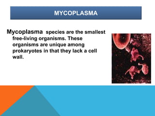
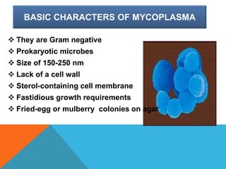
MYCOPLASMA
Prepared by:-
Jijo george
BSc MLT
PRESENTATION OUTLINE
• Discovery
• Morphology
• Biochemical reaction
• Antigenic properties
• pathogenicity
• Treatment
DISCOVERY
In 1896 Nocard and Roux reported the cultivation
of the causative agent of contagious bovine
pleuropneumonia (CBPP), which was at that
time a grave and widespread disease in cattle
herds. The work of Nocard and Roux
represented the first isolation of a mycoplasma
species
EDMOND NOCARD AND
EMILE ROUX
HISTORY
The name mycoplasma, from Greek Mykes (fungus) and
plasma (formed),was proposed in the 1950s, replacing
the term pleuro pneumonia-like organisms (PPLO) referring
to organisms similar to the causative agent CBPP. It
was later found that the fungus-like growth pattern of
M.mycoides is unique to that species.
MYCOPLASMA
Mycoplasma species are the smallest
free-living organisms. These
organisms are unique among
prokaryotes in that they lack a cell
wall.
OVERVIEW OF MYCOPLASMA INFECTIONS
Mycoplasma species are the smallest freeliving
organisms and are unique among and are
unique among prokaryotes in that they lack a
cell wall. This feature is largely responsible for
their biologic properties, including lack of a
Gram stain reaction and nonsusceptibility to
many commonly prescribed to antimicrobial
agents, including betalactams. Mycoplasma
organisms are usually associated with mucosa.
CAN PART OF NORMAL FLORA
They reside extracellularly in the respiratory and
urogenital tracts and rarely penetrate the sub
mucosa, except in the case of
immunosuppression or instrumentation, when
they may invade the bloodstream and
disseminate to numerous organs and tissues.
IMPORTANT MYCOPLASMA INFECTING
HUMAN
Family – Mycoplasmataceae – requiring
cholesterol or other sterols as an essential
growth factor.
a. Genus Mycoplasma – which utilize glucose or
arginine but do not split urea.
b. Genus Ureaplasma– which hydrolyze urea
BASIC CHARACTERS OF MYCOPLASMA
 They are Gram negative
 Prokaryotic microbes
 Size of 150-250 nm
 Lack of a cell wall
 Sterol-containing cell membrane
 Fastidious growth requirements
 Fried-egg or mulberry colonies on agar
MYCOPLASMA ARE CELL WALL DEFICIENT
MICROORGANISMS
Cross-section of
Mycoplasma bacteria,
a common cause of
atypical pneumonia.
This bacteria is
unusual in that it lacks
a cell wall.
CULTURING MYCOPLASMA
 Mycoplasma can be
cultured on liquid or solid
medium
 Growths optimally at 35 to
37c
 Medium of growth should
be enriched with 20% be
horse or human serum.
 The colonies appears as
fried egg appearance
CHARACTERS OF MYCOPLASMA
They are prokaryotes but lack a cell wall.
However, they have a unique cell membrane
that contains sterols, which are not present in
either bacteria or viruses. Mycoplasma
organisms are small (150-250 nm) and have
deformable membranes. The name Mycoplasma
refers to the plasticity of the bacterial forms
resembling fungal elements.
IMPORTANT SPECIES IN MYCOPLASMA
Scientists have isolated at least 17 species of
Mycoplasma from humans, 4 types of
organisms are responsible for most clinically
significant infections that may come to the
attention of practicing physicians. These
species are Mycoplasma pneumoniae,
Mycoplasma hominis, Mycoplasma genitalium,
and Ureaplasma species
HOW THEY DIFFER FROM OTHER
BACTERIA
 They have sterols in the cell membrane.
 They share no DNA homology with known
bacteria.
 They have low guanine levels plus cytosine
content.
 Their genome has a low molecular weight.
 They exhibit no reversion to walled forms.
HOW MYCOPLASMA DIFFER FROM OTHER
COMMON BACTERIA.
However, the term mycoplasma has been frequently
used to denote any species included in the class
Mollicutes .The common characteristics are the
complete lack of a bacterial cell wall, osmotic fragility,
colony shape and filterability through 450-nm pore
diameter membrane filters.The relatively close
Phylogenetic relationship of these genera was
measured by comparative sequence analysis of the
5S and 16S ribosomal RNA (rRNA)
SPECIFIC CHARACTERS OF
MYCOPLASMA
However, the term mycoplasma has been frequently used to
denote any species included in the class Mollicutes).The
common characteristics are the complete lack of a
bacterial cell wall, osmotic fragility, colony shape and
filterability through 450-nm pore diameter membrane
filters.The relatively close Phylogenetic relationship of
these genera was measured by comparative sequence
analysis of the 5S and 16S ribosomal RNA (rRNA). The
rRNA sequence analyses also revealed that the Mollicutes
are not at the root of the bacterial Phylogenetic tree, but
rather developed by degenerate evolution from gram-
positive bacteria with a low mol% G+C (guanine plus
cytosine) content of DNA
HOW THEY DIFFER FROM VIRUSES
 They grow on cell free
media in vitro.
 They contain both
RNA and DNA.
 They have both
intracellular and
extracellular
parasitism in vivo.
HOW RACE INFLUCE MYCOPLASMA
INFECTIONS
Patients with sickle cell disease or related
hemoglobinopathies are at increased risk for
severe M pneumonia infections and may
develop large pleural effusions and marked
respiratory distress. Since sickle cell disease
and other related hemoglobinopathies are most
common among common among African
Americans, severe complications of
mycoplasmal infections also occur most
frequently in this group of patients
MYCOPLASMA FOUND ON SURFACES OF
MUCOUS MEMBRANES
Mycoplasma are found most often on the surfaces
of mucous membranes.They can cause chronic
inflammatory diseases of the respiratory
system, urogenital tract, and joints. The most
common human illnesses caused by
Mycoplasma are due to infection with M.
pneumoniae, which is responsible for 10-20% of
all pneumonias.
ANTIGENIC PROPERTIES
 The surface antigens
are glycolipids and
proteins
 Glycolipids are
identified by
complement fixation.
 Proteins antigens
detected by ELISA
method.
RESISTANCE
 They are normally destroyed by heat at 45 c in
15 minutes.
 They are relatively resistant to penicillin, and
Cephalosporin
 Sensitive to Tetracycline, and several other
antibiotics
WHY MYCOPLASMA ARE PATHOGENIC
The lack of a cell wall most probably facilitates the
close contact between M. pneumoniae and its host
cell and guarantees the exchange of its
compounds, which support the growth of the
bacterium. As a consequence of this bacterial
surface-parasitism the host cell is severely
damaged. The exchange of toxic metabolic
compounds is discussed as a possible cause of cell
damage, however, at this stage not a single toxic
compound has been identified as a causative agent
of cell damage.
SPREAD OF MYCOPLASMA INFECTIONS
 The disease is world wide,
and found in all age groups,
 Transmission by drop let
infection of nasopharyngeal
secretions.
 Spread is associated with
close contact of infected
person
 Important infection in Military
personal.
 Even the persons recovered
from infection will harbor
the pathogens for 2 moths or
more
CLINICAL MANIFESTATIONS
 Generalized aches and pains
 Fever (usually 102°F)
 Cough - Usually non-productive
 Sore throat (no exudative Pharyngitis)
 Headache
 Chills but not rigors
 Nasal congestion with coryza
 Earache
 General malaise
RESPIRATORY SPREAD
Infection moves easily among people in close
contact because it is spread primarily when
infected droplets from the respiratory system
circulate in the air due to coughing, spitting or
sneezing
PNEUMONIA LEADING MANIFESTATION IN
MYCOPLASMA INFECTIONS
PNEUMONIA
Pneumonia caused by
Mycoplasma is also called
atypical pneumonia, walking
pneumonia, or community
acquired pneumonia.
MYCOPLASMA PNEUMONIA
Mycoplasma pneumonia is most often seen in
children and young people. Up to 15 % of all
cases of pneumonia in patients younger than 40
years are caused by mycoplasma pneumoniae.
Most mycoplasma infections are manifested
clinically as bronchitis and/or Pharyngitis.
Pneumonia develops in between 3 and 10% of
the patients.
MYCOPLASMA PRESENTS AS NON
SPECIFIC RESPIRATORY INFECTIONS
Infections commonly involve the oropharynx,
trachea, bronchi, and lungs, usually causing
unilateral pneumonia of the lower lobe. The
radiographic appearance can be distinguished
from that of other non bacterial pneumonias.
MYCOPLASMA CONTRIBUTES TO
SEVERAL RESPIRATORY INFECTIONS
M.Pneumonia infections leads to clinically
apparent disease involving the upper
respiratory tract. In 5-10% of patients(with the
rate depending on age), the infection
progresses to tracheobronchitis or pneumonia
and is usually self limited. Pleural
effusion(usually small) occurs in 5-20% of
patients. Mycoplasmas have also been
implicated in the pathogenesis of asthma,
leading to acute and chronic wheezing in some
individuals.
RADIOLOGICAL PRESENTATION
The radiological picture is
extremely variable, but one or
both lower lobes are usually
involved. The opacities
usually start as partly node-
like peribronchial opacities,
which may gradually develop
to involve whole segments or
lobes
OTHER SYSTEMIC INFECTIONS IN
MYCOPLASMA
In rare instances other organs may be involved
(central nervous system, pancreas, joints, skin,
heart, and pericardium), probably as a result of
haematogenous spread.
MYCOPLASMA IN CHILDREN
 In children younger than 3 years primarily
develop upper respiratory infection.
 M.pneumoniae infection is uncommon in the
first year of life; however in neonates, it may
cause severe disease.
 M.pneumoniae infection is common in school aged
children, with the highest rate of infection in
individuals aged 5-20 years, in whom the tendency
is to develop bronchitis and pneumonia.
MYCOPLASMA IN NEW BORN
Colonization of infants by
genital mycoplasma species
usually occurs during
passage through an
infected birth canal and
genital mycoplasmal
organisms have been
isolated from the upper
respiratory tract in 15% of
infants. Colonization usually
does not persist beyond 2
years
CARDIAC MANIFESTATIONS
 Arrhythmia and/or ECG abnormalities
(conduction defects)
 Congestive failure
 Pericarditis
 Myocarditis
 Endocarditis
NEUROLOGICAL MANIFESTATIONS
 Cranial neuropathy
 Aseptic meningitis or Meningoencephalitis
 Transverse myelitis
 Brainstem dysfunction
 Dysfunction of the pyramidal or extrapyramidal
tract
 Cerebellar dysfunction
 Cerebral infarction
 Guillain-Barre syndrome
 Peripheral neuropathy
MUSCULOSKELETAL MANIFESTATIONS
 Polyarthralgias
 Acute arthritis
(monoarticular or
migratory)
 Digital necrosis
UREAPLASMA SPECIES
IMPORTANT SPECIES IN UREAPLASMA
The ureaplasma genus now is subdivided into 2
species: U.urealyticum and U.parvum. For
clinical purposes, separating infections caused
by the different 2 species is not possible or
necessary. In both the clinical setting and in the
diagnostic laboratory, they are considered
ureaplasma species.
UREAPLASMA DIFFERS FROM
MYCOPLASMA
The Ureaplasma are the only
non fermentative
molecules i.e., they do not
ferment the growth
substrates such as
carbohydrates and amino
acids like other molecules
but they depend on the
hydrolysis of urea for their
energy.
UREASE TEST DIFFERENTIATES
MYCOPLASMA FROM UREAPLASMA
SPECIES
UREAPLASMA UREALYTICUM
 Some strains of Mycoplasma frequently
isolated from Urogenital tract of human beings
and animals
 They are also called T strains or T form
mycoplasma
 They are peculiar to hydrolyze urea, which is
essential growth factor in addition to
Cholesterol
 At present the have been reclassified as
Ureaplasma urealyticum.
UREAPLASMA CAN BE A NORMAL FLORA
IN SEXUALLY ACTIVE INDIVIDUALS
U.urealyticum is part of the normal genital flora
of both men and women. It is found in about
70% of sexually active humans.
 It had also been described to be associated
with a number of diseases in humans, including
non-specific urethritis (NSU), infertility,
chorioamnionitis, still birth, premature birth, and
in the perinatal period, pneumonia,
bronchopulmonary displasia, and meningitis
SPREAD OF UREAPLASMA INFECTIONS
 Major infections are produced by M.hominis
and M.urealyticum
Commonly spread by Sexual
contact
UREAPLASMA CAN BE OPPORTUNISTIC
PATHOGEN IN PREGNANCY
Ureaplasma urealyticum, a common commensals
of the urogenital tract of sexually mature
humans, is gaining recognition as an important
opportunistic pathogen during pregnancy.
While its etiologic significance in many aspects
of adverse pregnancy remains controversial,
recent evidence indicates that U. urealyticum in
the absence of other organisms is a cause of
chorioamnionitis.
UREAPLASMA CAN PRODUCE SEVERAL
DISSEMINATED COMPLICATIONS
Evidence indicates that U. urealyticum is a cause
of septicemia meningitis, and pneumonia in
newborn infants, particularly those born
prematurely. There is strong but not definitive
evidence that Ureaplasma infection of the lower
respiratory tract can lead to development of
chronic lung disease in very low birth-weight
infants. Although risk factors for colonization of
the lower genitourinary tract have been
identified
UREAPLASMA SPECIES: CAUSES
 Urethritis
 Pyelonephritis
 Pelvic inflammatory disease
 Endometritis or chorioamnionitis
 Infectious arthritis
 Surgical wound infections
 Neonatal pneumonia
 Neonatal meningitis
MYCOPLASMA AND STERILITY
Mycoplasma species do not cause vaginitis, but
they may proliferate in patients with bacterial
vaginosis and may contribute to the condition.
M hominis has been isolated from the
endometria and fallopian tubes of approximately
10% of women with salphingits; M.genitalium
may also be involved in pelvic inflammatory
disease and Cervicitis. Whether Ureaplasma
infection causes involuntary infertility remains
speculative.
MYCOPLASMA IN HIV INFECTIONS
 Mycoplasmas tend to cause
more severe infections in the
HIV infected persons and
cause prolonged infections.
 Other Immunosuppressed
patients are susceptible to
Mycoplasma infections
DIAGNOSIS IN IMMUNOSUPPRESSED A
PRIORITY
The correct microbiological
diagnosis takes on
greater importance in
patients who are
Immunosuppressed and
at greater risk for
disseminated infection
and a poor outcome.
BIOCHEMICAL REACTION
GROWTH OF BACTERIOLOGICAL MEDIUM
 For isolation swabs from
throat or respiratory
secretions inoculated not
Mycoplasma medium
 The growth is slow and takes
1 – 3 weeks
 The colonies appear as fried
egg, with central opaque
granular area surrounded by
flat translucent peripheral
zone
MYCOPLASMA ON PPLO AGAR
TYPICAL MYCOPLASMA COLONIES ON
ENRICHED MEDIUM
 The colonies showing
typical fried egg
appearance.
 The colonies appear 2-
6 days of incubation.
 The size of the
colonies can be from
10 – 600 microns in
size.
COLONY CHARACTERS OF MYCOPLASMA
ISOLATES
BIOCHEMICAL CHARACTERS OF
MYCOPLASMA
 The metabolism of
Mycoplasma are
fermentative
 Most species utilize
glucose or arginine
 Urea is hydrolyzed by
Ureaplasma only
M. PNEUMONIAE COLONIES
DEMONSTRATED IN DIENES METHOD
 The colonies can be
demonstrated by Dienes
method.
 In which a block of agar
containing the colony is
cut and placed on a slide,
covered with a cover slip
on which has been dried in
alcoholic solution of
methylene blue and azure.
DIAGNOSIS
MYCOPLASMA INFECTIONS
Majority of cases are Diagnosed with
Serological Tests
COLD AGGLUTINATION TEST
Cold Agglutination test is
associated with
macroglobulin antibodies
that antibodies that
agglutinate human o RBC at
low temperature
PROCEDURE
 The serial dilutions of patients
serum are mixed with an equal
volume of 0.2% washed human O
group erythrocytes at low
temperature
 The clumping is observed at 4c
overnight.
 However the clumping is
dissociated at 37c
 A titer of 1:32 or > is suggestive.
 A raised titer in paired serum
sample is more suggestive of
infection.
RBC SHOWING NON AGGLUTINATING AND
AGGLUTINATING RBC
STREPTOCOCCAL MG TEST
 The test is performed by
mixing serial dilutions of
patients serum with heat killed
suspension of Streptococcus
MG.
 The sample is incubated at 37c
 The agglutination titer of 1:20
or > is suggestive.
OTHER SEROLOGICAL TESTS
 Immunofluorescence
 Hemagglutination
inhibition test
 Complement fixation
test less sensitive .
DIAGNOSIS OF UROGENITAL INFECTIONS
 Material from urethra, cervical, or vaginal or
centrifuged deposit of urine is added to
separate vials with liquid mycoplasmal medium
containing phenol red and 0.1% glucose,
arginine or urea
 The Ureaplasmal urease also breaks down urea
to ammonia
NEWER METHODS IN DIAGNOSIS
Phylogeny based rapid
identification of urogenital
Mycoplasmas and
ureaplasmas based on
amplification of part 165rRNA
gene by PCR is available
EMERGING TRENDS IN PCR PRIMERS
Now several DNA primers
specific for several
Mycoplasmas are
available and useful for
specific identification of
species
DRUGS USED IN MYCOPLASMA
INFECTIONS
 Erythromycin (M. pneumoniae and Ureaplasma
spp.)
 Tetracycline.
 Clindamycin ( M. hominis )
 Levofloxacin
 Doxycycline
 Gentamycin
Prepared by:-
Jijo George
BSc MLT
Thanks…………………!!!!!!!!!!

Mycoplasma

  • 1.
  • 2.
    PRESENTATION OUTLINE • Discovery •Morphology • Biochemical reaction • Antigenic properties • pathogenicity • Treatment
  • 3.
    DISCOVERY In 1896 Nocardand Roux reported the cultivation of the causative agent of contagious bovine pleuropneumonia (CBPP), which was at that time a grave and widespread disease in cattle herds. The work of Nocard and Roux represented the first isolation of a mycoplasma species
  • 4.
  • 5.
    HISTORY The name mycoplasma,from Greek Mykes (fungus) and plasma (formed),was proposed in the 1950s, replacing the term pleuro pneumonia-like organisms (PPLO) referring to organisms similar to the causative agent CBPP. It was later found that the fungus-like growth pattern of M.mycoides is unique to that species.
  • 6.
    MYCOPLASMA Mycoplasma species arethe smallest free-living organisms. These organisms are unique among prokaryotes in that they lack a cell wall.
  • 7.
    OVERVIEW OF MYCOPLASMAINFECTIONS Mycoplasma species are the smallest freeliving organisms and are unique among and are unique among prokaryotes in that they lack a cell wall. This feature is largely responsible for their biologic properties, including lack of a Gram stain reaction and nonsusceptibility to many commonly prescribed to antimicrobial agents, including betalactams. Mycoplasma organisms are usually associated with mucosa.
  • 8.
    CAN PART OFNORMAL FLORA They reside extracellularly in the respiratory and urogenital tracts and rarely penetrate the sub mucosa, except in the case of immunosuppression or instrumentation, when they may invade the bloodstream and disseminate to numerous organs and tissues.
  • 9.
    IMPORTANT MYCOPLASMA INFECTING HUMAN Family– Mycoplasmataceae – requiring cholesterol or other sterols as an essential growth factor. a. Genus Mycoplasma – which utilize glucose or arginine but do not split urea. b. Genus Ureaplasma– which hydrolyze urea
  • 10.
    BASIC CHARACTERS OFMYCOPLASMA  They are Gram negative  Prokaryotic microbes  Size of 150-250 nm  Lack of a cell wall  Sterol-containing cell membrane  Fastidious growth requirements  Fried-egg or mulberry colonies on agar
  • 11.
    MYCOPLASMA ARE CELLWALL DEFICIENT MICROORGANISMS Cross-section of Mycoplasma bacteria, a common cause of atypical pneumonia. This bacteria is unusual in that it lacks a cell wall.
  • 12.
    CULTURING MYCOPLASMA  Mycoplasmacan be cultured on liquid or solid medium  Growths optimally at 35 to 37c  Medium of growth should be enriched with 20% be horse or human serum.  The colonies appears as fried egg appearance
  • 13.
    CHARACTERS OF MYCOPLASMA Theyare prokaryotes but lack a cell wall. However, they have a unique cell membrane that contains sterols, which are not present in either bacteria or viruses. Mycoplasma organisms are small (150-250 nm) and have deformable membranes. The name Mycoplasma refers to the plasticity of the bacterial forms resembling fungal elements.
  • 14.
    IMPORTANT SPECIES INMYCOPLASMA Scientists have isolated at least 17 species of Mycoplasma from humans, 4 types of organisms are responsible for most clinically significant infections that may come to the attention of practicing physicians. These species are Mycoplasma pneumoniae, Mycoplasma hominis, Mycoplasma genitalium, and Ureaplasma species
  • 15.
    HOW THEY DIFFERFROM OTHER BACTERIA  They have sterols in the cell membrane.  They share no DNA homology with known bacteria.  They have low guanine levels plus cytosine content.  Their genome has a low molecular weight.  They exhibit no reversion to walled forms.
  • 16.
    HOW MYCOPLASMA DIFFERFROM OTHER COMMON BACTERIA. However, the term mycoplasma has been frequently used to denote any species included in the class Mollicutes .The common characteristics are the complete lack of a bacterial cell wall, osmotic fragility, colony shape and filterability through 450-nm pore diameter membrane filters.The relatively close Phylogenetic relationship of these genera was measured by comparative sequence analysis of the 5S and 16S ribosomal RNA (rRNA)
  • 17.
    SPECIFIC CHARACTERS OF MYCOPLASMA However,the term mycoplasma has been frequently used to denote any species included in the class Mollicutes).The common characteristics are the complete lack of a bacterial cell wall, osmotic fragility, colony shape and filterability through 450-nm pore diameter membrane filters.The relatively close Phylogenetic relationship of these genera was measured by comparative sequence analysis of the 5S and 16S ribosomal RNA (rRNA). The rRNA sequence analyses also revealed that the Mollicutes are not at the root of the bacterial Phylogenetic tree, but rather developed by degenerate evolution from gram- positive bacteria with a low mol% G+C (guanine plus cytosine) content of DNA
  • 18.
    HOW THEY DIFFERFROM VIRUSES  They grow on cell free media in vitro.  They contain both RNA and DNA.  They have both intracellular and extracellular parasitism in vivo.
  • 19.
    HOW RACE INFLUCEMYCOPLASMA INFECTIONS Patients with sickle cell disease or related hemoglobinopathies are at increased risk for severe M pneumonia infections and may develop large pleural effusions and marked respiratory distress. Since sickle cell disease and other related hemoglobinopathies are most common among common among African Americans, severe complications of mycoplasmal infections also occur most frequently in this group of patients
  • 20.
    MYCOPLASMA FOUND ONSURFACES OF MUCOUS MEMBRANES Mycoplasma are found most often on the surfaces of mucous membranes.They can cause chronic inflammatory diseases of the respiratory system, urogenital tract, and joints. The most common human illnesses caused by Mycoplasma are due to infection with M. pneumoniae, which is responsible for 10-20% of all pneumonias.
  • 21.
    ANTIGENIC PROPERTIES  Thesurface antigens are glycolipids and proteins  Glycolipids are identified by complement fixation.  Proteins antigens detected by ELISA method.
  • 22.
    RESISTANCE  They arenormally destroyed by heat at 45 c in 15 minutes.  They are relatively resistant to penicillin, and Cephalosporin  Sensitive to Tetracycline, and several other antibiotics
  • 23.
    WHY MYCOPLASMA AREPATHOGENIC The lack of a cell wall most probably facilitates the close contact between M. pneumoniae and its host cell and guarantees the exchange of its compounds, which support the growth of the bacterium. As a consequence of this bacterial surface-parasitism the host cell is severely damaged. The exchange of toxic metabolic compounds is discussed as a possible cause of cell damage, however, at this stage not a single toxic compound has been identified as a causative agent of cell damage.
  • 24.
    SPREAD OF MYCOPLASMAINFECTIONS  The disease is world wide, and found in all age groups,  Transmission by drop let infection of nasopharyngeal secretions.  Spread is associated with close contact of infected person  Important infection in Military personal.  Even the persons recovered from infection will harbor the pathogens for 2 moths or more
  • 25.
    CLINICAL MANIFESTATIONS  Generalizedaches and pains  Fever (usually 102°F)  Cough - Usually non-productive  Sore throat (no exudative Pharyngitis)  Headache  Chills but not rigors  Nasal congestion with coryza  Earache  General malaise
  • 26.
    RESPIRATORY SPREAD Infection moveseasily among people in close contact because it is spread primarily when infected droplets from the respiratory system circulate in the air due to coughing, spitting or sneezing
  • 27.
    PNEUMONIA LEADING MANIFESTATIONIN MYCOPLASMA INFECTIONS
  • 28.
    PNEUMONIA Pneumonia caused by Mycoplasmais also called atypical pneumonia, walking pneumonia, or community acquired pneumonia.
  • 29.
    MYCOPLASMA PNEUMONIA Mycoplasma pneumoniais most often seen in children and young people. Up to 15 % of all cases of pneumonia in patients younger than 40 years are caused by mycoplasma pneumoniae. Most mycoplasma infections are manifested clinically as bronchitis and/or Pharyngitis. Pneumonia develops in between 3 and 10% of the patients.
  • 30.
    MYCOPLASMA PRESENTS ASNON SPECIFIC RESPIRATORY INFECTIONS Infections commonly involve the oropharynx, trachea, bronchi, and lungs, usually causing unilateral pneumonia of the lower lobe. The radiographic appearance can be distinguished from that of other non bacterial pneumonias.
  • 31.
    MYCOPLASMA CONTRIBUTES TO SEVERALRESPIRATORY INFECTIONS M.Pneumonia infections leads to clinically apparent disease involving the upper respiratory tract. In 5-10% of patients(with the rate depending on age), the infection progresses to tracheobronchitis or pneumonia and is usually self limited. Pleural effusion(usually small) occurs in 5-20% of patients. Mycoplasmas have also been implicated in the pathogenesis of asthma, leading to acute and chronic wheezing in some individuals.
  • 32.
    RADIOLOGICAL PRESENTATION The radiologicalpicture is extremely variable, but one or both lower lobes are usually involved. The opacities usually start as partly node- like peribronchial opacities, which may gradually develop to involve whole segments or lobes
  • 33.
    OTHER SYSTEMIC INFECTIONSIN MYCOPLASMA In rare instances other organs may be involved (central nervous system, pancreas, joints, skin, heart, and pericardium), probably as a result of haematogenous spread.
  • 34.
    MYCOPLASMA IN CHILDREN In children younger than 3 years primarily develop upper respiratory infection.  M.pneumoniae infection is uncommon in the first year of life; however in neonates, it may cause severe disease.  M.pneumoniae infection is common in school aged children, with the highest rate of infection in individuals aged 5-20 years, in whom the tendency is to develop bronchitis and pneumonia.
  • 35.
    MYCOPLASMA IN NEWBORN Colonization of infants by genital mycoplasma species usually occurs during passage through an infected birth canal and genital mycoplasmal organisms have been isolated from the upper respiratory tract in 15% of infants. Colonization usually does not persist beyond 2 years
  • 36.
    CARDIAC MANIFESTATIONS  Arrhythmiaand/or ECG abnormalities (conduction defects)  Congestive failure  Pericarditis  Myocarditis  Endocarditis
  • 37.
    NEUROLOGICAL MANIFESTATIONS  Cranialneuropathy  Aseptic meningitis or Meningoencephalitis  Transverse myelitis  Brainstem dysfunction  Dysfunction of the pyramidal or extrapyramidal tract  Cerebellar dysfunction  Cerebral infarction  Guillain-Barre syndrome  Peripheral neuropathy
  • 38.
    MUSCULOSKELETAL MANIFESTATIONS  Polyarthralgias Acute arthritis (monoarticular or migratory)  Digital necrosis
  • 39.
  • 40.
    IMPORTANT SPECIES INUREAPLASMA The ureaplasma genus now is subdivided into 2 species: U.urealyticum and U.parvum. For clinical purposes, separating infections caused by the different 2 species is not possible or necessary. In both the clinical setting and in the diagnostic laboratory, they are considered ureaplasma species.
  • 41.
    UREAPLASMA DIFFERS FROM MYCOPLASMA TheUreaplasma are the only non fermentative molecules i.e., they do not ferment the growth substrates such as carbohydrates and amino acids like other molecules but they depend on the hydrolysis of urea for their energy.
  • 42.
  • 43.
    UREAPLASMA UREALYTICUM  Somestrains of Mycoplasma frequently isolated from Urogenital tract of human beings and animals  They are also called T strains or T form mycoplasma  They are peculiar to hydrolyze urea, which is essential growth factor in addition to Cholesterol  At present the have been reclassified as Ureaplasma urealyticum.
  • 44.
    UREAPLASMA CAN BEA NORMAL FLORA IN SEXUALLY ACTIVE INDIVIDUALS U.urealyticum is part of the normal genital flora of both men and women. It is found in about 70% of sexually active humans.  It had also been described to be associated with a number of diseases in humans, including non-specific urethritis (NSU), infertility, chorioamnionitis, still birth, premature birth, and in the perinatal period, pneumonia, bronchopulmonary displasia, and meningitis
  • 45.
    SPREAD OF UREAPLASMAINFECTIONS  Major infections are produced by M.hominis and M.urealyticum Commonly spread by Sexual contact
  • 46.
    UREAPLASMA CAN BEOPPORTUNISTIC PATHOGEN IN PREGNANCY Ureaplasma urealyticum, a common commensals of the urogenital tract of sexually mature humans, is gaining recognition as an important opportunistic pathogen during pregnancy. While its etiologic significance in many aspects of adverse pregnancy remains controversial, recent evidence indicates that U. urealyticum in the absence of other organisms is a cause of chorioamnionitis.
  • 47.
    UREAPLASMA CAN PRODUCESEVERAL DISSEMINATED COMPLICATIONS Evidence indicates that U. urealyticum is a cause of septicemia meningitis, and pneumonia in newborn infants, particularly those born prematurely. There is strong but not definitive evidence that Ureaplasma infection of the lower respiratory tract can lead to development of chronic lung disease in very low birth-weight infants. Although risk factors for colonization of the lower genitourinary tract have been identified
  • 48.
    UREAPLASMA SPECIES: CAUSES Urethritis  Pyelonephritis  Pelvic inflammatory disease  Endometritis or chorioamnionitis  Infectious arthritis  Surgical wound infections  Neonatal pneumonia  Neonatal meningitis
  • 49.
    MYCOPLASMA AND STERILITY Mycoplasmaspecies do not cause vaginitis, but they may proliferate in patients with bacterial vaginosis and may contribute to the condition. M hominis has been isolated from the endometria and fallopian tubes of approximately 10% of women with salphingits; M.genitalium may also be involved in pelvic inflammatory disease and Cervicitis. Whether Ureaplasma infection causes involuntary infertility remains speculative.
  • 50.
    MYCOPLASMA IN HIVINFECTIONS  Mycoplasmas tend to cause more severe infections in the HIV infected persons and cause prolonged infections.  Other Immunosuppressed patients are susceptible to Mycoplasma infections
  • 51.
    DIAGNOSIS IN IMMUNOSUPPRESSEDA PRIORITY The correct microbiological diagnosis takes on greater importance in patients who are Immunosuppressed and at greater risk for disseminated infection and a poor outcome.
  • 52.
  • 53.
    GROWTH OF BACTERIOLOGICALMEDIUM  For isolation swabs from throat or respiratory secretions inoculated not Mycoplasma medium  The growth is slow and takes 1 – 3 weeks  The colonies appear as fried egg, with central opaque granular area surrounded by flat translucent peripheral zone
  • 54.
  • 55.
    TYPICAL MYCOPLASMA COLONIESON ENRICHED MEDIUM  The colonies showing typical fried egg appearance.  The colonies appear 2- 6 days of incubation.  The size of the colonies can be from 10 – 600 microns in size.
  • 56.
    COLONY CHARACTERS OFMYCOPLASMA ISOLATES
  • 57.
    BIOCHEMICAL CHARACTERS OF MYCOPLASMA The metabolism of Mycoplasma are fermentative  Most species utilize glucose or arginine  Urea is hydrolyzed by Ureaplasma only
  • 58.
    M. PNEUMONIAE COLONIES DEMONSTRATEDIN DIENES METHOD  The colonies can be demonstrated by Dienes method.  In which a block of agar containing the colony is cut and placed on a slide, covered with a cover slip on which has been dried in alcoholic solution of methylene blue and azure.
  • 59.
  • 60.
    Majority of casesare Diagnosed with Serological Tests
  • 61.
    COLD AGGLUTINATION TEST ColdAgglutination test is associated with macroglobulin antibodies that antibodies that agglutinate human o RBC at low temperature
  • 62.
    PROCEDURE  The serialdilutions of patients serum are mixed with an equal volume of 0.2% washed human O group erythrocytes at low temperature  The clumping is observed at 4c overnight.  However the clumping is dissociated at 37c  A titer of 1:32 or > is suggestive.  A raised titer in paired serum sample is more suggestive of infection.
  • 63.
    RBC SHOWING NONAGGLUTINATING AND AGGLUTINATING RBC
  • 64.
    STREPTOCOCCAL MG TEST The test is performed by mixing serial dilutions of patients serum with heat killed suspension of Streptococcus MG.  The sample is incubated at 37c  The agglutination titer of 1:20 or > is suggestive.
  • 65.
    OTHER SEROLOGICAL TESTS Immunofluorescence  Hemagglutination inhibition test  Complement fixation test less sensitive .
  • 66.
    DIAGNOSIS OF UROGENITALINFECTIONS  Material from urethra, cervical, or vaginal or centrifuged deposit of urine is added to separate vials with liquid mycoplasmal medium containing phenol red and 0.1% glucose, arginine or urea  The Ureaplasmal urease also breaks down urea to ammonia
  • 67.
    NEWER METHODS INDIAGNOSIS Phylogeny based rapid identification of urogenital Mycoplasmas and ureaplasmas based on amplification of part 165rRNA gene by PCR is available
  • 68.
    EMERGING TRENDS INPCR PRIMERS Now several DNA primers specific for several Mycoplasmas are available and useful for specific identification of species
  • 69.
    DRUGS USED INMYCOPLASMA INFECTIONS  Erythromycin (M. pneumoniae and Ureaplasma spp.)  Tetracycline.  Clindamycin ( M. hominis )  Levofloxacin  Doxycycline  Gentamycin
  • 70.
    Prepared by:- Jijo George BScMLT Thanks…………………!!!!!!!!!!