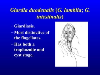
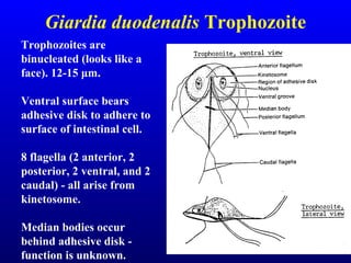
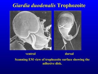
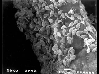
GIARDIA
Shilpa.k
Microbiology Tutor
AIMSRC
Giardia duodenalis (G. lamblia; G.
intestinalis)
– Giardiasis.
– Most distinctive of
the flagellates.
– Has both a
trophozoite and
cyst stage.
Giardia duodenalis Trophozoite
Trophozoites are
binucleated (looks like a
face). 12-15 μm.
Ventral surface bears
adhesive disk to adhere to
surface of intestinal cell.
8 flagella (2 anterior, 2
posterior, 2 ventral, and 2
caudal) - all arise from
kinetosome.
Median bodies occur
behind adhesive disk -
function is unknown.
Giardia duodenalis Trophozoite
Light microscope photos of trophozoites
Giardia duodenalis
• Lives in the upper part of the small
intestine (duodenum, jejunum, and
upper ileum).
• Here the trophozoites attach to the
epithelial cells.
Giardia duodenalis Trophozoite
Scanning EM view of trophozoite surface showing the
adhesive disk.
ventral dorsal
• Feeds on mucous that forms in response to irritation.
• Also absorbs vitamins and amino acids.
• Interferes with absorption in host especially lipids.
• Giardia can also interfere with vitamin/nutrient
absorption.
– Vitamin A vision
– Vitamin D rickets: Both of these are due to long
standing infections.
Cyst of Giardia duodenalis
The cyst forms as trophozoites become dehydrated
when they pass through the large intestine.
Morphology:
• ovoid in shape; 8-12 µm long x 7-10
µm wide
• thin cyst wall.
• Four nuclei present, often
concentrated at on end.
• Flagella shorten and are retracted
within cyst.
• Axonemes provide internal support.
Cyst of Giardia duodenalis
Cyst may remain viable in the external
environment (usually water) for many
months.
-14 billion cysts can be passed in
1 stool sample
-Moderate infections: 300
million cysts.
Cyst of Giardia duodenalis
Symptoms
• Range from none abdominal
discomfort causing acute or chronic
diarrhea and other GI signs.
• Gray, greasy, voluminous malodorous
diarrhea!
• Flatulence.
Giardia duodenalis
• Giardia trophozoites are attracted to bile
salts: so sometimes you can get infections
in bile ducts and gall bladder, causing
jaundice and colic.
• This is irritating but not life threatening
infection like E. histolytica.
Pathogenesis and Pathology
• Nutrient malabsorption and physical
blockage and damage to microvilli.
• Trophozoites attach to small intestine
cause damage (mechanical and toxins).
Pathogenesis and Pathology
1) Fat/CHO digestion decreases and causes maldigestion.
2) Absorption decreases due to villus blunting causing
malabsorption.
3) Malabsorption and maldigestion causes
diarrhea.
4) Physical damage: clubbing of villi; decreases
villus-to-crypt ratio; brush borders of cells are
irregular.
Giardia trophozoite
Trophozoite attaches to surface of epithelial cells with its
adhesive disk.
Epidemiology
• Get infected by ingesting cysts through
contaminated water.
• Most common intestinal flagellate of
people.
• World wide distribution; prevalence
ranges from 2.4-67.5%.
• Reservoir hosts can play a significant
role.
Reservoir Hosts
Transmission from animals to humans is
controversial; dependent on strain or type involved.
Human Infections
• There are hot spots: Vacations and Travels
Camping.
• Colorado ski resorts are notorious for
outbreaks drinking from Mountain Springs,
washing utensils/drinking water that is not
treated.
• Day care centers.
Diagnosis
• Trophozoites in diarrheic feces; cysts in
formed feces.
• At least 3 exams (one every other day)
before judge negative.
• ELISA tests: detect soluble antigen.
Treatment and Prognosis
• Drug of choice is Flagyl, Metronidazole:
15 mg/kg/day in 3 divided doses for 5–7
days
Giardia thrives in people not
necessarily hard to treat, but keeping
those who were infected from becoming
reinfected.
REFERENCES
• Centers for Disease Control and Prevention
1600 Clifton Rd. Atlanta, GA 30333, USA
• Garcia textbook of Medical parasitology
Giardia

Giardia

  • 1.
  • 2.
    Giardia duodenalis (G.lamblia; G. intestinalis) – Giardiasis. – Most distinctive of the flagellates. – Has both a trophozoite and cyst stage.
  • 3.
    Giardia duodenalis Trophozoite Trophozoitesare binucleated (looks like a face). 12-15 μm. Ventral surface bears adhesive disk to adhere to surface of intestinal cell. 8 flagella (2 anterior, 2 posterior, 2 ventral, and 2 caudal) - all arise from kinetosome. Median bodies occur behind adhesive disk - function is unknown.
  • 4.
    Giardia duodenalis Trophozoite Lightmicroscope photos of trophozoites
  • 5.
    Giardia duodenalis • Livesin the upper part of the small intestine (duodenum, jejunum, and upper ileum). • Here the trophozoites attach to the epithelial cells.
  • 6.
    Giardia duodenalis Trophozoite ScanningEM view of trophozoite surface showing the adhesive disk. ventral dorsal
  • 7.
    • Feeds onmucous that forms in response to irritation. • Also absorbs vitamins and amino acids. • Interferes with absorption in host especially lipids. • Giardia can also interfere with vitamin/nutrient absorption. – Vitamin A vision – Vitamin D rickets: Both of these are due to long standing infections.
  • 8.
    Cyst of Giardiaduodenalis The cyst forms as trophozoites become dehydrated when they pass through the large intestine. Morphology: • ovoid in shape; 8-12 µm long x 7-10 µm wide • thin cyst wall. • Four nuclei present, often concentrated at on end. • Flagella shorten and are retracted within cyst. • Axonemes provide internal support.
  • 9.
    Cyst of Giardiaduodenalis Cyst may remain viable in the external environment (usually water) for many months. -14 billion cysts can be passed in 1 stool sample -Moderate infections: 300 million cysts.
  • 10.
    Cyst of Giardiaduodenalis
  • 11.
    Symptoms • Range fromnone abdominal discomfort causing acute or chronic diarrhea and other GI signs. • Gray, greasy, voluminous malodorous diarrhea! • Flatulence.
  • 12.
    Giardia duodenalis • Giardiatrophozoites are attracted to bile salts: so sometimes you can get infections in bile ducts and gall bladder, causing jaundice and colic. • This is irritating but not life threatening infection like E. histolytica.
  • 13.
    Pathogenesis and Pathology •Nutrient malabsorption and physical blockage and damage to microvilli. • Trophozoites attach to small intestine cause damage (mechanical and toxins).
  • 14.
    Pathogenesis and Pathology 1)Fat/CHO digestion decreases and causes maldigestion. 2) Absorption decreases due to villus blunting causing malabsorption. 3) Malabsorption and maldigestion causes diarrhea. 4) Physical damage: clubbing of villi; decreases villus-to-crypt ratio; brush borders of cells are irregular.
  • 19.
    Giardia trophozoite Trophozoite attachesto surface of epithelial cells with its adhesive disk.
  • 20.
    Epidemiology • Get infectedby ingesting cysts through contaminated water. • Most common intestinal flagellate of people. • World wide distribution; prevalence ranges from 2.4-67.5%. • Reservoir hosts can play a significant role.
  • 21.
    Reservoir Hosts Transmission fromanimals to humans is controversial; dependent on strain or type involved.
  • 22.
    Human Infections • Thereare hot spots: Vacations and Travels Camping. • Colorado ski resorts are notorious for outbreaks drinking from Mountain Springs, washing utensils/drinking water that is not treated. • Day care centers.
  • 23.
    Diagnosis • Trophozoites indiarrheic feces; cysts in formed feces. • At least 3 exams (one every other day) before judge negative. • ELISA tests: detect soluble antigen.
  • 24.
    Treatment and Prognosis •Drug of choice is Flagyl, Metronidazole: 15 mg/kg/day in 3 divided doses for 5–7 days Giardia thrives in people not necessarily hard to treat, but keeping those who were infected from becoming reinfected.
  • 25.
    REFERENCES • Centers forDisease Control and Prevention 1600 Clifton Rd. Atlanta, GA 30333, USA • Garcia textbook of Medical parasitology