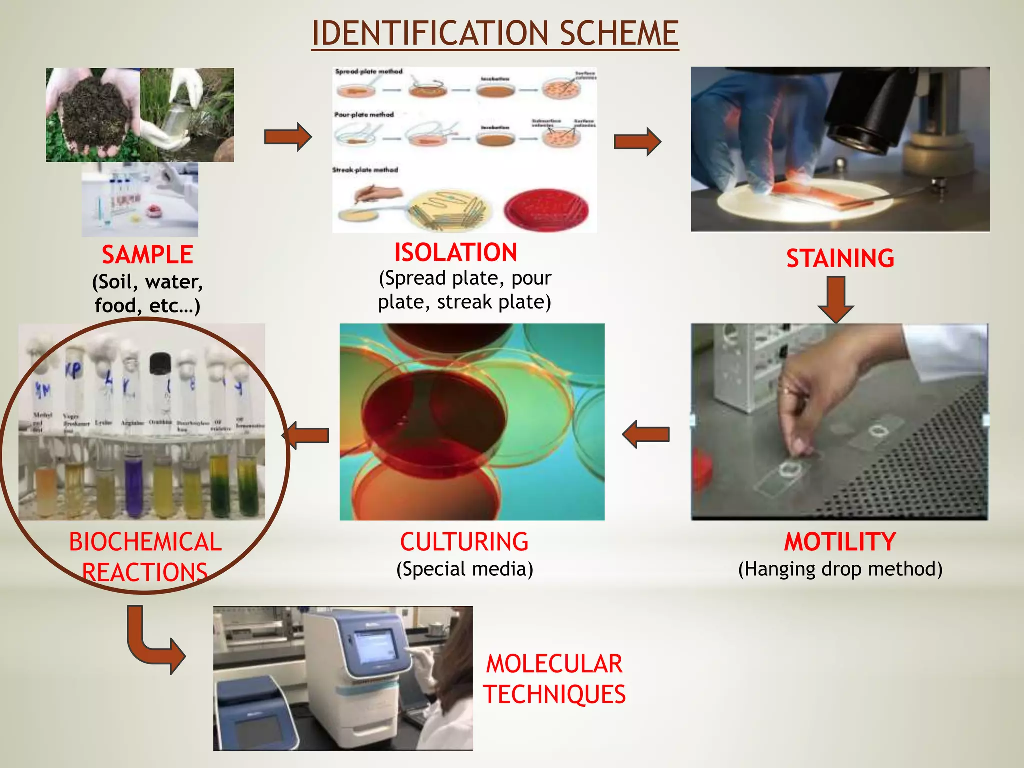
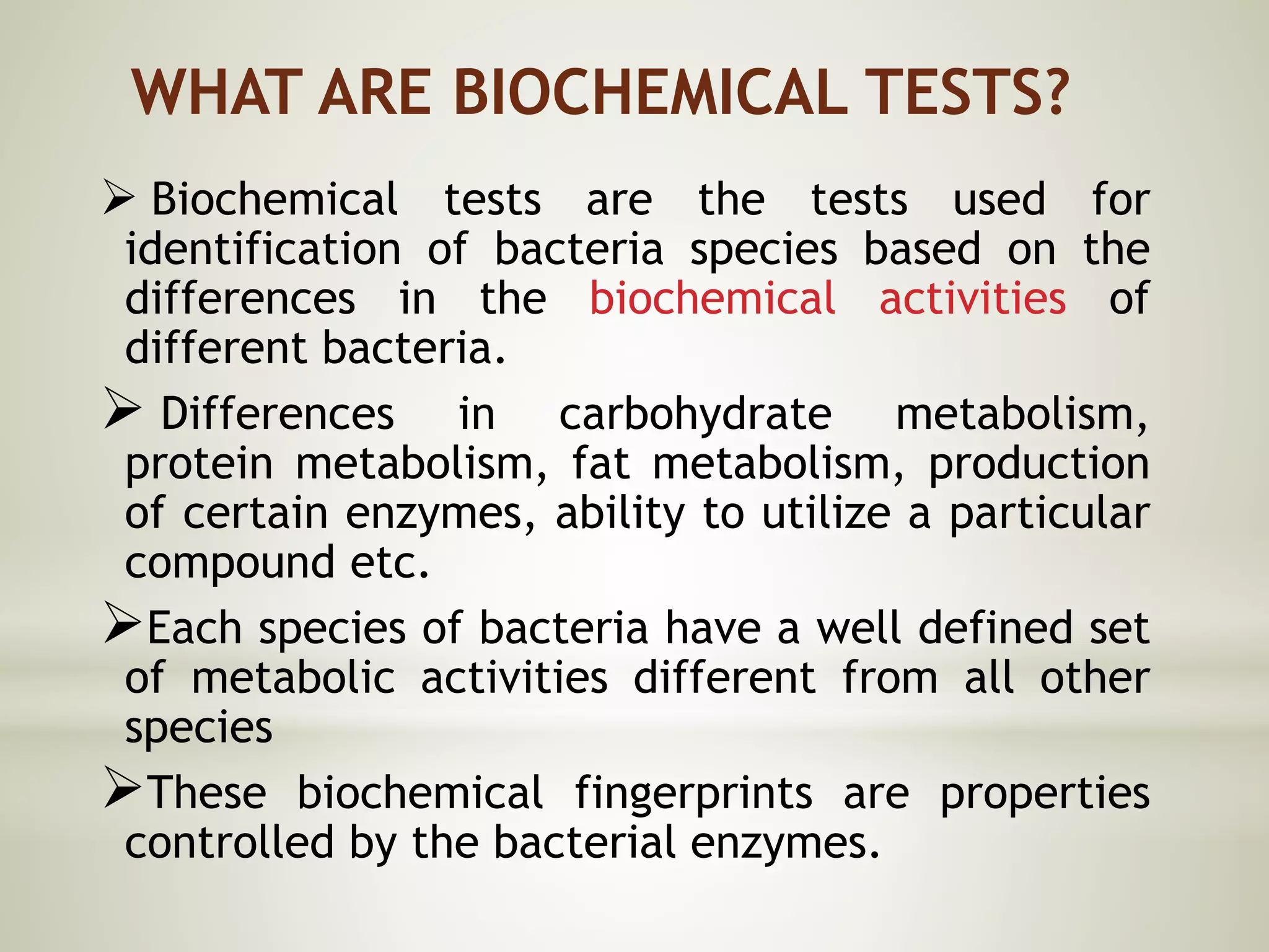
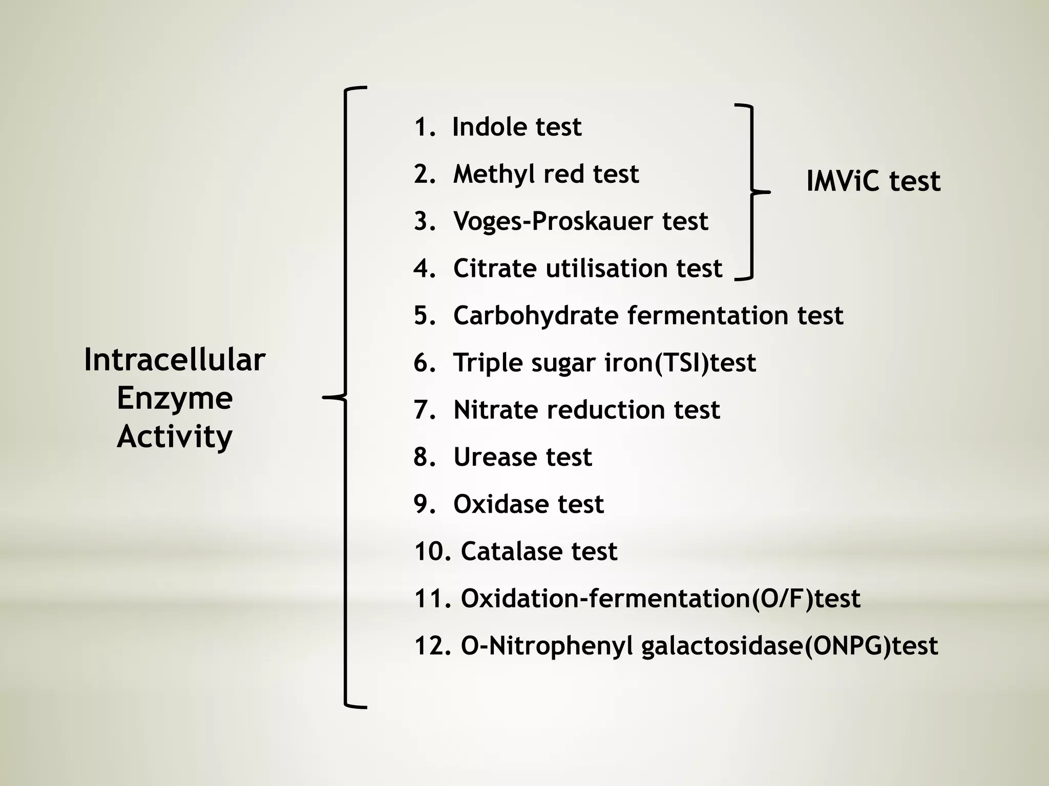
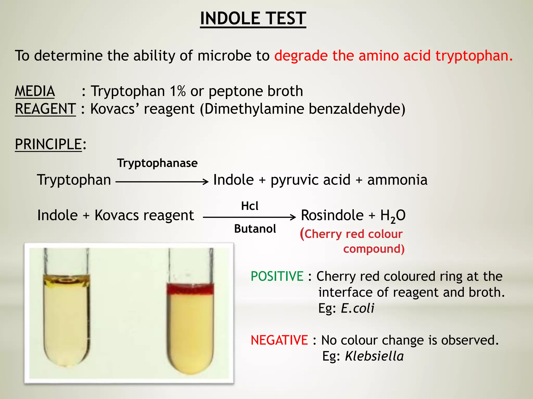
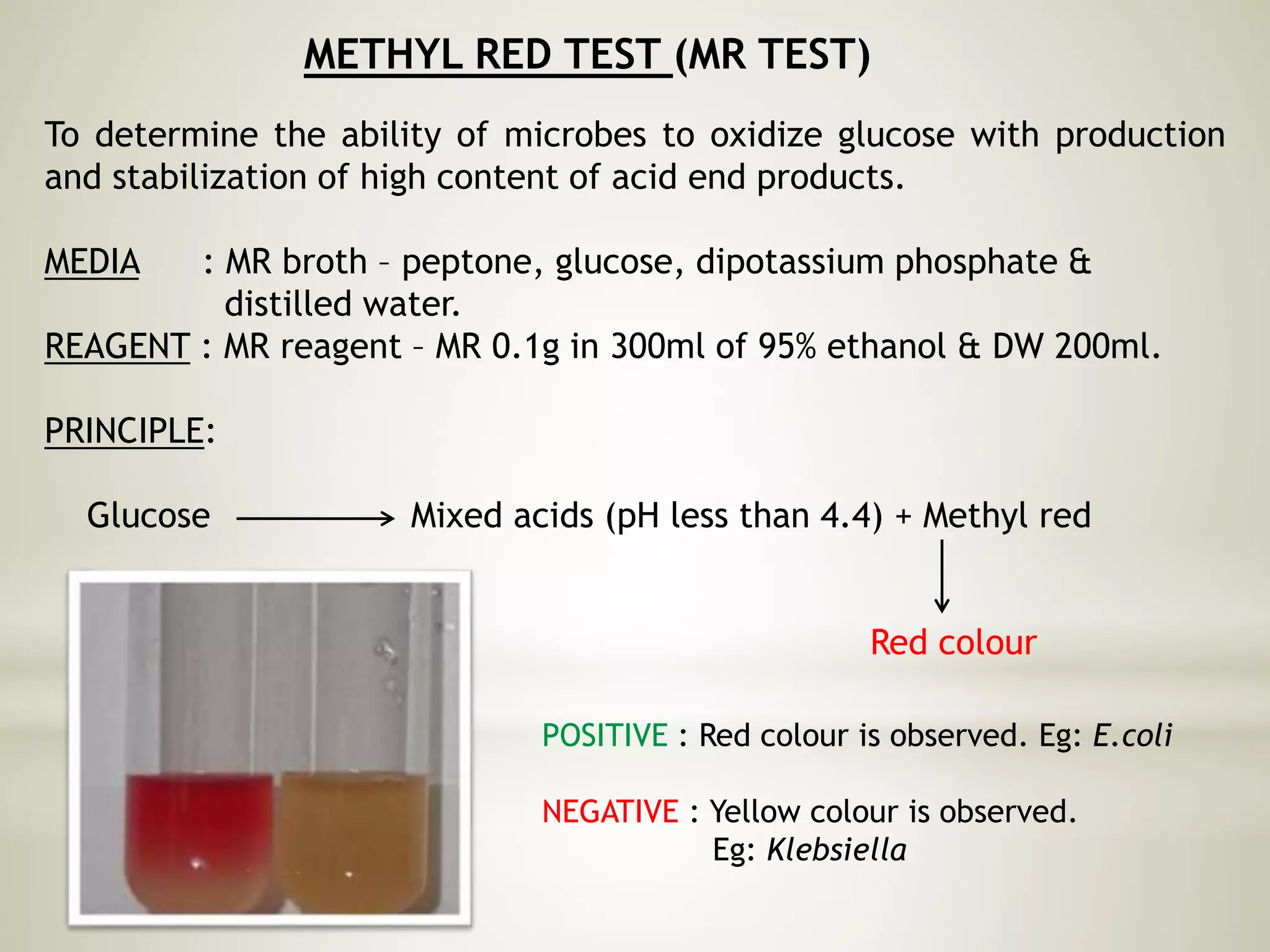
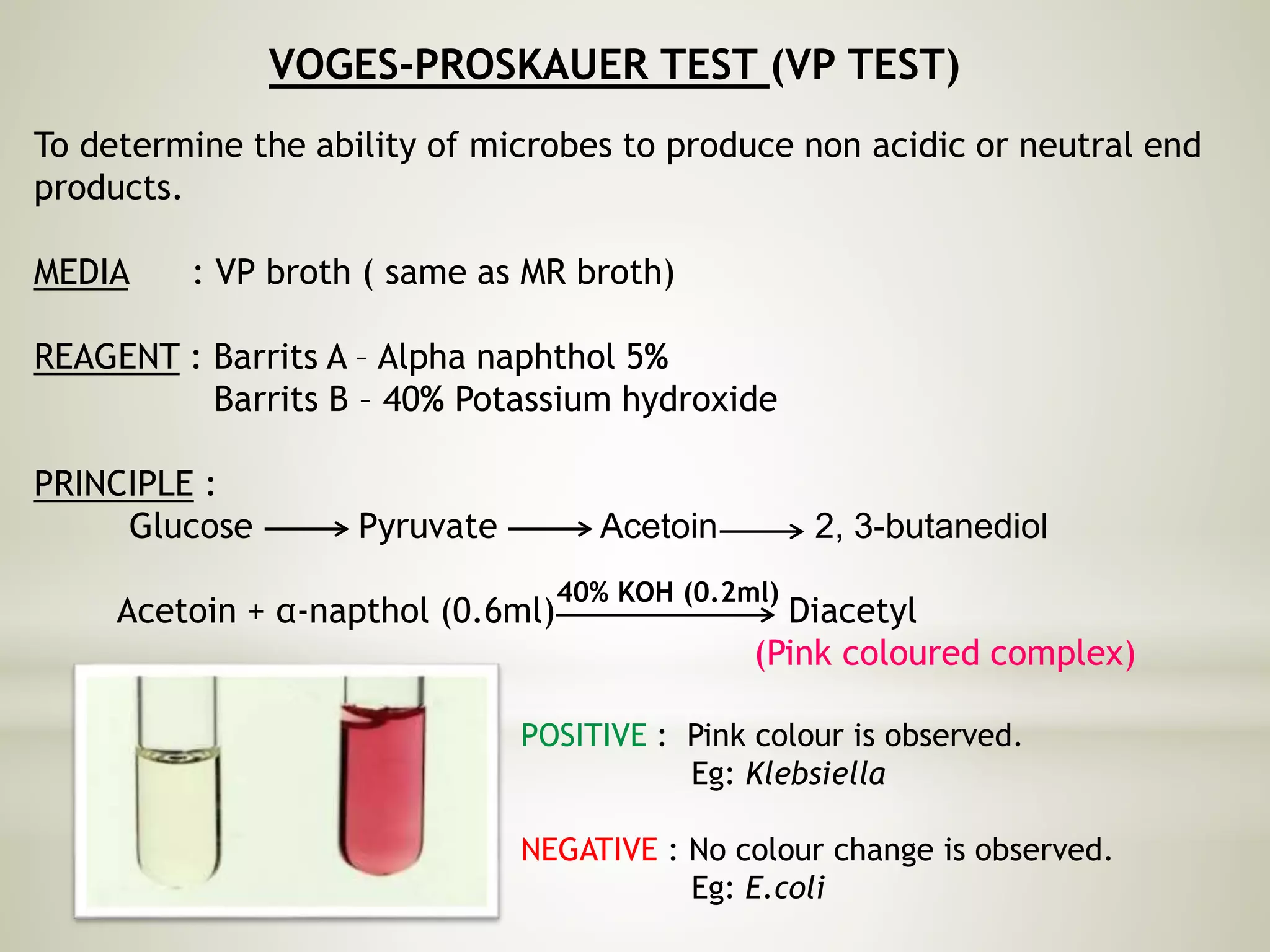
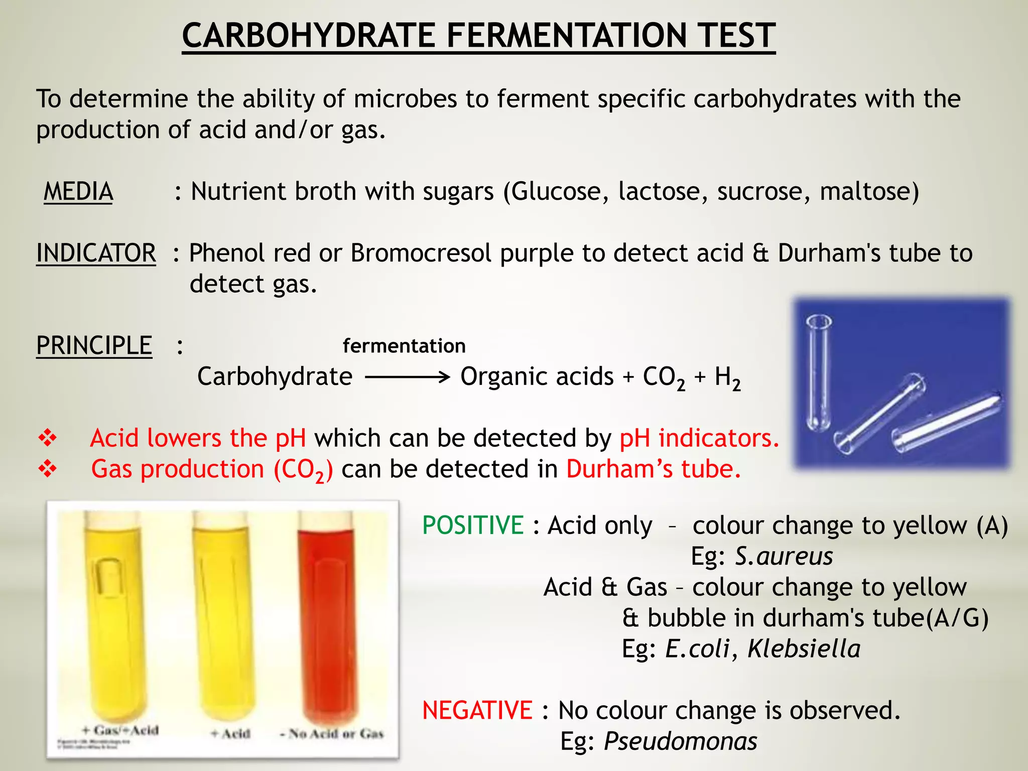
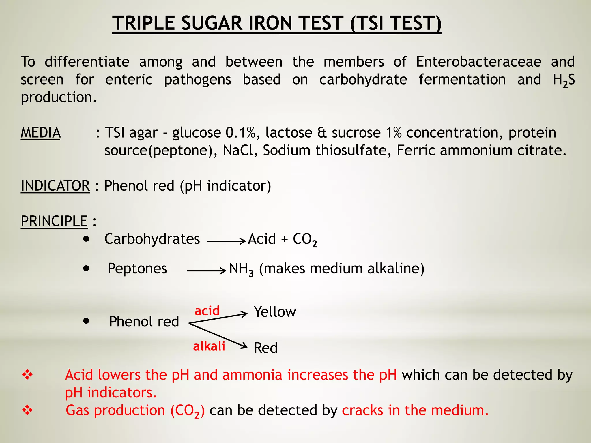
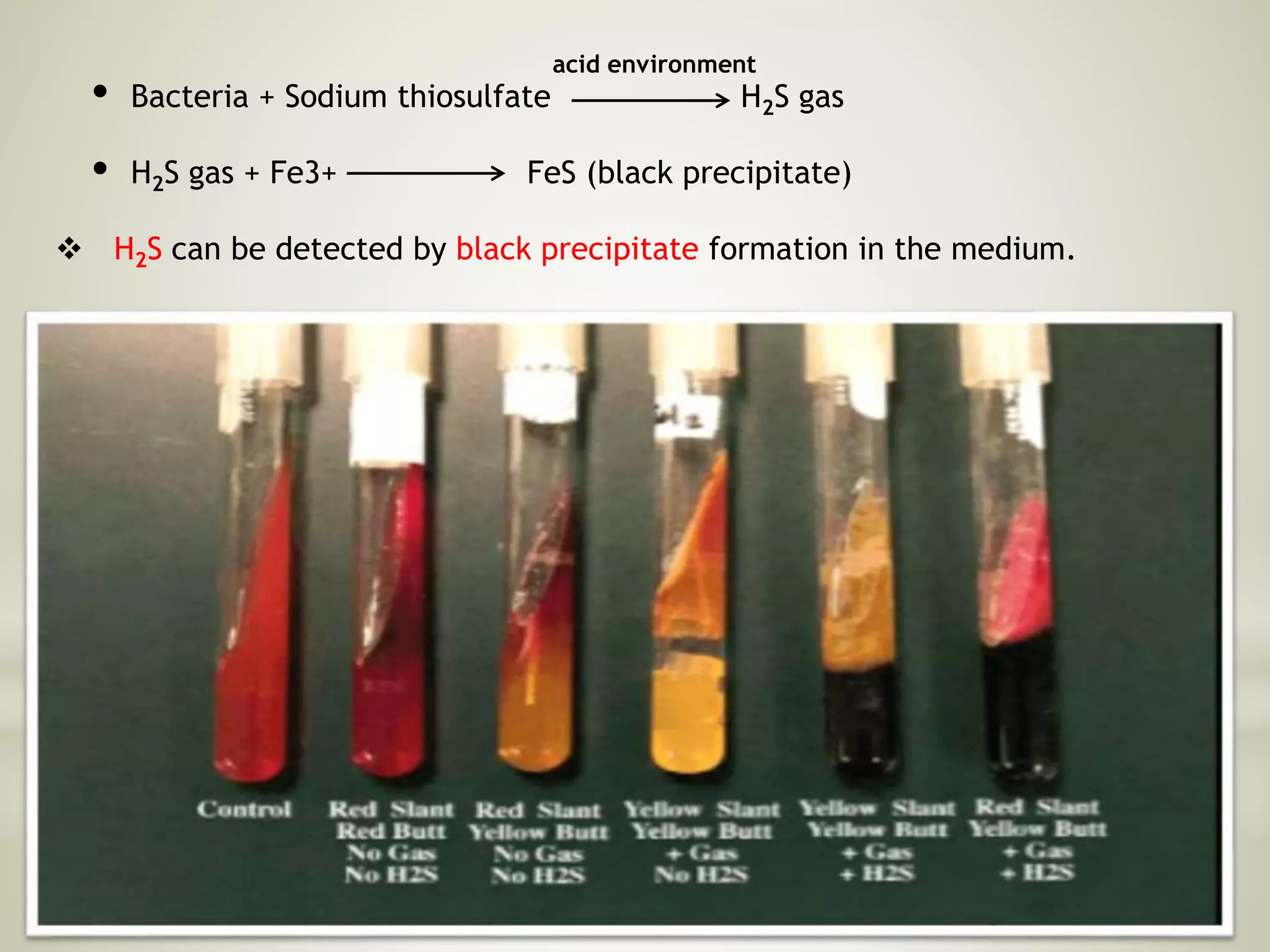
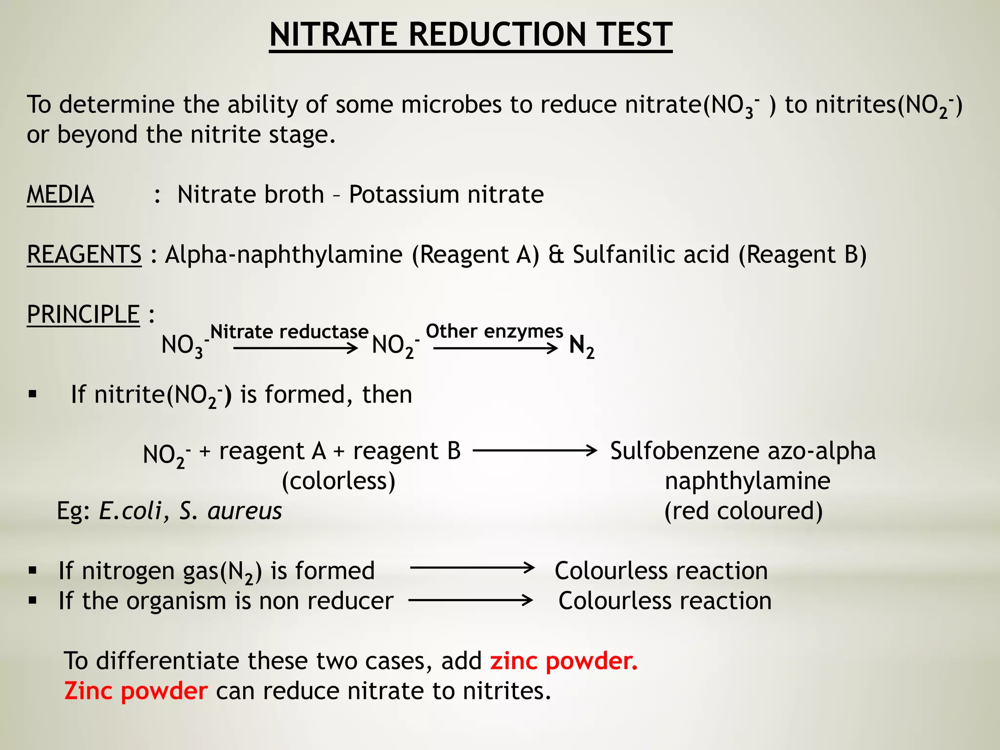
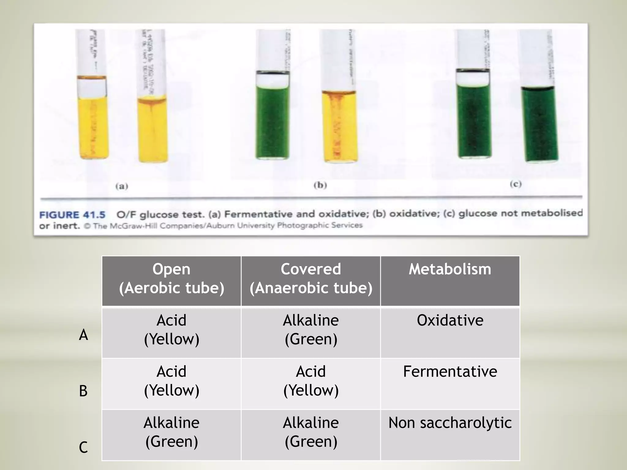
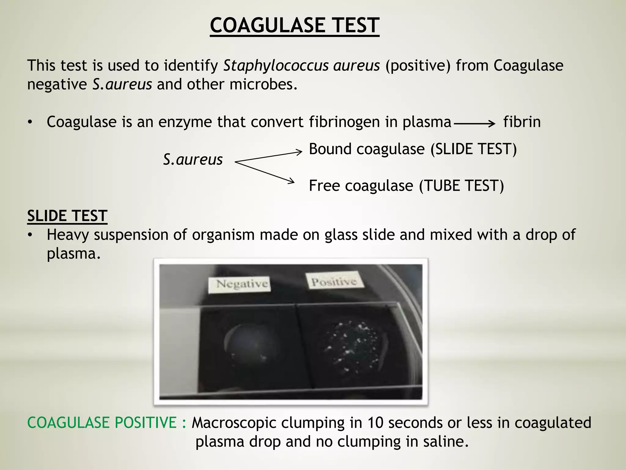
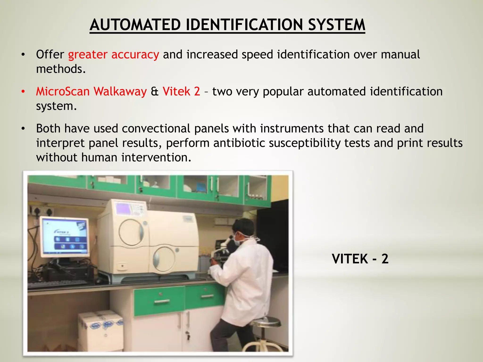
SAKEENA ASMI T
MAHATMA GANDHI UNIVERSITY
BIOCHEMICAL METHODS
FOR IDENTIFICATION OF
BACTERIA
• To distinguish harmless microbes from
pathogenic microbes.
• Characterize an outbreak of disease and
determine the source.
• Verify the authenticity of pathogenic strain
for quality control purposes.
• Determine appropriate antimicrobial therapy.
• Basically for the prevention, control and
treatment of a disease.
WHY IS IDENTIFICATION IMPORTANT?
IDENTIFICATION SCHEME
SAMPLE
(Soil, water,
food, etc…)
ISOLATION
(Spread plate, pour
plate, streak plate)
STAINING
MOTILITY
(Hanging drop method)
CULTURING
(Special media)
BIOCHEMICAL
REACTIONS
MOLECULAR
TECHNIQUES
 Biochemical tests are the tests used for
identification of bacteria species based on the
differences in the biochemical activities of
different bacteria.
 Differences in carbohydrate metabolism,
protein metabolism, fat metabolism, production
of certain enzymes, ability to utilize a particular
compound etc.
Each species of bacteria have a well defined set
of metabolic activities different from all other
species
These biochemical fingerprints are properties
controlled by the bacterial enzymes.
WHAT ARE BIOCHEMICAL TESTS?
1. Indole test
2. Methyl red test
3. Voges-Proskauer test
4. Citrate utilisation test
5. Carbohydrate fermentation test
6. Triple sugar iron(TSI)test
7. Nitrate reduction test
8. Urease test
9. Oxidase test
10. Catalase test
11. Oxidation-fermentation(O/F)test
12. O-Nitrophenyl galactosidase(ONPG)test
IMViC test
Intracellular
Enzyme
Activity
1. Coagulase test
2. Gelatinase test
3. Starch hydrolysis test
4. Lipid hydrolysis test
5. Deoxyribonuclease test(DNase
test)
Extracellular
Enzyme
Activity
INDOLE TEST
To determine the ability of microbe to degrade the amino acid tryptophan.
MEDIA : Tryptophan 1% or peptone broth
REAGENT : Kovacs’ reagent (Dimethylamine benzaldehyde)
PRINCIPLE:
Tryptophanase
Tryptophan Indole + pyruvic acid + ammonia
Indole + Kovacs reagent Rosindole + H2O
(Cherry red colour
compound)
Hcl
Butanol
POSITIVE : Cherry red coloured ring at the
interface of reagent and broth.
Eg: E.coli
NEGATIVE : No colour change is observed.
Eg: Klebsiella
SPOT INDOLE
To perform this test, a test colony is smeared in a
piece of filter paper saturated with Kovac’s
reagent. The appearance of red colour indicates a
positive test.
METHYL RED TEST (MR TEST)
To determine the ability of microbes to oxidize glucose with production
and stabilization of high content of acid end products.
MEDIA : MR broth – peptone, glucose, dipotassium phosphate &
distilled water.
REAGENT : MR reagent – MR 0.1g in 300ml of 95% ethanol & DW 200ml.
PRINCIPLE:
Glucose Mixed acids (pH less than 4.4) + Methyl red
Red colour
POSITIVE : Red colour is observed. Eg: E.coli
NEGATIVE : Yellow colour is observed.
Eg: Klebsiella
VOGES-PROSKAUER TEST (VP TEST)
To determine the ability of microbes to produce non acidic or neutral end
products.
MEDIA : VP broth ( same as MR broth)
REAGENT : Barrits A – Alpha naphthol 5%
Barrits B – 40% Potassium hydroxide
PRINCIPLE :
Glucose Pyruvate Acetoin 2, 3-butanediol
Acetoin + α-napthol (0.6ml) Diacetyl
(Pink coloured complex)
40% KOH (0.2ml)
POSITIVE : Pink colour is observed.
Eg: Klebsiella
NEGATIVE : No colour change is observed.
Eg: E.coli
CITRATE UTILISATION TEST
To determine the ability of the microbes to ferment citrate as sole carbon
source.
MEDIA : Simmons citrate medium
INDICATOR : Bromothymol blue
PRINCIPLE:
Sodium citrate Pyruvic acid + Oxaloacetic acid+ CO2
Excess sodium from + CO2 + H2O Na2CO3 (pH ) (green blue)
sodium citrate
Citrate permease
POSITIVE : Change of colour from green to blue
Eg: Klebsiella
NEGATIVE : No colour change is observed.
Eg: E.coli
CARBOHYDRATE FERMENTATION TEST
To determine the ability of microbes to ferment specific carbohydrates with the
production of acid and/or gas.
MEDIA : Nutrient broth with sugars (Glucose, lactose, sucrose, maltose)
INDICATOR : Phenol red or Bromocresol purple to detect acid & Durham's tube to
detect gas.
PRINCIPLE :
Carbohydrate Organic acids + CO2 + H2
 Acid lowers the pH which can be detected by pH indicators.
 Gas production (CO2) can be detected in Durham’s tube.
POSITIVE : Acid only – colour change to yellow (A)
Eg: S.aureus
Acid & Gas – colour change to yellow
& bubble in durham's tube(A/G)
Eg: E.coli, Klebsiella
NEGATIVE : No colour change is observed.
Eg: Pseudomonas
fermentation
TRIPLE SUGAR IRON TEST (TSI TEST)
To differentiate among and between the members of Enterobacteraceae and
screen for enteric pathogens based on carbohydrate fermentation and H2S
production.
MEDIA : TSI agar - glucose 0.1%, lactose & sucrose 1% concentration, protein
source(peptone), NaCl, Sodium thiosulfate, Ferric ammonium citrate.
INDICATOR : Phenol red (pH indicator)
PRINCIPLE :
Carbohydrates Acid + CO2
Peptones NH3 (makes medium alkaline)
Phenol red
 Acid lowers the pH and ammonia increases the pH which can be detected by
pH indicators.
 Gas production (CO2) can be detected by cracks in the medium.
Yellow
Red
acid
alkali
Bacteria + Sodium thiosulfate H2S gas
H2S gas + Fe3+ FeS (black precipitate)
 H2S can be detected by black precipitate formation in the medium.
acid environment
Result interpretation:
• Red slant, red butt, no gas, no H2S - K/K no H2S
• Red slant, yellow butt, no gas, no H2S - K/A no H2S
• Yellow slant, yellow butt, gas, no H2s - A/ A no H2S
• Yellow slant, yellow butt, gas, H2s - A/ A H2S
• Red slant, yellow butt, gas, H2S - K/ A H2S
 Alkaline slant /acidic butt only glucose is fermented
 Acidic slant / acidic butt glucose, sucrose, lactose all 3 sugars
are fermented
 Bubbles or cracks present gas production
 Black precipitate present H2S production
NITRATE REDUCTION TEST
To determine the ability of some microbes to reduce nitrate(NO3
- ) to nitrites(NO2
-)
or beyond the nitrite stage.
MEDIA : Nitrate broth – Potassium nitrate
REAGENTS : Alpha-naphthylamine (Reagent A) & Sulfanilic acid (Reagent B)
PRINCIPLE :
NO3
- NO2
- N2
 If nitrite(NO2
-) is formed, then
NO2
-
Nitrate reductase Other enzymes
+ reagent A + reagent B Sulfobenzene azo-alpha
(colorless) naphthylamine
Eg: E.coli, S. aureus (red coloured)
 If nitrogen gas(N2) is formed Colourless reaction
 If the organism is non reducer Colourless reaction
To differentiate these two cases, add zinc powder.
Zinc powder can reduce nitrate to nitrites.
• If organism reduced nitrate to nitrogen gas, then there are no nitrates present
and the addition of zinc dust will have no effect. Hence the test is positive and
gives colourless reaction the organism is a REDUCER
• If nitrates were not reduced then zinc will reduce them to nitrites. Nitrites will
react with 2 reagents and give a red colour. Test is negative and organism is NON
REDUCER. Eg: N.gonorrhoeae
Nitrate broth
Zinc powder
Nitrogen gas
/non reducer
Nitrogen gas
Reducer
Non reducer
UREASE TEST
To determine the ability of microbes to degrade urea by urease.
MEDIA : Christensen’s Urea agar
REAGENTS : Phenol red
PRINCIPLE :
Urea + 2 H2O CO2 + H2O + 2 NH3
Ammonia increases the pH which can be detected by pH indicator
Urease
POSITIVE : Pink colour is observed
Eg: Proteus
NEGATIVE : No colour change
Eg: E.coli
OXIDASE TEST
To determine the ability of microbes to produce oxidase enzyme.
REAGENT : 1% Kovacs reagent (tetra methyl para phenylenediamine dihydrochloride)
PRINCIPLE :
• Cytochrome C oxidase is an enzyme that facilitates transfer of electrons to
oxygen in aerobic bacterial respiratory transport system.
• Here the oxidase reagent substitutes as the electron acceptor.
Kovacs reagent Indophenol
(colourless oxidase reagent) (purple colour)
Cytochrome oxidase
POSITIVE : Purple colour is observed
Eg: Pseudomonas
NEGATIVE : No colour change
Eg: E.coli, S. aureus
CATALASE TEST
To determine the ability of microbes to produce catalase enzyme
REAGENT : Hydrogen peroxide
PRINCIPLE:
• During respiration many microbes produce byproducts that are toxic to cells
like Hydrogen peroxide(H2O2)
• To neutralize this toxin microbes produce an enzyme CATALASE
2 H2O2 2 H2O + 02 (gas bubbles)
POSITIVE : Presence of effervescence
Eg: E.coli, S.aureus etc
NEGATIVE : No effervescence
Eg: Streptococcus
Catalase
OXIDATIVE – FERMENTATIVE TEST (OF TEST)
To differentiate between oxidative and fermentative bacteria
MEDIA : Hugh Leifson’s OF media (high glucose & low peptone)
INDICATOR : Bromothymol blue
PRINCIPLE :
• Under anaerobic condition(overlay with a layer of oil), fermentative organisms
covert glucose mixed acids.
• Under aerobic condition, non-fermenting(oxidative) organisms produce small
amount of weak acids.
• The decrease amount of peptones and increase amount of glucose facilitates
detection of weak acids.
• Acid decreases the pH which can be detected by pH indicator which changes
the colour from green to yellow.
Examples: Oxidative – Pseudomonas aeruginosa
Fermentative - E.coli
Non saccharolytic - Alcaligens faecalis
Open
(Aerobic tube)
Covered
(Anaerobic tube)
Metabolism
Acid
(Yellow)
Alkaline
(Green)
Oxidative
Acid
(Yellow)
Acid
(Yellow)
Fermentative
Alkaline
(Green)
Alkaline
(Green)
Non saccharolytic
A
B
C
ONPG TEST FOR β-GALACTOSIDASE
To determine the presence or absence of the enzyme β-galactosidase
MEDIA : ONPG broth
PRINCIPLE :
• β – galactosidase is an enzyme that converts
Lactose Galactose + Glucose
• O – Nitro – β – D – galactopyranoside is structurally similar to lactose except that
orthonitro phenol has been substituted for glucose.
Onpg(colourless) Galactose + orthonitro phenol(yellow)
galactosidase
galactosidase
POSITIVE : Yellow colour observed
Eg: E.coli
NEGATIVE : No colour change
Eg: Proteus vulgaris
COAGULASE TEST
This test is used to identify Staphylococcus aureus (positive) from Coagulase
negative S.aureus and other microbes.
• Coagulase is an enzyme that convert fibrinogen in plasma fibrin
S.aureus
SLIDE TEST
• Heavy suspension of organism made on glass slide and mixed with a drop of
plasma.
COAGULASE POSITIVE : Macroscopic clumping in 10 seconds or less in coagulated
plasma drop and no clumping in saline.
Bound coagulase (SLIDE TEST)
Free coagulase (TUBE TEST)
COAGULASE NEGATIVE : No clumping in either drop.
• Negative results should be confirmed with tube coagulase test.
TUBE TEST
• A suspension of organism is suspended and incubated with plasma at 370C
COAGULASE POSITIVE : Clot of any size is observed.
Eg: S.aureus
COAGULASE NEGATIVE : No clot (plasma remains wholly liquid or shows only a
flocculant or ropy precipitate)
Eg: S.epidermidis, E.coli
GELATINASE TEST
To determine the ability of an enzyme to produce the enzyme gelatinase that
hydrolyze gelatin
MEDIA : Gelatin medium – gelatin, peptone, beef extract
PRINCIPLE :
Gelatin Polypeptides Amino acids
• If the organism produce gelatinase, then it will liquefy the medium.
gelatinase gelatinase
POSITIVE : Partial or complete
liquefaction
Eg: Aeromonas hydrophila
NEGATIVE : No liquefaction
Eg: E.coli
STARCH HYDROLYSIS TEST
To determine the ability of an organism to produce the enzyme amylase and
hydrolyze starch.
MEDIA : Starch agar – nutrient agar + starch
INDICATOR : Iodine solution (Grams iodine)
PRINCIPLE :
 The starch molecule consists of two constituents:
Amylose, an unbranched glucose polymer
Amylopectin, a large branched polymer
 Both amylose and amylopectin are rapidly hydrolyzed by certain bacteria using
enzymes called α-amylases.
Starch Dextrin + Maltose + Glucose
(Amylose + Amylopectin)
α - amylase
POSITIVE : If the organism has produced amylase, will hydrolyze starch and a
clear area appears after adding grams iodine.
Eg: Bacillus subtilis, B.megaterium
NEGATIVE: Absence of clearing after adding grams iodine, no starch hydrolysis.
Eg: Staphylococcus epidermidis, S.agalactiae
API STRIPS – RAPID TESTS
(Analytical Profile Index)
• Commercial miniaturized biochemical test panels – cover a significant number
of clinically important groups of bacteria as well as food and water associated
microorganisms.
• Different test panels are prepared in dehydrated forms which are reconstituted
upon use by action of bacterial suspensions.
• After incubation, positive test results are scored as a 7 digit number (profile).
• Identity of the bacterium is then easily derived from the database with the
relevant cumulative profile code book or software.
MOST USED API SYSTEM
AUTOMATED IDENTIFICATION SYSTEM
• Offer greater accuracy and increased speed identification over manual
methods.
• MicroScan Walkaway & Vitek 2 – two very popular automated identification
system.
• Both have used convectional panels with instruments that can read and
interpret panel results, perform antibiotic susceptibility tests and print results
without human intervention.
VITEK - 2
MICROSCAN WALKAWAY
.
.
.
THANK

Biochemical reactions

  • 1.
    SAKEENA ASMI T MAHATMAGANDHI UNIVERSITY BIOCHEMICAL METHODS FOR IDENTIFICATION OF BACTERIA
  • 2.
    • To distinguishharmless microbes from pathogenic microbes. • Characterize an outbreak of disease and determine the source. • Verify the authenticity of pathogenic strain for quality control purposes. • Determine appropriate antimicrobial therapy. • Basically for the prevention, control and treatment of a disease. WHY IS IDENTIFICATION IMPORTANT?
  • 3.
    IDENTIFICATION SCHEME SAMPLE (Soil, water, food,etc…) ISOLATION (Spread plate, pour plate, streak plate) STAINING MOTILITY (Hanging drop method) CULTURING (Special media) BIOCHEMICAL REACTIONS MOLECULAR TECHNIQUES
  • 4.
     Biochemical testsare the tests used for identification of bacteria species based on the differences in the biochemical activities of different bacteria.  Differences in carbohydrate metabolism, protein metabolism, fat metabolism, production of certain enzymes, ability to utilize a particular compound etc. Each species of bacteria have a well defined set of metabolic activities different from all other species These biochemical fingerprints are properties controlled by the bacterial enzymes. WHAT ARE BIOCHEMICAL TESTS?
  • 5.
    1. Indole test 2.Methyl red test 3. Voges-Proskauer test 4. Citrate utilisation test 5. Carbohydrate fermentation test 6. Triple sugar iron(TSI)test 7. Nitrate reduction test 8. Urease test 9. Oxidase test 10. Catalase test 11. Oxidation-fermentation(O/F)test 12. O-Nitrophenyl galactosidase(ONPG)test IMViC test Intracellular Enzyme Activity
  • 6.
    1. Coagulase test 2.Gelatinase test 3. Starch hydrolysis test 4. Lipid hydrolysis test 5. Deoxyribonuclease test(DNase test) Extracellular Enzyme Activity
  • 7.
    INDOLE TEST To determinethe ability of microbe to degrade the amino acid tryptophan. MEDIA : Tryptophan 1% or peptone broth REAGENT : Kovacs’ reagent (Dimethylamine benzaldehyde) PRINCIPLE: Tryptophanase Tryptophan Indole + pyruvic acid + ammonia Indole + Kovacs reagent Rosindole + H2O (Cherry red colour compound) Hcl Butanol POSITIVE : Cherry red coloured ring at the interface of reagent and broth. Eg: E.coli NEGATIVE : No colour change is observed. Eg: Klebsiella
  • 8.
    SPOT INDOLE To performthis test, a test colony is smeared in a piece of filter paper saturated with Kovac’s reagent. The appearance of red colour indicates a positive test.
  • 9.
    METHYL RED TEST(MR TEST) To determine the ability of microbes to oxidize glucose with production and stabilization of high content of acid end products. MEDIA : MR broth – peptone, glucose, dipotassium phosphate & distilled water. REAGENT : MR reagent – MR 0.1g in 300ml of 95% ethanol & DW 200ml. PRINCIPLE: Glucose Mixed acids (pH less than 4.4) + Methyl red Red colour POSITIVE : Red colour is observed. Eg: E.coli NEGATIVE : Yellow colour is observed. Eg: Klebsiella
  • 10.
    VOGES-PROSKAUER TEST (VPTEST) To determine the ability of microbes to produce non acidic or neutral end products. MEDIA : VP broth ( same as MR broth) REAGENT : Barrits A – Alpha naphthol 5% Barrits B – 40% Potassium hydroxide PRINCIPLE : Glucose Pyruvate Acetoin 2, 3-butanediol Acetoin + α-napthol (0.6ml) Diacetyl (Pink coloured complex) 40% KOH (0.2ml) POSITIVE : Pink colour is observed. Eg: Klebsiella NEGATIVE : No colour change is observed. Eg: E.coli
  • 11.
    CITRATE UTILISATION TEST Todetermine the ability of the microbes to ferment citrate as sole carbon source. MEDIA : Simmons citrate medium INDICATOR : Bromothymol blue PRINCIPLE: Sodium citrate Pyruvic acid + Oxaloacetic acid+ CO2 Excess sodium from + CO2 + H2O Na2CO3 (pH ) (green blue) sodium citrate Citrate permease POSITIVE : Change of colour from green to blue Eg: Klebsiella NEGATIVE : No colour change is observed. Eg: E.coli
  • 12.
    CARBOHYDRATE FERMENTATION TEST Todetermine the ability of microbes to ferment specific carbohydrates with the production of acid and/or gas. MEDIA : Nutrient broth with sugars (Glucose, lactose, sucrose, maltose) INDICATOR : Phenol red or Bromocresol purple to detect acid & Durham's tube to detect gas. PRINCIPLE : Carbohydrate Organic acids + CO2 + H2  Acid lowers the pH which can be detected by pH indicators.  Gas production (CO2) can be detected in Durham’s tube. POSITIVE : Acid only – colour change to yellow (A) Eg: S.aureus Acid & Gas – colour change to yellow & bubble in durham's tube(A/G) Eg: E.coli, Klebsiella NEGATIVE : No colour change is observed. Eg: Pseudomonas fermentation
  • 13.
    TRIPLE SUGAR IRONTEST (TSI TEST) To differentiate among and between the members of Enterobacteraceae and screen for enteric pathogens based on carbohydrate fermentation and H2S production. MEDIA : TSI agar - glucose 0.1%, lactose & sucrose 1% concentration, protein source(peptone), NaCl, Sodium thiosulfate, Ferric ammonium citrate. INDICATOR : Phenol red (pH indicator) PRINCIPLE : Carbohydrates Acid + CO2 Peptones NH3 (makes medium alkaline) Phenol red  Acid lowers the pH and ammonia increases the pH which can be detected by pH indicators.  Gas production (CO2) can be detected by cracks in the medium. Yellow Red acid alkali
  • 14.
    Bacteria + Sodiumthiosulfate H2S gas H2S gas + Fe3+ FeS (black precipitate)  H2S can be detected by black precipitate formation in the medium. acid environment
  • 15.
    Result interpretation: • Redslant, red butt, no gas, no H2S - K/K no H2S • Red slant, yellow butt, no gas, no H2S - K/A no H2S • Yellow slant, yellow butt, gas, no H2s - A/ A no H2S • Yellow slant, yellow butt, gas, H2s - A/ A H2S • Red slant, yellow butt, gas, H2S - K/ A H2S  Alkaline slant /acidic butt only glucose is fermented  Acidic slant / acidic butt glucose, sucrose, lactose all 3 sugars are fermented  Bubbles or cracks present gas production  Black precipitate present H2S production
  • 16.
    NITRATE REDUCTION TEST Todetermine the ability of some microbes to reduce nitrate(NO3 - ) to nitrites(NO2 -) or beyond the nitrite stage. MEDIA : Nitrate broth – Potassium nitrate REAGENTS : Alpha-naphthylamine (Reagent A) & Sulfanilic acid (Reagent B) PRINCIPLE : NO3 - NO2 - N2  If nitrite(NO2 -) is formed, then NO2 - Nitrate reductase Other enzymes + reagent A + reagent B Sulfobenzene azo-alpha (colorless) naphthylamine Eg: E.coli, S. aureus (red coloured)  If nitrogen gas(N2) is formed Colourless reaction  If the organism is non reducer Colourless reaction To differentiate these two cases, add zinc powder. Zinc powder can reduce nitrate to nitrites.
  • 17.
    • If organismreduced nitrate to nitrogen gas, then there are no nitrates present and the addition of zinc dust will have no effect. Hence the test is positive and gives colourless reaction the organism is a REDUCER • If nitrates were not reduced then zinc will reduce them to nitrites. Nitrites will react with 2 reagents and give a red colour. Test is negative and organism is NON REDUCER. Eg: N.gonorrhoeae Nitrate broth Zinc powder Nitrogen gas /non reducer Nitrogen gas Reducer Non reducer
  • 18.
    UREASE TEST To determinethe ability of microbes to degrade urea by urease. MEDIA : Christensen’s Urea agar REAGENTS : Phenol red PRINCIPLE : Urea + 2 H2O CO2 + H2O + 2 NH3 Ammonia increases the pH which can be detected by pH indicator Urease POSITIVE : Pink colour is observed Eg: Proteus NEGATIVE : No colour change Eg: E.coli
  • 19.
    OXIDASE TEST To determinethe ability of microbes to produce oxidase enzyme. REAGENT : 1% Kovacs reagent (tetra methyl para phenylenediamine dihydrochloride) PRINCIPLE : • Cytochrome C oxidase is an enzyme that facilitates transfer of electrons to oxygen in aerobic bacterial respiratory transport system. • Here the oxidase reagent substitutes as the electron acceptor. Kovacs reagent Indophenol (colourless oxidase reagent) (purple colour) Cytochrome oxidase POSITIVE : Purple colour is observed Eg: Pseudomonas NEGATIVE : No colour change Eg: E.coli, S. aureus
  • 20.
    CATALASE TEST To determinethe ability of microbes to produce catalase enzyme REAGENT : Hydrogen peroxide PRINCIPLE: • During respiration many microbes produce byproducts that are toxic to cells like Hydrogen peroxide(H2O2) • To neutralize this toxin microbes produce an enzyme CATALASE 2 H2O2 2 H2O + 02 (gas bubbles) POSITIVE : Presence of effervescence Eg: E.coli, S.aureus etc NEGATIVE : No effervescence Eg: Streptococcus Catalase
  • 21.
    OXIDATIVE – FERMENTATIVETEST (OF TEST) To differentiate between oxidative and fermentative bacteria MEDIA : Hugh Leifson’s OF media (high glucose & low peptone) INDICATOR : Bromothymol blue PRINCIPLE : • Under anaerobic condition(overlay with a layer of oil), fermentative organisms covert glucose mixed acids. • Under aerobic condition, non-fermenting(oxidative) organisms produce small amount of weak acids. • The decrease amount of peptones and increase amount of glucose facilitates detection of weak acids. • Acid decreases the pH which can be detected by pH indicator which changes the colour from green to yellow. Examples: Oxidative – Pseudomonas aeruginosa Fermentative - E.coli Non saccharolytic - Alcaligens faecalis
  • 22.
  • 23.
    ONPG TEST FORβ-GALACTOSIDASE To determine the presence or absence of the enzyme β-galactosidase MEDIA : ONPG broth PRINCIPLE : • β – galactosidase is an enzyme that converts Lactose Galactose + Glucose • O – Nitro – β – D – galactopyranoside is structurally similar to lactose except that orthonitro phenol has been substituted for glucose. Onpg(colourless) Galactose + orthonitro phenol(yellow) galactosidase galactosidase POSITIVE : Yellow colour observed Eg: E.coli NEGATIVE : No colour change Eg: Proteus vulgaris
  • 24.
    COAGULASE TEST This testis used to identify Staphylococcus aureus (positive) from Coagulase negative S.aureus and other microbes. • Coagulase is an enzyme that convert fibrinogen in plasma fibrin S.aureus SLIDE TEST • Heavy suspension of organism made on glass slide and mixed with a drop of plasma. COAGULASE POSITIVE : Macroscopic clumping in 10 seconds or less in coagulated plasma drop and no clumping in saline. Bound coagulase (SLIDE TEST) Free coagulase (TUBE TEST)
  • 25.
    COAGULASE NEGATIVE :No clumping in either drop. • Negative results should be confirmed with tube coagulase test. TUBE TEST • A suspension of organism is suspended and incubated with plasma at 370C COAGULASE POSITIVE : Clot of any size is observed. Eg: S.aureus COAGULASE NEGATIVE : No clot (plasma remains wholly liquid or shows only a flocculant or ropy precipitate) Eg: S.epidermidis, E.coli
  • 26.
    GELATINASE TEST To determinethe ability of an enzyme to produce the enzyme gelatinase that hydrolyze gelatin MEDIA : Gelatin medium – gelatin, peptone, beef extract PRINCIPLE : Gelatin Polypeptides Amino acids • If the organism produce gelatinase, then it will liquefy the medium. gelatinase gelatinase POSITIVE : Partial or complete liquefaction Eg: Aeromonas hydrophila NEGATIVE : No liquefaction Eg: E.coli
  • 27.
    STARCH HYDROLYSIS TEST Todetermine the ability of an organism to produce the enzyme amylase and hydrolyze starch. MEDIA : Starch agar – nutrient agar + starch INDICATOR : Iodine solution (Grams iodine) PRINCIPLE :  The starch molecule consists of two constituents: Amylose, an unbranched glucose polymer Amylopectin, a large branched polymer  Both amylose and amylopectin are rapidly hydrolyzed by certain bacteria using enzymes called α-amylases. Starch Dextrin + Maltose + Glucose (Amylose + Amylopectin) α - amylase
  • 28.
    POSITIVE : Ifthe organism has produced amylase, will hydrolyze starch and a clear area appears after adding grams iodine. Eg: Bacillus subtilis, B.megaterium NEGATIVE: Absence of clearing after adding grams iodine, no starch hydrolysis. Eg: Staphylococcus epidermidis, S.agalactiae
  • 29.
    API STRIPS –RAPID TESTS (Analytical Profile Index) • Commercial miniaturized biochemical test panels – cover a significant number of clinically important groups of bacteria as well as food and water associated microorganisms. • Different test panels are prepared in dehydrated forms which are reconstituted upon use by action of bacterial suspensions. • After incubation, positive test results are scored as a 7 digit number (profile). • Identity of the bacterium is then easily derived from the database with the relevant cumulative profile code book or software.
  • 30.
  • 31.
    AUTOMATED IDENTIFICATION SYSTEM •Offer greater accuracy and increased speed identification over manual methods. • MicroScan Walkaway & Vitek 2 – two very popular automated identification system. • Both have used convectional panels with instruments that can read and interpret panel results, perform antibiotic susceptibility tests and print results without human intervention. VITEK - 2
  • 32.
  • 33.