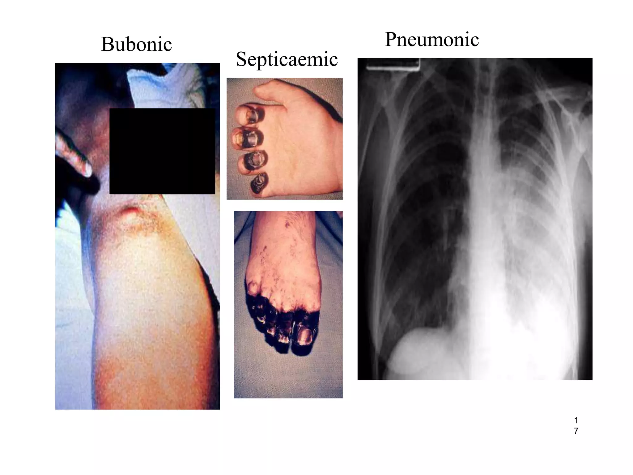
Yersinia
MICROBIOLOGY
 Yersinia belongs to family
enterbacteriaceae.
 Gram negative rods
 Motile (except Y. pestis)
 Bipolar staining- Wayson’s stain
or Giemsa stain
 (resembling to safety pin)
 The three medically important
species of Yersinia are:
 Y.pestis
 Y. pseudotuberculosis
 Y. entrocolitica
INTRODUCTION
Plague is a zoonotic disease caused by highly virulent bacteria Yersinia
pestis.
Distribution of plague is worldwide maintained by wild rodents and
their fleas causing localized infection.
Disease takes three main forms: Bubonic, Septicemic and Pneumonic.
Untreated bubonic plague can lead to secondary pneumonic stage
with highmortality.
Plague has lead to three pandemics.
 Yersinia pestis bacteria can also be used as biological weapon.
WHO identify plague as emerging disease.
Gram negative pleomorphic
bacteria that
causes Plague
Yersenia pestis
 Y. pestis survives in the stomach of the
flea and in the soil of animal burrows
 RESERVIOR:
rodents mainly,prairie dogs,field
mice,chipmunks,rabbits,cats etc.
 VECTOR:
flea
 Human are accidental host
 Plague is a classic rodent zoonosis.
 One of the most devastating diseases of
human history.
HISTORY
•Importance
•one of three WHO quarantinable
diseases
•Estimated 200 million deaths recorded
•Three prior pandemics
•Justinian 541 ad
•Black death 1346
•China 1855
Cases and Deaths due to Human Plague in India
Year Place Cases Deaths
1963 Throughout India 197 22
1964 Penninsular India 106 13
1965 -do- 1 0
1966 -do- 46 2
1967-1993 - Nil Nil
1994 Maharashtra/Gujarat 876 54
1995-2001 Nil Nil
2002 Simla Dist.Himachal
Pradesh
16 4
2003 Nil Nil
2004 Uttar Kashi
Dist.Uttarakhand
8 3
2005-2015 - Nil Nil
morphology of Yersinia pestis
• Gram –negative Coccobacillus
• Non-motile
• Non spore forming, capsulated
• Size : 0.5-0.8 μm(diameter)
1-3 μm(long)
• Optimum pH : 7.2-7.6 (extreme 5-9.6)
• On methylene blue staining: bipolar staining (safety pin appearance)
• With two end poles darkly stained and central area is clear seen
safety pin
appearance
culture
Its aerobic and facultative anareobic. Grows mostly at 27°C.
• Nutreint agar: colonies are small, delicate, transparent colonies
are seen.
• Blood agar: non- hemolytic colonies are seen.
• Macconkey agar: colourless colonies are found.
Biochemical reactions
• Y. pestis ferments glucose, mannitol and maltose
• With acid production and no gas
• Lactose and sucrose not fermented
• Catalase + ve
• Indole –ve, citrate –ve
• MR +ve, VP –ve
• Getaltin not liquifed
Resistance
• Destroyed by heat at 55°C
• Destroyed by sunlight, drying
• Sensitive to chemical disinfectants(0.5% phenol in 15 minutes)
• Survive for months in soil of rodent burrows.
• Viable for long periods in cold and moist environment.
Fleaborne transmission
Direct/physical contact with infected animal/human
Ingestion of infected animal
Inhalation of infectious droplets from animals or humans
Different
routes of
transmission
of
Y. Pestis
to humans
TRANSMISSION
Bubonic Plague is an
infection of the lymph
nodes in the Lymphatic
system which includes the
tonsils, adenoids, spleen and
thymus
Glands that swell are called
buboes, which is how Bubonic
Plague got it’s name. Buboes
are found in the groin, armpit
or neck and most often will
occur close to the site of
the initial infection.
Bubonic Plague is the
most common.
Transmitted from the
bite of an infected flea or
rodent.
Symptoms usually occur
within 3 to 7 days of
exposure.
Pain may occur before
the actual swelling begins.
If bacteria is left
untreated it can spread
into the bloodstream and
symptoms of septicemic
plague may begin.
Infection in the
blood.
Bacterium
replicate in the
bloodstream.
 Transmission from a
flea, rodent bite, or if bubonic
plague is left untreated.
Symptoms include
fever, chills, abdominal
pain, rapid heart
rate, vomiting, delirium, shock,
and bleeding into the skin or
organs.
Massive involvement of
blood vessels results in
haemmorage in the skin
& mucosa. “Black death”
Symptoms of Septicemic
Plague
The “Black Death”
• Pneumonic Plague is an
infection of the lungs and is
the most serious of the three
plagues yet the least
common. Yersinia pestis infects
a person’s lungs and leads to
pneumonia.
• Infection happens if a person
inhales the plague bacteria
from an infected person or
animal and is highly
contagious.
Symptoms usually occur
2 to 3 days after the
exposure to the airborne
particles of the bacteria.
Death usually occurs
within 2 to 6 days after
symptoms begin.
Early treatment of
pneumonic plague is
necessary to reduce the
chances of death.
About 10% of people
with bubonic plague will get
pneumonic plague.
CLINICAL FEATURES
• OTHER
COMPLICATIONS
• MENINGITIS
• CUTANEOUS DISEASE
• PHARYNGEAL DISEASE
• ENTERIC DISEASE
• SUSTAINED OCCULT FEVER
FROM ABSCESSED
INTRAABDOMINAL BUBOES
1
7
Bubonic
Septicaemic
Pneumonic
Specimen collection:
- Pus or fluid aspirated form bubos in case of Bubonic
plague.
- Sputum or blood in pneumonic plague.
- Blood in Septicemic plague.
- CSf in meningeal plague.
Direct Microscopy
 Gram’s staining:
 Gram negative coccobacilli are seen
 Methylene blue staining:
 Typical bipolar staining is seen.
Culture Characterstics
 ON BLOOD AGAR:
• non-hemolytic
• at 27° C
• opaque with a gray to yellow color in the center; they
remain transparent and gray to white in color on the
periphery.
 ON MACCONKEY AGAR:
 Colourless colonies are formed.
X
Serological tests
• Antibodies and Antigen can be detected by:
•Passive haemagglutination
•Complement fixation test
•ELISA
Molecular tests
•Polymerase chain reaction (PCR)
TREATMENT
• ANTIBIOTICS FOR CONTAINED CASUALTIES
• (FOR MASS CASUALTIES)
• 1ST CHOICES
• STREPTOMYCIN - FDA-APPROVED
• 30 MG/KG IM DIVIDED Q8-12 KIDS (MAX 2G/DAY)
• 1G IM BID ADULT
• BACTERICIDAL
• GENTAMICIN –AS EFFECTIVE, MORE AVAIL, QD
DOSING
• 5MG/KG IV QD, W/LEVELS OR LOAD 2MG/KG THEN
1.7MG/KG Q8
• 2.5MG/KG IM/IV Q8H KIDS (Q12HR FOR <1WK OR
PREMATURE)
TREATMENT
• 2ND CHOICES
• TETRACYCLINE'S - AS GOOD IN VITRO
• DOXYCYCLINE
• SINGLE 200MG IV LOADING DOSE (SOME
SOURCES)
• 100MG IV BID OR 200 MG IV QD ADULTS& KIDS
>45KG
• 2.2MG/KG IV Q12HR (MAX 200MG) KIDS <45KG
• BETTER ABSORPTION, DISTRIBUTION, HALF-LIFE
THAN TCN
• 1ST CHOICE PO THERAPY FOR MASS CASUALTIES
• TETRACYCLINE
• 500 MG PO QID ADULTS
TREATMENT
• 2ND CHOICES
• FLUOROQUINOLONES–BETTER IN VITRO
• CIPROFLOXACIN
• 400 MG IV Q12HRADULTS
• 15 MG/KG IV Q12HR KIDS (MAX 1G/DAY)
• LEVOFLOXACIN
• OFLOXACIN
• CHLORAMPHENICOL
• 1ST CHOICE FOR MENINGITIS +/-AMINOGLYCOSIDE
• CROSSES BLOOD-BRAIN BARRIER
• 25MG/KG IV Q6HR ADULTS & KIDS, KEEP LEVEL 5-20 ΜG/ML
• AVOID IN KIDS <2 YO (GREY BABY SYNDROME)
PREVENTION
• VACCINATION - BUBONIC
ONLY
• KILLED VIRULENT STRAIN –
USED IN U.S.
• FORMALIN-FIXED, NO LONGER
COMMERCIALLYAVAILABLE
• FUTURE PRODUCTION
AND LICENSURE
UNKNOWN
• SERIES
• 3 PRIMARY (1.0CC, 0.2 CC AT1-
3 MO AND 5-6 MOLATER)
• 2 BOOSTERS 0.2CC AT 6 MO
BIOWEAPON POTENTIAL
• AEROSOL
• BIOWEAPONS
PROGRAMS DEVELOPED
TECHNIQUES TO
AEROSOLIZE PLAGUE
DIRECTLY
• PNEUMONIC FORM WOULD
BE EXPECTED
• PROVEN INFECTIVITY
OF PRIMATES
INFECTION CONTROL
• MECHANISM FOR PERSON-PERSON SPREAD
• RESPIRATORY DROPLETS MOST LIKELY, NOT
DROPLET NUCLEI
• HISTORICALLY PREVENTED BY MASKS
• RESPIRATORY DROPLET PRECAUTIONS
• WEAR MASK, GOWN, GLOVES, EYE PROTECTION
• SUSPECTED CASES - ISOLATE
• IMMEDIATELY RESPIRATORY (EVEN FOR BUBONIC)
• AVOID UNNECESSARY CLOSE CONTACT 1ST 48 HRS OF
ISOLATION.
• DURATION
• 2 DAYS AFTER INITIATING ANTIBIOTICS AND CLINICALLY
IMPROVED
• AFTER SPUTUM CULTURES NEGATIVE
INFECTION CONTROL
• NATIONAL CONTROL
PROGRAMS
• SURVEILLANCE
• EARLY
DIAGNOSIS, TREATMENT &
ISOLATION OF CASES
• ENVIRONMENTAL
SANITATION & EXPOSURE
AVOIDANCE
• PUBLIC EDUCATION
Yersenia

Yersenia

  • 1.
  • 2.
    MICROBIOLOGY  Yersinia belongsto family enterbacteriaceae.  Gram negative rods  Motile (except Y. pestis)  Bipolar staining- Wayson’s stain or Giemsa stain  (resembling to safety pin)  The three medically important species of Yersinia are:  Y.pestis  Y. pseudotuberculosis  Y. entrocolitica
  • 3.
    INTRODUCTION Plague is azoonotic disease caused by highly virulent bacteria Yersinia pestis. Distribution of plague is worldwide maintained by wild rodents and their fleas causing localized infection. Disease takes three main forms: Bubonic, Septicemic and Pneumonic. Untreated bubonic plague can lead to secondary pneumonic stage with highmortality. Plague has lead to three pandemics.  Yersinia pestis bacteria can also be used as biological weapon. WHO identify plague as emerging disease.
  • 4.
    Gram negative pleomorphic bacteriathat causes Plague Yersenia pestis
  • 5.
     Y. pestissurvives in the stomach of the flea and in the soil of animal burrows  RESERVIOR: rodents mainly,prairie dogs,field mice,chipmunks,rabbits,cats etc.  VECTOR: flea  Human are accidental host
  • 6.
     Plague isa classic rodent zoonosis.  One of the most devastating diseases of human history.
  • 7.
    HISTORY •Importance •one of threeWHO quarantinable diseases •Estimated 200 million deaths recorded •Three prior pandemics •Justinian 541 ad •Black death 1346 •China 1855
  • 9.
    Cases and Deathsdue to Human Plague in India Year Place Cases Deaths 1963 Throughout India 197 22 1964 Penninsular India 106 13 1965 -do- 1 0 1966 -do- 46 2 1967-1993 - Nil Nil 1994 Maharashtra/Gujarat 876 54 1995-2001 Nil Nil 2002 Simla Dist.Himachal Pradesh 16 4 2003 Nil Nil 2004 Uttar Kashi Dist.Uttarakhand 8 3 2005-2015 - Nil Nil
  • 10.
    morphology of Yersiniapestis • Gram –negative Coccobacillus • Non-motile • Non spore forming, capsulated • Size : 0.5-0.8 μm(diameter) 1-3 μm(long) • Optimum pH : 7.2-7.6 (extreme 5-9.6) • On methylene blue staining: bipolar staining (safety pin appearance) • With two end poles darkly stained and central area is clear seen safety pin appearance
  • 11.
    culture Its aerobic andfacultative anareobic. Grows mostly at 27°C. • Nutreint agar: colonies are small, delicate, transparent colonies are seen. • Blood agar: non- hemolytic colonies are seen. • Macconkey agar: colourless colonies are found.
  • 12.
    Biochemical reactions • Y.pestis ferments glucose, mannitol and maltose • With acid production and no gas • Lactose and sucrose not fermented • Catalase + ve • Indole –ve, citrate –ve • MR +ve, VP –ve • Getaltin not liquifed
  • 13.
    Resistance • Destroyed byheat at 55°C • Destroyed by sunlight, drying • Sensitive to chemical disinfectants(0.5% phenol in 15 minutes) • Survive for months in soil of rodent burrows. • Viable for long periods in cold and moist environment.
  • 14.
    Fleaborne transmission Direct/physical contactwith infected animal/human Ingestion of infected animal Inhalation of infectious droplets from animals or humans Different routes of transmission of Y. Pestis to humans TRANSMISSION
  • 16.
    Bubonic Plague isan infection of the lymph nodes in the Lymphatic system which includes the tonsils, adenoids, spleen and thymus Glands that swell are called buboes, which is how Bubonic Plague got it’s name. Buboes are found in the groin, armpit or neck and most often will occur close to the site of the initial infection.
  • 17.
    Bubonic Plague isthe most common. Transmitted from the bite of an infected flea or rodent. Symptoms usually occur within 3 to 7 days of exposure. Pain may occur before the actual swelling begins. If bacteria is left untreated it can spread into the bloodstream and symptoms of septicemic plague may begin.
  • 18.
    Infection in the blood. Bacterium replicatein the bloodstream.  Transmission from a flea, rodent bite, or if bubonic plague is left untreated. Symptoms include fever, chills, abdominal pain, rapid heart rate, vomiting, delirium, shock, and bleeding into the skin or organs. Massive involvement of blood vessels results in haemmorage in the skin & mucosa. “Black death”
  • 19.
  • 20.
    • Pneumonic Plagueis an infection of the lungs and is the most serious of the three plagues yet the least common. Yersinia pestis infects a person’s lungs and leads to pneumonia. • Infection happens if a person inhales the plague bacteria from an infected person or animal and is highly contagious.
  • 21.
    Symptoms usually occur 2to 3 days after the exposure to the airborne particles of the bacteria. Death usually occurs within 2 to 6 days after symptoms begin. Early treatment of pneumonic plague is necessary to reduce the chances of death. About 10% of people with bubonic plague will get pneumonic plague.
  • 22.
    CLINICAL FEATURES • OTHER COMPLICATIONS •MENINGITIS • CUTANEOUS DISEASE • PHARYNGEAL DISEASE • ENTERIC DISEASE • SUSTAINED OCCULT FEVER FROM ABSCESSED INTRAABDOMINAL BUBOES
  • 23.
  • 24.
    Specimen collection: - Pusor fluid aspirated form bubos in case of Bubonic plague. - Sputum or blood in pneumonic plague. - Blood in Septicemic plague. - CSf in meningeal plague.
  • 25.
    Direct Microscopy  Gram’sstaining:  Gram negative coccobacilli are seen  Methylene blue staining:  Typical bipolar staining is seen.
  • 26.
    Culture Characterstics  ONBLOOD AGAR: • non-hemolytic • at 27° C • opaque with a gray to yellow color in the center; they remain transparent and gray to white in color on the periphery.  ON MACCONKEY AGAR:  Colourless colonies are formed. X
  • 27.
    Serological tests • Antibodiesand Antigen can be detected by: •Passive haemagglutination •Complement fixation test •ELISA Molecular tests •Polymerase chain reaction (PCR)
  • 28.
    TREATMENT • ANTIBIOTICS FORCONTAINED CASUALTIES • (FOR MASS CASUALTIES) • 1ST CHOICES • STREPTOMYCIN - FDA-APPROVED • 30 MG/KG IM DIVIDED Q8-12 KIDS (MAX 2G/DAY) • 1G IM BID ADULT • BACTERICIDAL • GENTAMICIN –AS EFFECTIVE, MORE AVAIL, QD DOSING • 5MG/KG IV QD, W/LEVELS OR LOAD 2MG/KG THEN 1.7MG/KG Q8 • 2.5MG/KG IM/IV Q8H KIDS (Q12HR FOR <1WK OR PREMATURE)
  • 29.
    TREATMENT • 2ND CHOICES •TETRACYCLINE'S - AS GOOD IN VITRO • DOXYCYCLINE • SINGLE 200MG IV LOADING DOSE (SOME SOURCES) • 100MG IV BID OR 200 MG IV QD ADULTS& KIDS >45KG • 2.2MG/KG IV Q12HR (MAX 200MG) KIDS <45KG • BETTER ABSORPTION, DISTRIBUTION, HALF-LIFE THAN TCN • 1ST CHOICE PO THERAPY FOR MASS CASUALTIES • TETRACYCLINE • 500 MG PO QID ADULTS
  • 30.
    TREATMENT • 2ND CHOICES •FLUOROQUINOLONES–BETTER IN VITRO • CIPROFLOXACIN • 400 MG IV Q12HRADULTS • 15 MG/KG IV Q12HR KIDS (MAX 1G/DAY) • LEVOFLOXACIN • OFLOXACIN • CHLORAMPHENICOL • 1ST CHOICE FOR MENINGITIS +/-AMINOGLYCOSIDE • CROSSES BLOOD-BRAIN BARRIER • 25MG/KG IV Q6HR ADULTS & KIDS, KEEP LEVEL 5-20 ΜG/ML • AVOID IN KIDS <2 YO (GREY BABY SYNDROME)
  • 31.
    PREVENTION • VACCINATION -BUBONIC ONLY • KILLED VIRULENT STRAIN – USED IN U.S. • FORMALIN-FIXED, NO LONGER COMMERCIALLYAVAILABLE • FUTURE PRODUCTION AND LICENSURE UNKNOWN • SERIES • 3 PRIMARY (1.0CC, 0.2 CC AT1- 3 MO AND 5-6 MOLATER) • 2 BOOSTERS 0.2CC AT 6 MO
  • 32.
    BIOWEAPON POTENTIAL • AEROSOL •BIOWEAPONS PROGRAMS DEVELOPED TECHNIQUES TO AEROSOLIZE PLAGUE DIRECTLY • PNEUMONIC FORM WOULD BE EXPECTED • PROVEN INFECTIVITY OF PRIMATES
  • 33.
    INFECTION CONTROL • MECHANISMFOR PERSON-PERSON SPREAD • RESPIRATORY DROPLETS MOST LIKELY, NOT DROPLET NUCLEI • HISTORICALLY PREVENTED BY MASKS • RESPIRATORY DROPLET PRECAUTIONS • WEAR MASK, GOWN, GLOVES, EYE PROTECTION • SUSPECTED CASES - ISOLATE • IMMEDIATELY RESPIRATORY (EVEN FOR BUBONIC) • AVOID UNNECESSARY CLOSE CONTACT 1ST 48 HRS OF ISOLATION. • DURATION • 2 DAYS AFTER INITIATING ANTIBIOTICS AND CLINICALLY IMPROVED • AFTER SPUTUM CULTURES NEGATIVE
  • 34.
    INFECTION CONTROL • NATIONALCONTROL PROGRAMS • SURVEILLANCE • EARLY DIAGNOSIS, TREATMENT & ISOLATION OF CASES • ENVIRONMENTAL SANITATION & EXPOSURE AVOIDANCE • PUBLIC EDUCATION