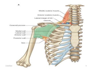
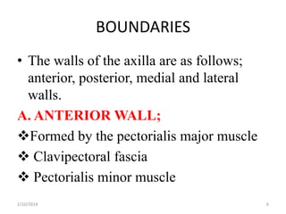
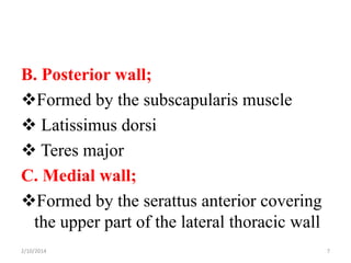
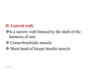
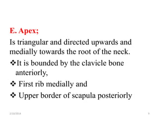
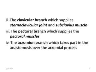
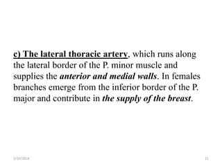
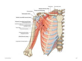
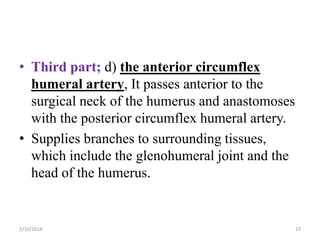
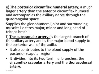
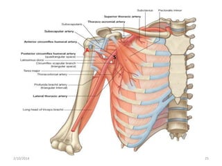
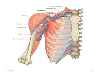
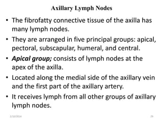
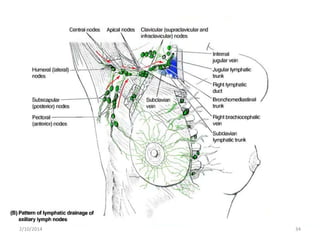
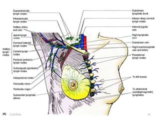
The axilla is a pyramidal space bounded by bones and muscles that provides a passage for vessels and nerves to the upper limb. It contains the brachial plexus, axillary artery and vein, lymph nodes, and connective tissue. The axillary artery gives off branches including the thoracoacromial, lateral thoracic, anterior and posterior circumflex humeral, and subscapular arteries. The axillary vein receives tributaries that generally follow the arterial branches. Axillary lymph nodes are arranged in five groups - apical, pectoral, subscapular, humeral, and central - that drain lymph from different regions.


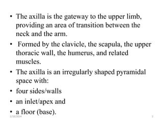


![Lecture 25 Intermuscular sapces and axilla [Autosaved].pptx](https://cdn.slidesharecdn.com/ss_thumbnails/lecture25intermuscularsapcesandaxillaautosaved-251110002658-47b36c78-thumbnail.jpg?width=640&height=640&fit=bounds)


![CTEV [ clubfoot] DR ARUN LAL ,DR MOHAMED ASHRAF travancore medical college k...](https://cdn.slidesharecdn.com/ss_thumbnails/ctevclubfootdrarunlaldrmohamedashraftravancoremedicalcollegekollamkeralaindia-260208063247-18fc466c-thumbnail.jpg?width=640&height=640&fit=bounds)


