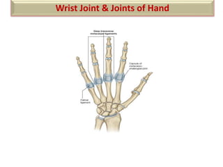
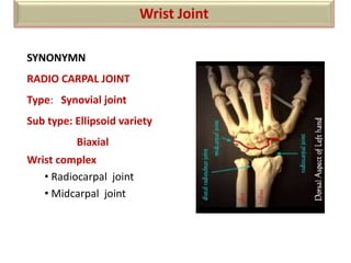
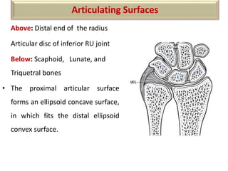
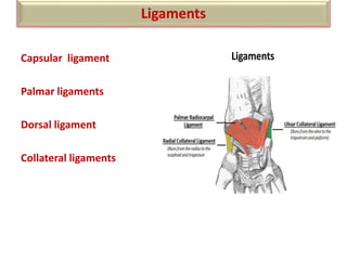
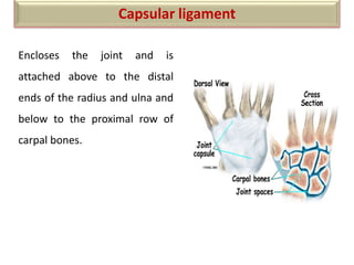
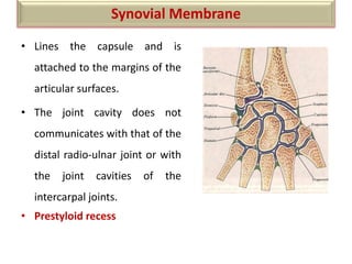
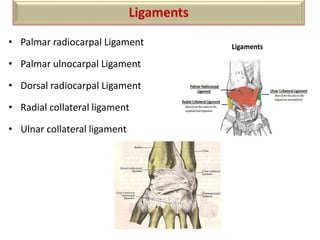
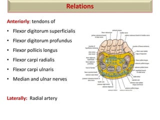
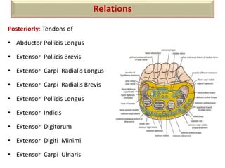
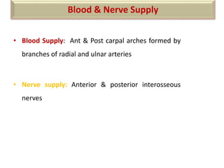
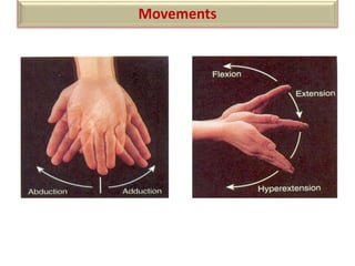
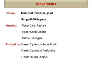
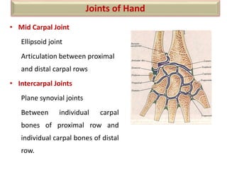
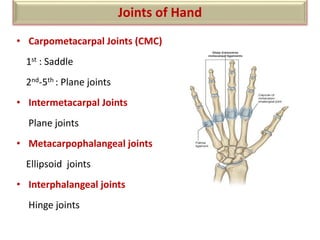
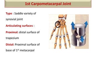
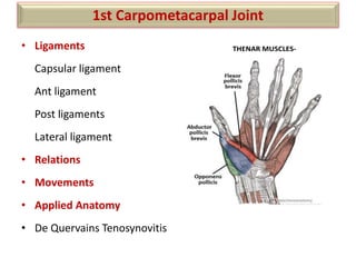
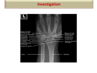
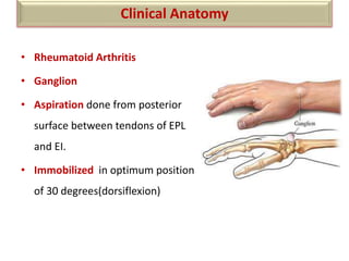
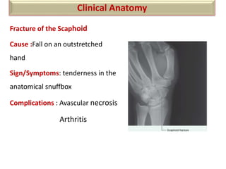
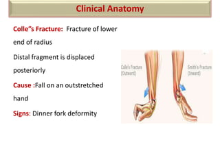
Wrist Joint & Joints of Hand
Wrist Joint
SYNONYMN
RADIO CARPAL JOINT
Type: Synovial joint
Sub type: Ellipsoid variety
Biaxial
Wrist complex
• Radiocarpal joint
• Midcarpal joint
Above: Distal end of the radius
Articular disc of inferior RU joint
Below: Scaphoid, Lunate, and
Triquetral bones
• The proximal articular surface
forms an ellipsoid concave surface,
in which fits the distal ellipsoid
convex surface.
Articulating Surfaces
Capsular ligament
Palmar ligaments
Dorsal ligament
Collateral ligaments
Ligaments
Ligaments
Encloses the joint and is
attached above to the distal
ends of the radius and ulna and
below to the proximal row of
carpal bones.
Capsular ligament
• Lines the capsule and is
attached to the margins of the
articular surfaces.
• The joint cavity does not
communicates with that of the
distal radio-ulnar joint or with
the joint cavities of the
intercarpal joints.
• Prestyloid recess
Synovial Membrane
• Palmar radiocarpal Ligament
• Palmar ulnocarpal Ligament
• Dorsal radiocarpal Ligament
• Radial collateral ligament
• Ulnar collateral ligament
Ligaments
Ligaments
Anteriorly: tendons of
• Flexor digitorum superficialis
• Flexor digitorum profundus
• Flexor pollicis longus
• Flexor carpi radialis
• Flexor carpi ulnaris
• Median and ulnar nerves
Laterally: Radial artery
Relations
Posteriorly: Tendons of
• Abductor Pollicis Longus
• Extensor Pollicis Brevis
• Extensor Carpi Radialis Longus
• Extensor Carpi Radialis Brevis
• Extensor Pollicis Longus
• Extensor Indicis
• Extensor Digitorum
• Extensor Digiti Minimi
• Extensor Carpi Ulnaris
Relations
• Blood Supply: Ant & Post carpal arches formed by
branches of radial and ulnar arteries
• Nerve supply: Anterior & posterior interosseous
nerves
Blood & Nerve Supply
Movements
Flexion: Mainly at midcarpal joint
Range-0-60 degrees
Muscles: Flexor Carpi Radialis
Flexor Carpi Ulnaris
Palmaris Longus.
Assisted by: Flexor Digitorum Superficialis
Flexor Digitorum Profundus
Flexor Pollicis Longus.
Movements
Extension: Mainly at wrist joint
Range-0-50 degrees
Muscles: Extensor Carpi Radialis Longus
Extensor Carpi Radialis Brevis
Extensor Carpi Ulnaris
Assisted by: Extensor Digitorum
Extensor Indicis
Extensor Digiti Minimi
Extensor Pollicis Longus
Movements
Abduction: At Midcarpal joint
Range:0-15 degrees
Muscles : Flexor Carpi Radialis
Extensor Carpi Radialis Longus
Extensor Carpi Radialis Brevis
Assisted by: Abductor Pollicis Longus
Extensor Pollicis Longus
Extensor Pollicis Brevis
Movements
Adduction: Mainly at wrist joint
Range:0-50 degree
Muscles: Flexor Carpi Ulnaris
Extensor Carpi Ulnaris
Circumduction: Combination of flexion adduction, extension and
abduction
Movements
• Mid Carpal Joint
Ellipsoid joint
Articulation between proximal
and distal carpal rows
• Intercarpal Joints
Plane synovial joints
Between individual carpal
bones of proximal row and
individual carpal bones of distal
row.
Joints of Hand
• Carpometacarpal Joints (CMC)
1st : Saddle
2nd-5th : Plane joints
• Intermetacarpal Joints
Plane joints
• Metacarpophalangeal joints
Ellipsoid joints
• Interphalangeal joints
Hinge joints
Joints of Hand
Type : Saddle variety of
synovial joint
Articulating surfaces :
Proximal: distal surface of
trapezium
Distal: Proximal surface of
base of 1st metacarpal
1st Carpometacarpal Joint
• Ligaments
Capsular ligament
Ant ligament
Post ligaments
Lateral ligament
• Relations
• Movements
• Applied Anatomy
• De Quervains Tenosynovitis
1st Carpometacarpal Joint
THENAR MUSCLES-
Investigation
X-RAY
• Rheumatoid Arthritis
• Ganglion
• Aspiration done from posterior
surface between tendons of EPL
and EI.
• Immobilized in optimum position
of 30 degrees(dorsiflexion)
Clinical Anatomy
Fracture of the Scaphoid
Cause :Fall on an outstretched
hand
Sign/Symptoms: tenderness in the
anatomical snuffbox
Complications : Avascular necrosis
Arthritis
Clinical Anatomy
Colle”s Fracture: Fracture of lower
end of radius
Distal fragment is displaced
posteriorly
Cause :Fall on an outstretched
hand
Signs: Dinner fork deformity
Clinical Anatomy
THANKYOU

Wrist joint

  • 1.
    Wrist Joint &Joints of Hand
  • 2.
    Wrist Joint SYNONYMN RADIO CARPALJOINT Type: Synovial joint Sub type: Ellipsoid variety Biaxial Wrist complex • Radiocarpal joint • Midcarpal joint
  • 3.
    Above: Distal endof the radius Articular disc of inferior RU joint Below: Scaphoid, Lunate, and Triquetral bones • The proximal articular surface forms an ellipsoid concave surface, in which fits the distal ellipsoid convex surface. Articulating Surfaces
  • 4.
    Capsular ligament Palmar ligaments Dorsalligament Collateral ligaments Ligaments Ligaments
  • 5.
    Encloses the jointand is attached above to the distal ends of the radius and ulna and below to the proximal row of carpal bones. Capsular ligament
  • 6.
    • Lines thecapsule and is attached to the margins of the articular surfaces. • The joint cavity does not communicates with that of the distal radio-ulnar joint or with the joint cavities of the intercarpal joints. • Prestyloid recess Synovial Membrane
  • 7.
    • Palmar radiocarpalLigament • Palmar ulnocarpal Ligament • Dorsal radiocarpal Ligament • Radial collateral ligament • Ulnar collateral ligament Ligaments Ligaments
  • 8.
    Anteriorly: tendons of •Flexor digitorum superficialis • Flexor digitorum profundus • Flexor pollicis longus • Flexor carpi radialis • Flexor carpi ulnaris • Median and ulnar nerves Laterally: Radial artery Relations
  • 9.
    Posteriorly: Tendons of •Abductor Pollicis Longus • Extensor Pollicis Brevis • Extensor Carpi Radialis Longus • Extensor Carpi Radialis Brevis • Extensor Pollicis Longus • Extensor Indicis • Extensor Digitorum • Extensor Digiti Minimi • Extensor Carpi Ulnaris Relations
  • 10.
    • Blood Supply:Ant & Post carpal arches formed by branches of radial and ulnar arteries • Nerve supply: Anterior & posterior interosseous nerves Blood & Nerve Supply
  • 11.
  • 12.
    Flexion: Mainly atmidcarpal joint Range-0-60 degrees Muscles: Flexor Carpi Radialis Flexor Carpi Ulnaris Palmaris Longus. Assisted by: Flexor Digitorum Superficialis Flexor Digitorum Profundus Flexor Pollicis Longus. Movements
  • 13.
    Extension: Mainly atwrist joint Range-0-50 degrees Muscles: Extensor Carpi Radialis Longus Extensor Carpi Radialis Brevis Extensor Carpi Ulnaris Assisted by: Extensor Digitorum Extensor Indicis Extensor Digiti Minimi Extensor Pollicis Longus Movements
  • 14.
    Abduction: At Midcarpaljoint Range:0-15 degrees Muscles : Flexor Carpi Radialis Extensor Carpi Radialis Longus Extensor Carpi Radialis Brevis Assisted by: Abductor Pollicis Longus Extensor Pollicis Longus Extensor Pollicis Brevis Movements
  • 15.
    Adduction: Mainly atwrist joint Range:0-50 degree Muscles: Flexor Carpi Ulnaris Extensor Carpi Ulnaris Circumduction: Combination of flexion adduction, extension and abduction Movements
  • 16.
    • Mid CarpalJoint Ellipsoid joint Articulation between proximal and distal carpal rows • Intercarpal Joints Plane synovial joints Between individual carpal bones of proximal row and individual carpal bones of distal row. Joints of Hand
  • 17.
    • Carpometacarpal Joints(CMC) 1st : Saddle 2nd-5th : Plane joints • Intermetacarpal Joints Plane joints • Metacarpophalangeal joints Ellipsoid joints • Interphalangeal joints Hinge joints Joints of Hand
  • 18.
    Type : Saddlevariety of synovial joint Articulating surfaces : Proximal: distal surface of trapezium Distal: Proximal surface of base of 1st metacarpal 1st Carpometacarpal Joint
  • 19.
    • Ligaments Capsular ligament Antligament Post ligaments Lateral ligament • Relations • Movements • Applied Anatomy • De Quervains Tenosynovitis 1st Carpometacarpal Joint THENAR MUSCLES-
  • 20.
  • 21.
    • Rheumatoid Arthritis •Ganglion • Aspiration done from posterior surface between tendons of EPL and EI. • Immobilized in optimum position of 30 degrees(dorsiflexion) Clinical Anatomy
  • 22.
    Fracture of theScaphoid Cause :Fall on an outstretched hand Sign/Symptoms: tenderness in the anatomical snuffbox Complications : Avascular necrosis Arthritis Clinical Anatomy
  • 23.
    Colle”s Fracture: Fractureof lower end of radius Distal fragment is displaced posteriorly Cause :Fall on an outstretched hand Signs: Dinner fork deformity Clinical Anatomy
  • 24.