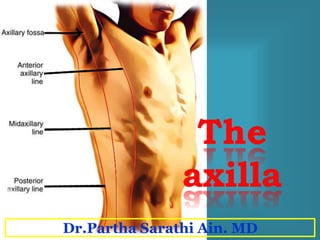
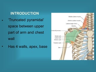
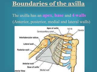
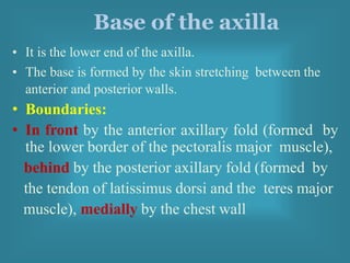
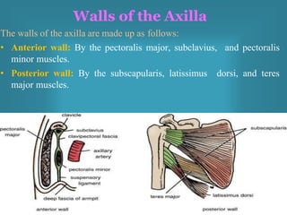
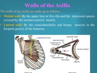
Dr.Partha Sarathi Ain. MD
The axilla
pyramid-shaped
• Definition: The axilla
space
(armpit) is a
between the
upper part of the arm and the side of the
chest
• Functions: It forms an important
passage for nerves, blood, and lymph
vessels as they travel from the root of the
neck to the upper limb.
INTRODUCTION
‘Truncated pyramidal’
space between upper
part of arm and chest
wall
Has 4 walls, apex, base
•
•
Boundaries of the axilla
The axilla has an apex, base and 4 walls
(Anterior, posterior, medial and lateral walls)
Apex of the axilla
• It is the upper end of the
axilla
• It continues into the root
of the neck (posterior
triangle of the neck)
through the cervico-
axillary canal.
• Boundraies of the
cervico-axillary canal
• In front by the clavicle,
• Behind by the upper
border of the scapula,
• Medially by the outer
border of the first rib
Base of the axilla
• It is the lower end of the axilla.
• The base is formed by the skin stretching between the
anterior and posterior walls.
• Boundaries:
• In front by the anterior axillary fold (formed by
the lower border of the pectoralis major muscle),
behind by the posterior axillary fold (formed by
the tendon of latissimus dorsi and the teres major
muscle), medially by the chest wall
Walls of the Axilla
The walls of the axilla are made up as follows:
• Anterior wall: By the pectoralis major, subclavius, and pectoralis
minor muscles.
• Posterior wall: By the subscapularis, latissimus dorsi, and teres
major muscles.
The walls of the axilla are made up as follows:
• Medial wall: By the upper four or five ribs and the intercostal spaces
covered by the serratus anterior muscle.
• Lateral wall: By the coracobrachialis and biceps muscles in the
bicipital groove of the humerus.
Walls of the Axilla
• Anterior wall: By the pectoralis major, subclavius,
and pectoralis minor muscles, clavipectoral fascia.
SP DEPT OF ANATOMY AFMC 9
cervico axillary canal
10
Structures passing through
apex
• Axillary vessels
• Cords of BP
• Long thoracic Nerve
• LN efferents
• Posterior wall: By the subscapularis, latissimus
dorsi, and teres major muscles.
• Medial wall:
By the upper four or
five ribs and the
intercostal spaces
covered by the
serratus anterior
muscle
Contents of the axilla
•Axillary artery
•Axillary vein
•Brachial plexus
•Axillary lymph nodes
11
Base
•Concavity directed below
•Boundaries
• Ant : anterior axillary fold
• Post: posterior axillary fold
• Medial: Chest wall
• Lateral: arm
•Formed by: skin, superficial fasciae, & deep fascia
•Deep fascia: axillary fascia extending between Pectoralis
Major (ant fold) and Lat Dorsi & Teres Major (post fold)
•Supported by Suspensory lig of Axilla
Axillary Artery
• Beginning: The axillary artery begins at the lateral
border of the first rib as a continuation of the
subclavian artery
• Termination: at the lower border of the teres major
muscle, where it continues as the brachial artery.
• The artery is closely related to the cords of the
brachial plexus and their branches and is enclosed
with them in a connective tissue sheath called the
axillary sheath. If this sheath is traced upward into
the root of the neck, it is seen to be continuous with
the prevertebral fascia
Parts of the axillary artery
• The pectoralis minor
muscle crosses in front
of the axillary artery and
divides it into three parts:
• First part above the
pectoralis minor muscle
• Second part behind the
pectoralis minor muscle
• Third part below the
pectoralis
minor muscle
First Part of the Axillary Artery
• This extends from the lateral border of the first rib to the
upper border of the pectoralis minor.
Relations
• Anteriorly: The pectoralis major and the skin.
• Posteriorly: The long t
thoracic nerve (nerve to
the serratus anterior)
• Laterally: The three cords
• of the brachial plexus
• Medially: The axillary vein
• This lies behind the pectoralis minor muscle.
Relations
• Anteriorly: The pectoralis minor, the pectoralis major, and the
skin
• Posteriorly: The posterior
cord of the brachial plexus,
the subscapularis muscle,
and the shoulder joint.
• Laterally: The lateral cord
of the brachial plexus.
• Medially: The medial cord
of the brachial plexus and
the axillary vein.
Second Part of the Axillary Artery
• This extends from the lower border of the pectoralis minor to the
lower border of the teres major.
• Relations
• Anteriorly: The pectoralis major for a short distance; lower down the artery is
crossed by the medial root of the median nerve.
• Posteriorly: The subscapularis,
the latissimus dorsi, and the teres
major. The axillary and radial nerves
also lie behind the artery.
• Laterally: The coracobrachialis,
the biceps, and the humerus.
The lateral root of the median
and the musculocutaneous nerves
also lie on the lateral side.
• Medially: The ulnar nerve,
the axillary vein, and the medial
cutaneous nerve of the arm.
Third Part of the Axillary Artery
Branches of the Axillary Artery
From the first part:
• The superior (highest) thoracic artery is small and runs
along the upper border of the pectoralis minor.
From the second part:
• 1.The thoracoacromial artery immediately divides into
terminal branches.
• 2.The lateral thoracic artery runs along the lower border of the
pectoralis minor
From the third part:
• 1. The subscapular artery runs along the lower border of
the subscapularis muscle.
• 2. The anterior circumflex humeral artery wind around the
front of the surgical neck of the humerus.
• 3. posterior circumflex humeral artery wind around the
back of the surgical neck of the humerus
Axillary Vein
• The axillary vein is formed at the lower border
of the teres major muscle by the union of the
venae comitantes of the brachial artery and the
basilic vein.
• It runs upward on the medial side of the axillary
artery and ends at the lateral border of the first
rib by becoming the subclavian vein.
• The vein receives tributaries, which correspond
to the branches of the axillary artery, and the
cephalic vein
Lymph
drainage of
upper limb.
Axillary lymph
nodes
20-30 in number, embedded in the fibro fatty tissue of Axilla Drain
 Lateral part of breast.
 Superficial lymphatic vessels from thoraco lumbar region above
level of umbilicus. Approx 75% of lymph from breast is drained
to axillary group of LNs, making them important for diagnosis
of cancer by biopsy. Axillary LNs are of 05 groups.
 Whole of upper limb except lateral side of arm, forearm, hand.
 Due to the surgical removal of metastasized axillary LNs in case of Ca-
breast chance of lymphoedema is very high.
 In case of surgical management of Ca-breast draining LNs are usually
removed to prevent recurrence of further metastasis.
25
Axillary lymph
nodes
ANTERIOR; (pectoral) along medial wall with Lateral Thoracic vein
POSTERIOR: (Subscapular ) along medial wall with Subscapular vein
LATERAL: along medial side of axillary vein CENTRAL:in
Centre of Axillary fat, Related to Intercostobrachial Nv.
APICAL: at apex of Axilla.
INFRACLAVICULAR: on Clavipectoral fascia in deltopectoral groove.
SP DEPT OF ANATOMY AFMC 23
Axilla.pptx
Axilla.pptx
Axilla.pptx
Axilla.pptx
Axilla.pptx
Axilla.pptx
Axilla.pptx
Axilla.pptx
Axilla.pptx

Axilla.pptx

  • 1.
  • 2.
    The axilla pyramid-shaped • Definition:The axilla space (armpit) is a between the upper part of the arm and the side of the chest • Functions: It forms an important passage for nerves, blood, and lymph vessels as they travel from the root of the neck to the upper limb.
  • 3.
    INTRODUCTION ‘Truncated pyramidal’ space betweenupper part of arm and chest wall Has 4 walls, apex, base • •
  • 4.
    Boundaries of theaxilla The axilla has an apex, base and 4 walls (Anterior, posterior, medial and lateral walls)
  • 5.
    Apex of theaxilla • It is the upper end of the axilla • It continues into the root of the neck (posterior triangle of the neck) through the cervico- axillary canal. • Boundraies of the cervico-axillary canal • In front by the clavicle, • Behind by the upper border of the scapula, • Medially by the outer border of the first rib
  • 6.
    Base of theaxilla • It is the lower end of the axilla. • The base is formed by the skin stretching between the anterior and posterior walls. • Boundaries: • In front by the anterior axillary fold (formed by the lower border of the pectoralis major muscle), behind by the posterior axillary fold (formed by the tendon of latissimus dorsi and the teres major muscle), medially by the chest wall
  • 8.
    Walls of theAxilla The walls of the axilla are made up as follows: • Anterior wall: By the pectoralis major, subclavius, and pectoralis minor muscles. • Posterior wall: By the subscapularis, latissimus dorsi, and teres major muscles.
  • 9.
    The walls ofthe axilla are made up as follows: • Medial wall: By the upper four or five ribs and the intercostal spaces covered by the serratus anterior muscle. • Lateral wall: By the coracobrachialis and biceps muscles in the bicipital groove of the humerus. Walls of the Axilla
  • 10.
    • Anterior wall:By the pectoralis major, subclavius, and pectoralis minor muscles, clavipectoral fascia.
  • 11.
    SP DEPT OFANATOMY AFMC 9 cervico axillary canal
  • 12.
    10 Structures passing through apex •Axillary vessels • Cords of BP • Long thoracic Nerve • LN efferents
  • 13.
    • Posterior wall:By the subscapularis, latissimus dorsi, and teres major muscles.
  • 14.
    • Medial wall: Bythe upper four or five ribs and the intercostal spaces covered by the serratus anterior muscle
  • 15.
    Contents of theaxilla •Axillary artery •Axillary vein •Brachial plexus •Axillary lymph nodes
  • 16.
    11 Base •Concavity directed below •Boundaries •Ant : anterior axillary fold • Post: posterior axillary fold • Medial: Chest wall • Lateral: arm •Formed by: skin, superficial fasciae, & deep fascia •Deep fascia: axillary fascia extending between Pectoralis Major (ant fold) and Lat Dorsi & Teres Major (post fold) •Supported by Suspensory lig of Axilla
  • 17.
    Axillary Artery • Beginning:The axillary artery begins at the lateral border of the first rib as a continuation of the subclavian artery • Termination: at the lower border of the teres major muscle, where it continues as the brachial artery. • The artery is closely related to the cords of the brachial plexus and their branches and is enclosed with them in a connective tissue sheath called the axillary sheath. If this sheath is traced upward into the root of the neck, it is seen to be continuous with the prevertebral fascia
  • 19.
    Parts of theaxillary artery • The pectoralis minor muscle crosses in front of the axillary artery and divides it into three parts: • First part above the pectoralis minor muscle • Second part behind the pectoralis minor muscle • Third part below the pectoralis minor muscle
  • 20.
    First Part ofthe Axillary Artery • This extends from the lateral border of the first rib to the upper border of the pectoralis minor. Relations • Anteriorly: The pectoralis major and the skin. • Posteriorly: The long t thoracic nerve (nerve to the serratus anterior) • Laterally: The three cords • of the brachial plexus • Medially: The axillary vein
  • 21.
    • This liesbehind the pectoralis minor muscle. Relations • Anteriorly: The pectoralis minor, the pectoralis major, and the skin • Posteriorly: The posterior cord of the brachial plexus, the subscapularis muscle, and the shoulder joint. • Laterally: The lateral cord of the brachial plexus. • Medially: The medial cord of the brachial plexus and the axillary vein. Second Part of the Axillary Artery
  • 22.
    • This extendsfrom the lower border of the pectoralis minor to the lower border of the teres major. • Relations • Anteriorly: The pectoralis major for a short distance; lower down the artery is crossed by the medial root of the median nerve. • Posteriorly: The subscapularis, the latissimus dorsi, and the teres major. The axillary and radial nerves also lie behind the artery. • Laterally: The coracobrachialis, the biceps, and the humerus. The lateral root of the median and the musculocutaneous nerves also lie on the lateral side. • Medially: The ulnar nerve, the axillary vein, and the medial cutaneous nerve of the arm. Third Part of the Axillary Artery
  • 23.
    Branches of theAxillary Artery From the first part: • The superior (highest) thoracic artery is small and runs along the upper border of the pectoralis minor. From the second part: • 1.The thoracoacromial artery immediately divides into terminal branches. • 2.The lateral thoracic artery runs along the lower border of the pectoralis minor From the third part: • 1. The subscapular artery runs along the lower border of the subscapularis muscle. • 2. The anterior circumflex humeral artery wind around the front of the surgical neck of the humerus. • 3. posterior circumflex humeral artery wind around the back of the surgical neck of the humerus
  • 25.
    Axillary Vein • Theaxillary vein is formed at the lower border of the teres major muscle by the union of the venae comitantes of the brachial artery and the basilic vein. • It runs upward on the medial side of the axillary artery and ends at the lateral border of the first rib by becoming the subclavian vein. • The vein receives tributaries, which correspond to the branches of the axillary artery, and the cephalic vein
  • 28.
  • 29.
    Axillary lymph nodes 20-30 innumber, embedded in the fibro fatty tissue of Axilla Drain  Lateral part of breast.  Superficial lymphatic vessels from thoraco lumbar region above level of umbilicus. Approx 75% of lymph from breast is drained to axillary group of LNs, making them important for diagnosis of cancer by biopsy. Axillary LNs are of 05 groups.  Whole of upper limb except lateral side of arm, forearm, hand.  Due to the surgical removal of metastasized axillary LNs in case of Ca- breast chance of lymphoedema is very high.  In case of surgical management of Ca-breast draining LNs are usually removed to prevent recurrence of further metastasis.
  • 30.
    25 Axillary lymph nodes ANTERIOR; (pectoral)along medial wall with Lateral Thoracic vein POSTERIOR: (Subscapular ) along medial wall with Subscapular vein LATERAL: along medial side of axillary vein CENTRAL:in Centre of Axillary fat, Related to Intercostobrachial Nv. APICAL: at apex of Axilla. INFRACLAVICULAR: on Clavipectoral fascia in deltopectoral groove.
  • 32.
    SP DEPT OFANATOMY AFMC 23