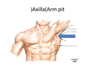
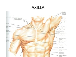
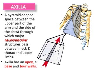
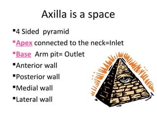
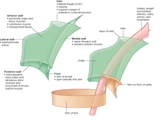
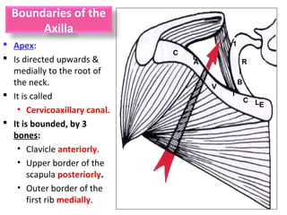
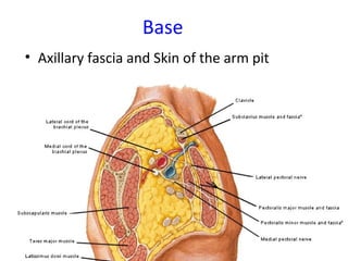
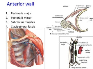
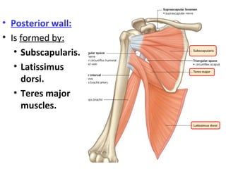
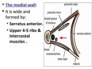
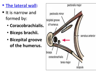
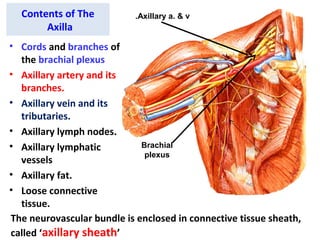
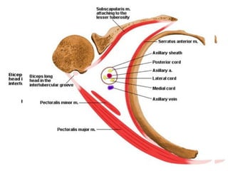
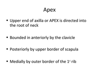
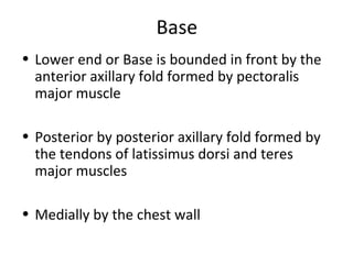
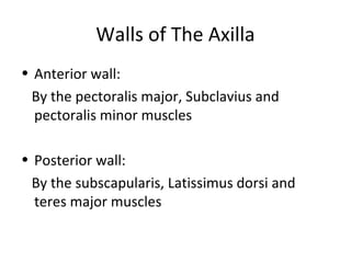
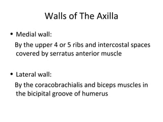
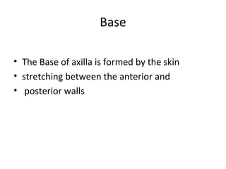
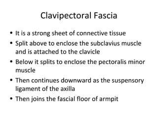
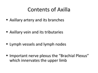
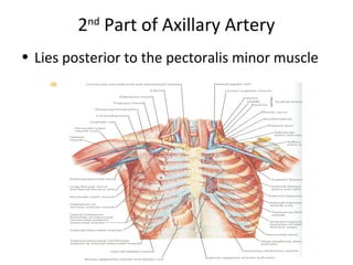
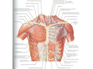
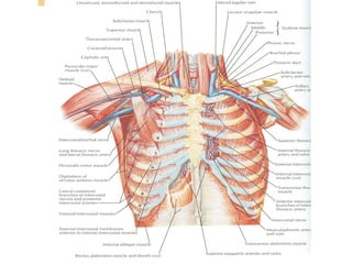
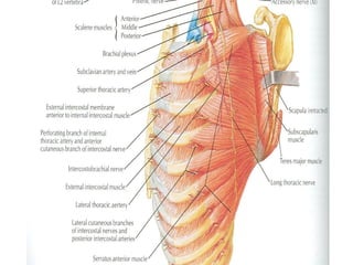
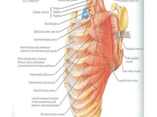
Anatomy of the Axilla
KALIISA EDWARD
BDS YEAR 3
Objectives
 Define axilla.
 Give its boundaries and contents.
Axilla(Arm pit(
AXILLA
Boundaries
Contents
Axillary Vessels
AXILLA
Definition
• It is a pyramid shaped space between the
upper part of the arm and the lateral wall of
the chest
• Important Nerves, Blood and Lymph vessels
travel through it from root of the neck to the
upper limb
AXILLA
• A pyramid-shaped
space between the
upper part of the
arm and the side of
the chest through
which major
neurovascularneurovascular
structures pass
between neck &
thorax and upper
limbs.
• Axilla has an apex, a
base and four walls.
Axilla is a space
4 Sided pyramid
Apex connected to the neck=Inlet
Base Arm pit= Outlet
Anterior wall
Posterior wall
Medial wall
Lateral wall
Boundaries of the
Axilla
 Apex:
 Is directed upwards &
medially to the root of
the neck.
 It is called
• Cervicoaxillary canal.
 It is bounded, by 3
bones:
• Clavicle anteriorly.
• Upper border of the
scapula posteriorly.
• Outer border of the
first rib medially.
C
L
A
V
I
C LE
1
R
I
B
Base
• Axillary fascia and Skin of the arm pit
Anterior wall
1. Pectoralis major
2. Pectoralis minor
3. Subclavius muscles
4. Clavipectoral fascia
• Posterior wall:
• Is formed by:
• Subscapularis.
• Latissimus
dorsi.
• Teres major
muscles.
 The medial wall:
 It is wide and
formed by:
• Serratus anterior.
• Upper 4-5 ribs &
Intercostal
muscles .
 The lateral wall:
 It is narrow and
formed by:
• Coracobrachialis.
• Biceps brachii.
• Bicepital groove
of the humerus.
Contents of The
Axilla
• Cords and branches of
the brachial plexus
• Axillary artery and its
branches.
• Axillary vein and its
tributaries.
• Axillary lymph nodes.
• Axillary lymphatic
vessels
• Axillary fat.
• Loose connective
tissue.
The neurovascular bundle is enclosed in connective tissue sheath,
called ‘axillary sheath’
Axillary a. & v.
Brachial
plexus
Apex
• Upper end of axilla or APEX is directed into
the root of neck
• Bounded in anteriorly by the clavicle
• Posteriorly by upper border of scapula
• Medially by outer border of the 1st
rib
Base
• Lower end or Base is bounded in front by the
anterior axillary fold formed by pectoralis
major muscle
• Posterior by posterior axillary fold formed by
the tendons of latissimus dorsi and teres
major muscles
• Medially by the chest wall
Walls of The Axilla
• Anterior wall:
By the pectoralis major, Subclavius and
pectoralis minor muscles
• Posterior wall:
By the subscapularis, Latissimus dorsi and
teres major muscles
Walls of The Axilla
• Medial wall:
By the upper 4 or 5 ribs and intercostal spaces
covered by serratus anterior muscle
• Lateral wall:
By the coracobrachialis and biceps muscles in
the bicipital groove of humerus
Base
• The Base of axilla is formed by the skin
• stretching between the anterior and
• posterior walls
Clavipectoral Fascia
• It is a strong sheet of connective tissue
• Split above to enclose the subclavius muscle
and is attached to the clavicle
• Below it splits to enclose the pectoralis minor
muscle
• Then continues downward as the suspensory
ligament of the axilla
• Then joins the fascial floor of armpit
Contents of Axilla
• Axillary artery and its branches
• Axillary vein and its tributaries
• Lymph vessels and lymph nodes
• Important nerve plexus the “Brachial Plexus”
which innervates the upper limb
Axillary Artery
• Is a continuation of subclavian artery
• Begins at the outer border of the 1st
rib
• Ends at the lower border of teres major
• It continues as the brachial artery
• Closely related to brachial plexus cords
• Enclosed with them in the axillary sheath
• Axillary sheath is continuous with the
prevertebral fascia
• Pectoralis minor divides it into 3 parts
1st
Part of Axillary Artery
• Extends from the outer border of the 1st
rib to the upper border of pectoralis minor
Relation
• Anterior: Pectoralis major, covering fascia,
skin, cephalic vein
• Posterior: Long thoracic nerve
• Lateral: Three cords of brachial plexus
• Medial: Axillary vein
2nd
Part of Axillary Artery
• Lies posterior to the pectoralis minor muscle
Relation
• Anterior: Pectoralis minor and major, covering
fascia and skin
• Posterior: Posterior cord of brachial plexus
• Lateral: Lateral cord of brachial plexus
• Medial: medial cord of brachial plexus and
axillary vein
3rd
Part of Axillary Artery
• Extends from lower border of pectoralis minor
to the lower border of teres major
Relation
• Anterior: Pectoralis major, medial root of the
median nerve
• Posterior: subscapularis, latissimus dorsi and
teres major
• Lateral: Coracobrachialis, biceps, humerus
• Medial: Ulnar nerve, axillary vein, medial
cutaneous nerve of the arm
Branches
• Branches of axillary artery supply the thoracic
wall and the shoulder region
• 1st
Part: Highest thoracic artery
• 2nd
Part: Thoracoacromial and lateral thoracic
arteries
• 3rd
Part: Subscapular artery, anterior and
posterior circumflex humeral arteries
Pectoralis Major
• Origin: Medial half of clavicle, sternum, upper
6 costal cartilages
• Insertion: Lateral lip of bicipital groove of the
humerus
• NS: Medial and Lateral pectoral Nerve from
medial and lateral pectoral cords of brachial
plexus
• Action: Adducts the arm and rotates it
medially, some fibers also cause flexion of arm
Subclavius
• Origin: From the first costal cartilage
• Insertion: Fibers move upward and laterally
into the inferior surface of clavicle
• NS: Nerve to the subclavius from upper trunk
of brachial plexus
• Action: Depresses the clavicle and steadies the
bone
Pectoralis Minor
• Origin: From 3rd
, 4th
and 5th
ribs
• Insertion: Coracoid Process
• NS: Medial pectoral nerve, a branch of the
medial cord of brachial plexus
• Action: Pulls the shoulder downward and
forward. Elevates the ribs of origin
Subscapularis
• Origin: Subscapular fossa on the anterior
surface of scapula
• Insertion: On the lesser tuberosity of the
humerus
• NS: Upper and Lower subscapular nerves
• Action: Medially rotates the arm and stabilizes
the shoulder joint
Latissimus Dorsi
• Origin: Posterior part of the iliac crest, lumbar fascia
and spines of lower 6 thoracic vertebrae, lower 3 ribs
• Insertion: Floor of the bicipital groove of humerus
with teres major
• NS: Thoracodorsal nerve from posterior cord of
brachial plexus
• Action: Extends, adducts and medially rotates the
arm
Teres Major
• Origin: lower third of the lateral border of
scapula
• Insertion: Medial lip of bicipital groove of
humerus
• NS: Lower subscapular nerve from posterior
cord of brachial plexus
• Action: Adducts and medially rotates the arm
Serratus Anterior
• Origin: From Outer surface of upper 8 ribs
• Insertion: Medial border of scapula in the
region of inferior angle
• NS: Long thoracic nerve
• Action: Draws the scapula forward, rotates it
The axilla
The axilla

The axilla

  • 1.
    Anatomy of theAxilla KALIISA EDWARD BDS YEAR 3
  • 2.
    Objectives  Define axilla. Give its boundaries and contents.
  • 3.
  • 4.
  • 5.
  • 6.
    Definition • It isa pyramid shaped space between the upper part of the arm and the lateral wall of the chest • Important Nerves, Blood and Lymph vessels travel through it from root of the neck to the upper limb
  • 7.
    AXILLA • A pyramid-shaped spacebetween the upper part of the arm and the side of the chest through which major neurovascularneurovascular structures pass between neck & thorax and upper limbs. • Axilla has an apex, a base and four walls.
  • 8.
    Axilla is aspace 4 Sided pyramid Apex connected to the neck=Inlet Base Arm pit= Outlet Anterior wall Posterior wall Medial wall Lateral wall
  • 10.
    Boundaries of the Axilla Apex:  Is directed upwards & medially to the root of the neck.  It is called • Cervicoaxillary canal.  It is bounded, by 3 bones: • Clavicle anteriorly. • Upper border of the scapula posteriorly. • Outer border of the first rib medially. C L A V I C LE 1 R I B
  • 11.
    Base • Axillary fasciaand Skin of the arm pit
  • 12.
    Anterior wall 1. Pectoralismajor 2. Pectoralis minor 3. Subclavius muscles 4. Clavipectoral fascia
  • 13.
    • Posterior wall: •Is formed by: • Subscapularis. • Latissimus dorsi. • Teres major muscles.
  • 14.
     The medialwall:  It is wide and formed by: • Serratus anterior. • Upper 4-5 ribs & Intercostal muscles .
  • 15.
     The lateralwall:  It is narrow and formed by: • Coracobrachialis. • Biceps brachii. • Bicepital groove of the humerus.
  • 16.
    Contents of The Axilla •Cords and branches of the brachial plexus • Axillary artery and its branches. • Axillary vein and its tributaries. • Axillary lymph nodes. • Axillary lymphatic vessels • Axillary fat. • Loose connective tissue. The neurovascular bundle is enclosed in connective tissue sheath, called ‘axillary sheath’ Axillary a. & v. Brachial plexus
  • 18.
    Apex • Upper endof axilla or APEX is directed into the root of neck • Bounded in anteriorly by the clavicle • Posteriorly by upper border of scapula • Medially by outer border of the 1st rib
  • 19.
    Base • Lower endor Base is bounded in front by the anterior axillary fold formed by pectoralis major muscle • Posterior by posterior axillary fold formed by the tendons of latissimus dorsi and teres major muscles • Medially by the chest wall
  • 20.
    Walls of TheAxilla • Anterior wall: By the pectoralis major, Subclavius and pectoralis minor muscles • Posterior wall: By the subscapularis, Latissimus dorsi and teres major muscles
  • 21.
    Walls of TheAxilla • Medial wall: By the upper 4 or 5 ribs and intercostal spaces covered by serratus anterior muscle • Lateral wall: By the coracobrachialis and biceps muscles in the bicipital groove of humerus
  • 22.
    Base • The Baseof axilla is formed by the skin • stretching between the anterior and • posterior walls
  • 23.
    Clavipectoral Fascia • Itis a strong sheet of connective tissue • Split above to enclose the subclavius muscle and is attached to the clavicle • Below it splits to enclose the pectoralis minor muscle • Then continues downward as the suspensory ligament of the axilla • Then joins the fascial floor of armpit
  • 24.
    Contents of Axilla •Axillary artery and its branches • Axillary vein and its tributaries • Lymph vessels and lymph nodes • Important nerve plexus the “Brachial Plexus” which innervates the upper limb
  • 25.
    Axillary Artery • Isa continuation of subclavian artery • Begins at the outer border of the 1st rib • Ends at the lower border of teres major • It continues as the brachial artery • Closely related to brachial plexus cords • Enclosed with them in the axillary sheath • Axillary sheath is continuous with the prevertebral fascia • Pectoralis minor divides it into 3 parts
  • 26.
    1st Part of AxillaryArtery • Extends from the outer border of the 1st rib to the upper border of pectoralis minor
  • 28.
    Relation • Anterior: Pectoralismajor, covering fascia, skin, cephalic vein • Posterior: Long thoracic nerve • Lateral: Three cords of brachial plexus • Medial: Axillary vein
  • 29.
    2nd Part of AxillaryArtery • Lies posterior to the pectoralis minor muscle
  • 30.
    Relation • Anterior: Pectoralisminor and major, covering fascia and skin • Posterior: Posterior cord of brachial plexus • Lateral: Lateral cord of brachial plexus • Medial: medial cord of brachial plexus and axillary vein
  • 31.
    3rd Part of AxillaryArtery • Extends from lower border of pectoralis minor to the lower border of teres major
  • 32.
    Relation • Anterior: Pectoralismajor, medial root of the median nerve • Posterior: subscapularis, latissimus dorsi and teres major • Lateral: Coracobrachialis, biceps, humerus • Medial: Ulnar nerve, axillary vein, medial cutaneous nerve of the arm
  • 33.
    Branches • Branches ofaxillary artery supply the thoracic wall and the shoulder region • 1st Part: Highest thoracic artery • 2nd Part: Thoracoacromial and lateral thoracic arteries • 3rd Part: Subscapular artery, anterior and posterior circumflex humeral arteries
  • 34.
    Pectoralis Major • Origin:Medial half of clavicle, sternum, upper 6 costal cartilages • Insertion: Lateral lip of bicipital groove of the humerus • NS: Medial and Lateral pectoral Nerve from medial and lateral pectoral cords of brachial plexus • Action: Adducts the arm and rotates it medially, some fibers also cause flexion of arm
  • 36.
    Subclavius • Origin: Fromthe first costal cartilage • Insertion: Fibers move upward and laterally into the inferior surface of clavicle • NS: Nerve to the subclavius from upper trunk of brachial plexus • Action: Depresses the clavicle and steadies the bone
  • 37.
    Pectoralis Minor • Origin:From 3rd , 4th and 5th ribs • Insertion: Coracoid Process • NS: Medial pectoral nerve, a branch of the medial cord of brachial plexus • Action: Pulls the shoulder downward and forward. Elevates the ribs of origin
  • 39.
    Subscapularis • Origin: Subscapularfossa on the anterior surface of scapula • Insertion: On the lesser tuberosity of the humerus • NS: Upper and Lower subscapular nerves • Action: Medially rotates the arm and stabilizes the shoulder joint
  • 41.
    Latissimus Dorsi • Origin:Posterior part of the iliac crest, lumbar fascia and spines of lower 6 thoracic vertebrae, lower 3 ribs • Insertion: Floor of the bicipital groove of humerus with teres major • NS: Thoracodorsal nerve from posterior cord of brachial plexus • Action: Extends, adducts and medially rotates the arm
  • 43.
    Teres Major • Origin:lower third of the lateral border of scapula • Insertion: Medial lip of bicipital groove of humerus • NS: Lower subscapular nerve from posterior cord of brachial plexus • Action: Adducts and medially rotates the arm
  • 44.
    Serratus Anterior • Origin:From Outer surface of upper 8 ribs • Insertion: Medial border of scapula in the region of inferior angle • NS: Long thoracic nerve • Action: Draws the scapula forward, rotates it