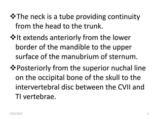
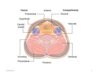
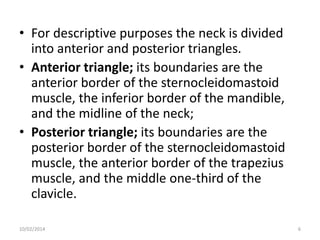
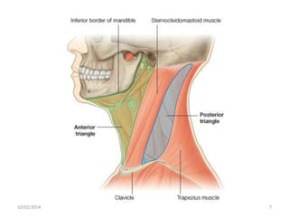
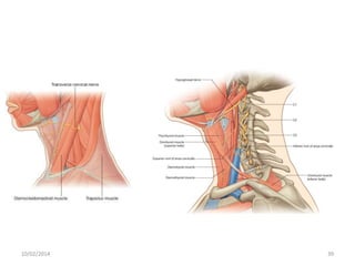
The neck contains four compartments that provide organization: the visceral, vertebral, and two vascular compartments. For descriptive purposes, the neck is divided into anterior and posterior triangles. The anterior triangle contains structures like muscles, blood vessels, and nerves. It is further divided into smaller triangles. The document describes the boundaries and contents of these triangles in detail. It provides information on the muscles, blood vessels, nerves, and other structures found in the anterior triangle of the neck.


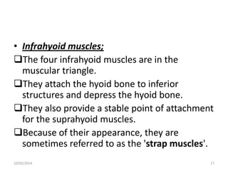
![• Within the tube four compartments provide
longitudinal organization.
the visceral compartment is anterior and
contains parts of the digestive and respiratory
systems, and several endocrine glands;
the vertebral compartment is posterior and
contains the cervical vertebrae, spinal cord,
cervical nerves, and muscles associated with the
vertebral column;
the two vascular compartments are lateral and
contain the major blood vessels and the vagus
nerve [X].
10/02/2014
4](https://image.slidesharecdn.com/neck-copy-140210053949-phpapp01/85/Neck-copy-4-320.jpg)
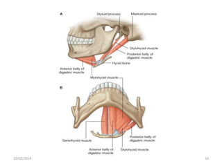
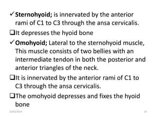
![1. Stylohyoid; innervated by the facial nerve
[VII], it pulls the hyoid bone
posterosuperiorly during swallowing.
2. Digastric; has 2 bellies (anterior and
posterior) connected by a tendon which
attaches to the body of the hyoid bone.
Innervation of the digastric muscle is from
two different cranial nerves.
• Posterior belly is by facial nerve (CN VII)
• Anterior belly is by trigeminal nerve (CN V)
10/02/2014
14](https://image.slidesharecdn.com/neck-copy-140210053949-phpapp01/85/Neck-copy-14-320.jpg)
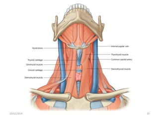
![3. Mylohyoid; It is innervated by the trigeminal
nerve [CN V]. It supports and elevates the floor of
the mouth and elevates the hyoid bone.
4. Geniohyoid; is innervated by a branch from the
anterior ramus of C1 carried along the hypoglossal
nerve [XII].
• It has two functions depending on which bone is
fixed:
fixation of the mandible elevates and pulls the
hyoid bone forward;
fixation of the hyoid bone pulls the mandible
downward and inward.
10/02/2014
15](https://image.slidesharecdn.com/neck-copy-140210053949-phpapp01/85/Neck-copy-15-320.jpg)
![Thyrohyoid; is located deep to the superior
parts of the omohyoid muscle. It is innervated
by fibers from the anterior ramus of C1 that
travel with the hypoglossal nerve [XII].
Sternothyroid; the last of the infrahyoid group
of muscles. is innervated by the anterior rami
of C1 to C3 through the ansa cervicalis.
The sternohyoid muscle draws the larynx
(thyroid cartilage) downward
10/02/2014
19](https://image.slidesharecdn.com/neck-copy-140210053949-phpapp01/85/Neck-copy-19-320.jpg)
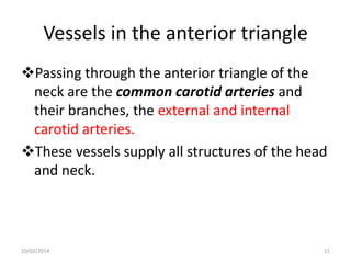
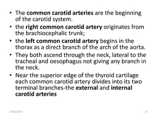
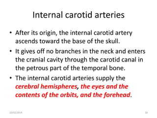
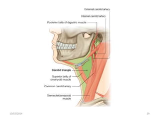
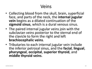
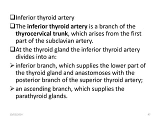
![• The superior part of each common carotid
artery and its division into external and
internal carotid arteries occurs in the carotid
triangle.
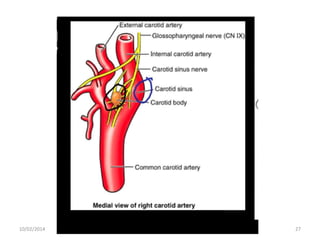
• At the bifurcation, the common carotid artery
and the beginning of the internal carotid
artery are dilated.
• This dilation is the carotid sinus and contains
receptors that monitor changes in blood
pressure and are innervated by a branch of
the glossopharyngeal nerve [IX].
10/02/2014
24](https://image.slidesharecdn.com/neck-copy-140210053949-phpapp01/85/Neck-copy-24-320.jpg)
![• Another accumulation of receptors in the area
of the bifurcation is responsible for detecting
changes in blood chemistry, primarily oxygen
content.
• This is the carotid body and is innervated by
branches from both the glossopharyngeal [IX]
and vagus [X] nerves.
10/02/2014
26](https://image.slidesharecdn.com/neck-copy-140210053949-phpapp01/85/Neck-copy-26-320.jpg)
![The cranial nerves in these categories include:
the facial [VII], glossopharyngeal [IX], vagus
[X], accessory [XI], and hypoglossal [XII].
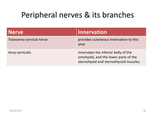
The peripheral nerves in these categories
include the transverse cervical nerve from the
cervical plexus and the upper and lower roots
of the ansa cervicalis.
10/02/2014
34](https://image.slidesharecdn.com/neck-copy-140210053949-phpapp01/85/Neck-copy-34-320.jpg)
![Cranial nerves & its branches
Nerve
Innervation
Facial nerve [VII]
the posterior belly of the digastric;
stylohyoid.
Glossopharyngeal nerve [IX]
stylopharyngeus muscle, sends a branch
to the carotid sinus, and supplies sensory
branches to the pharynx.
Vagus nerve [X]
Gives a motor branch to the pharynx, a
branch to the carotid body, the superior
laryngeal nerve (which divides into
external and internal laryngeal branches),
and possibly a cardiac branch.
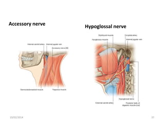
Accessory nerve [XI]
No branches in the anterior triangle but
innervates the trapezius
Hypoglossal nerve [XII]
No branches in the anterior triangle but
innervates the tongue
10/02/2014
35](https://image.slidesharecdn.com/neck-copy-140210053949-phpapp01/85/Neck-copy-35-320.jpg)
![Glossopharyngeal nerve [IX]
10/02/2014
Vagus nerve
36](https://image.slidesharecdn.com/neck-copy-140210053949-phpapp01/85/Neck-copy-36-320.jpg)
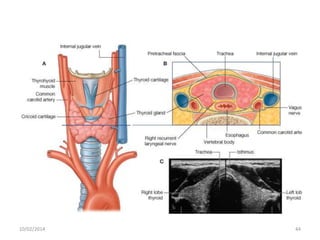

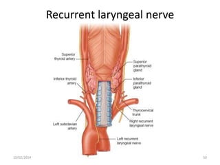
![Nerve supply
• The thyroid gland is closely related to and
supplied by the recurrent laryngeal nerves.
• After branching from the vagus nerve [X] and
looping around the subclavian artery on the
right and the arch of the aorta on the left, the
recurrent laryngeal nerves ascend in a groove
between the trachea and esophagus.
10/02/2014
49](https://image.slidesharecdn.com/neck-copy-140210053949-phpapp01/85/Neck-copy-49-320.jpg)


![CASE_PRESENTATION_ON_subdural_hematoma(SDH)[1 FINAL PPT]-1.pptx](https://cdn.slidesharecdn.com/ss_thumbnails/casepresentationonsubduralhematomasdh1finalppt-1-260129172522-d405d375-thumbnail.jpg?width=640&height=640&fit=bounds)