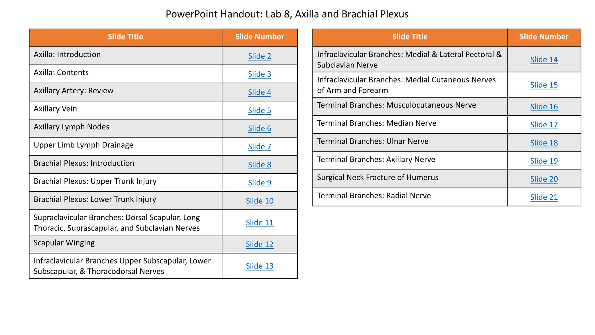
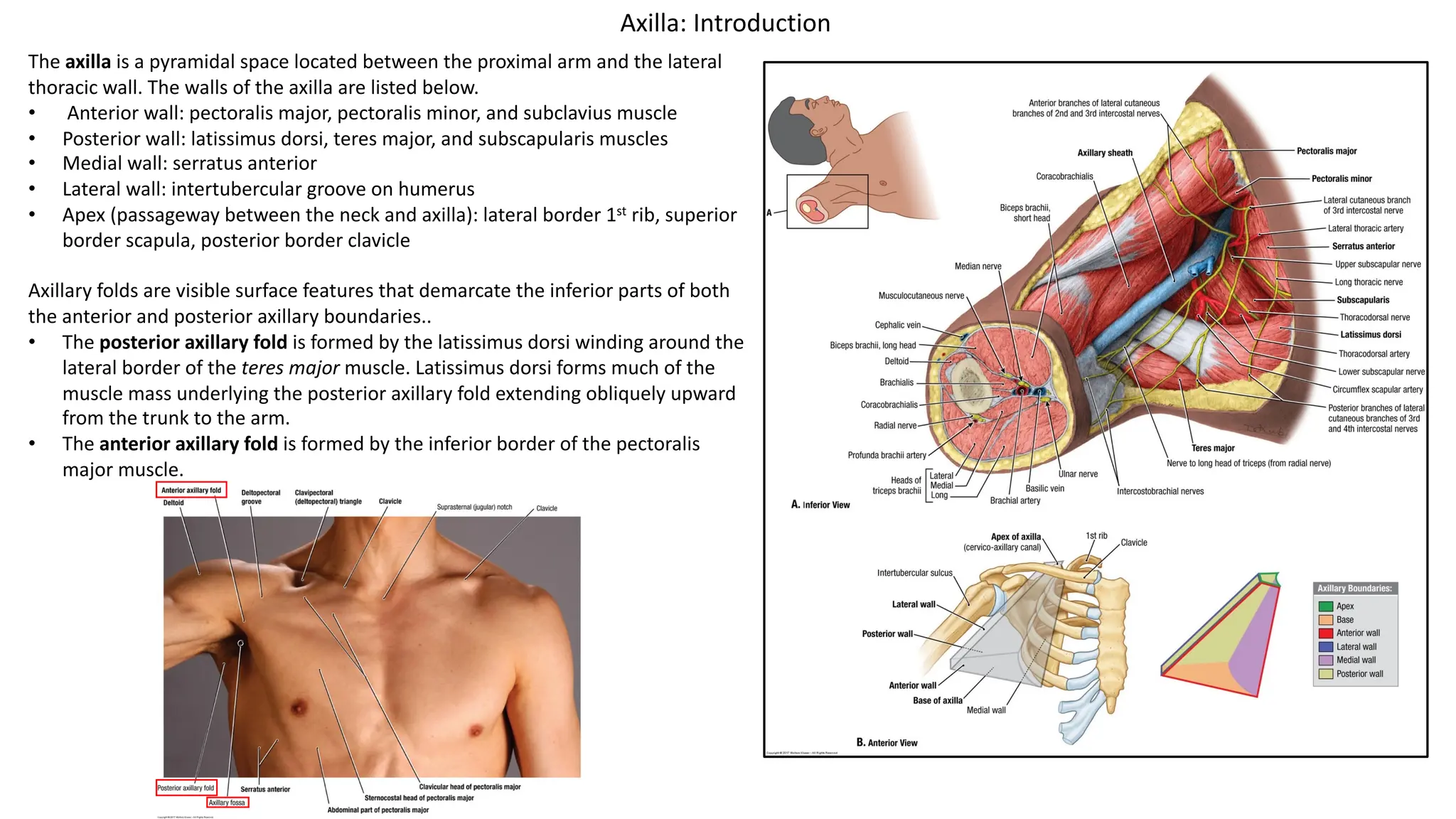
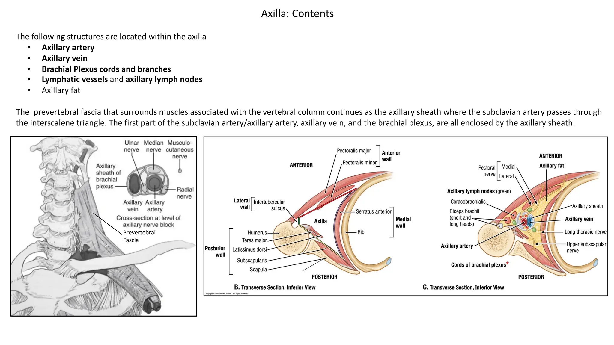
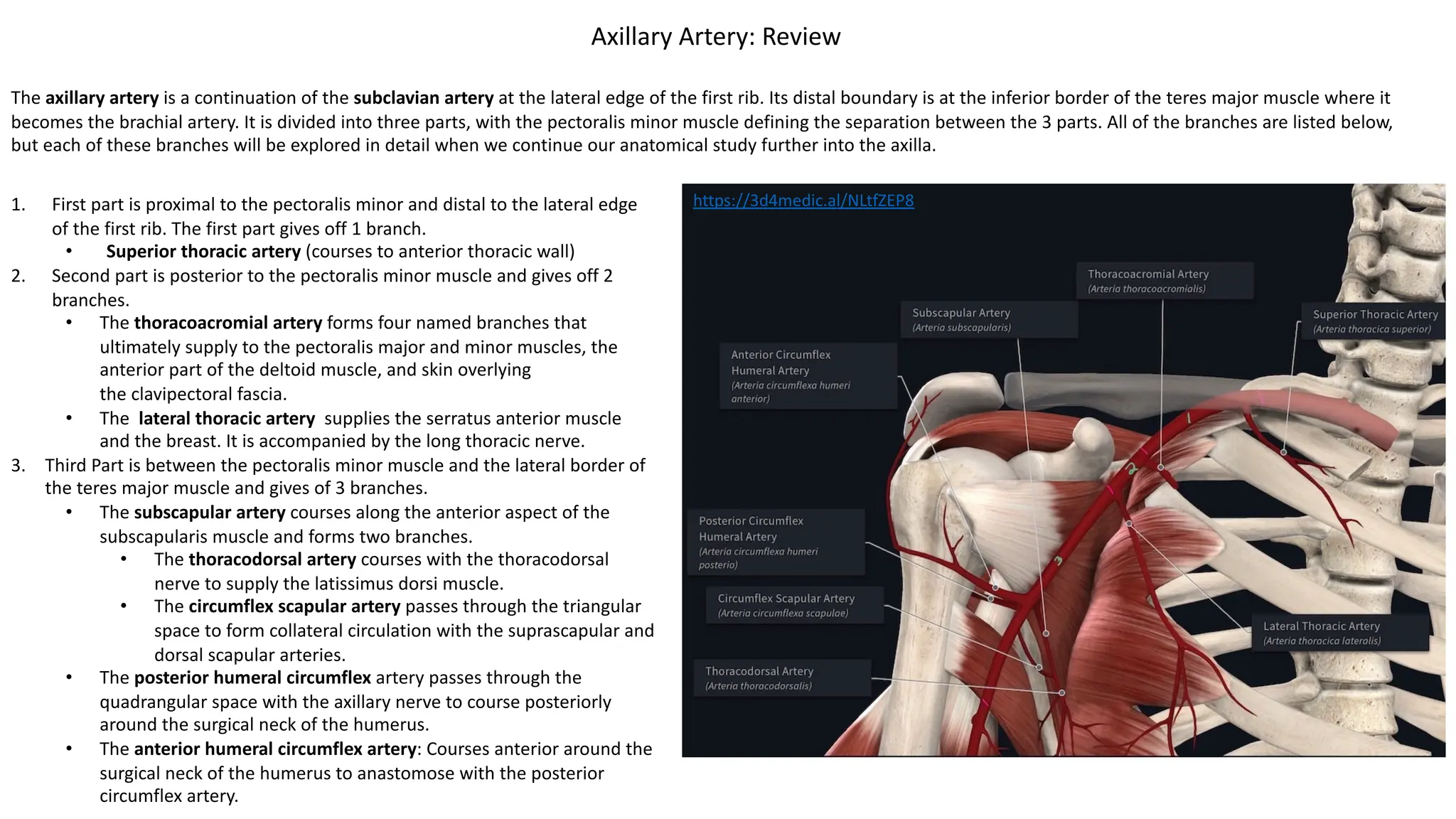
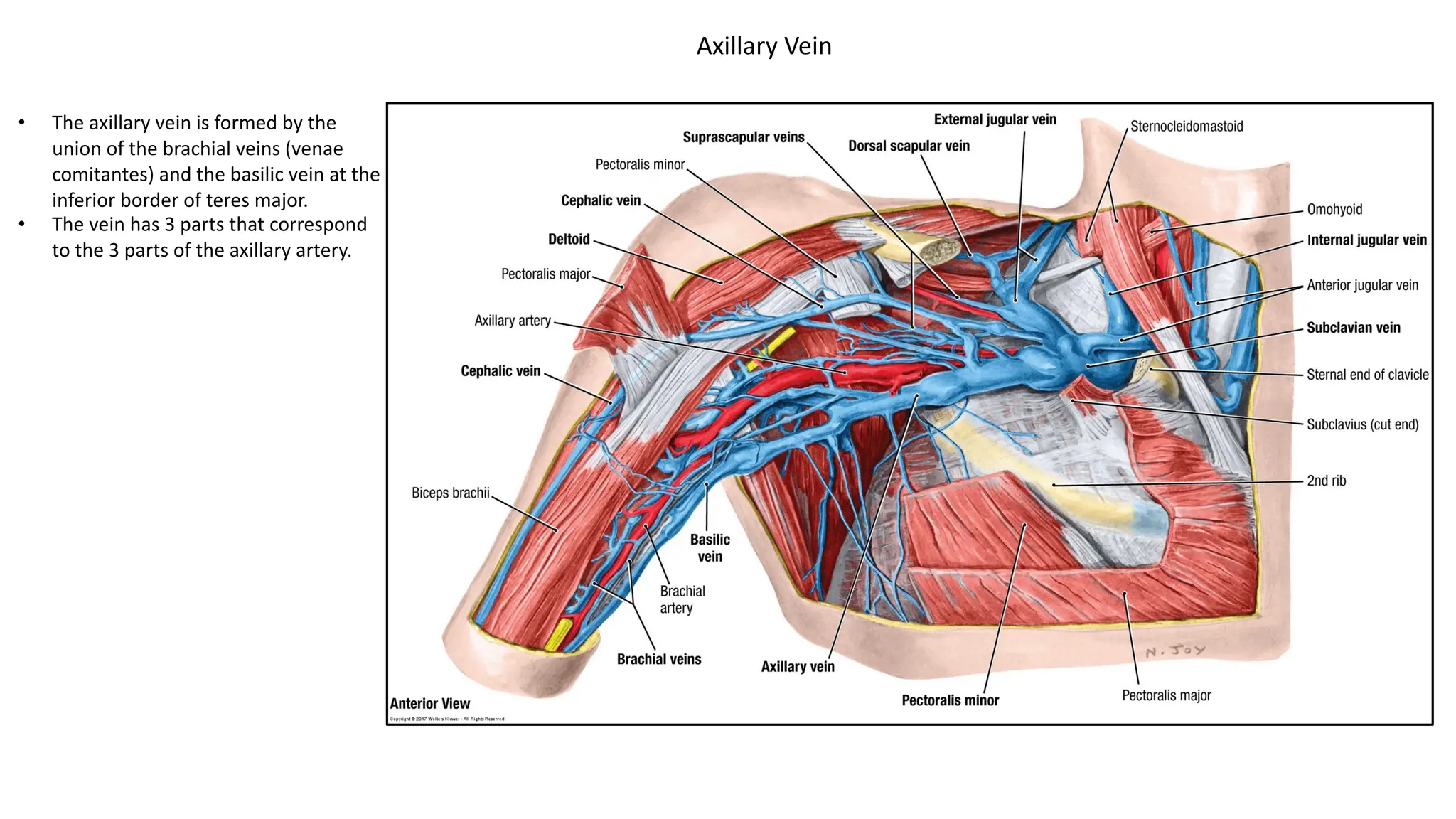
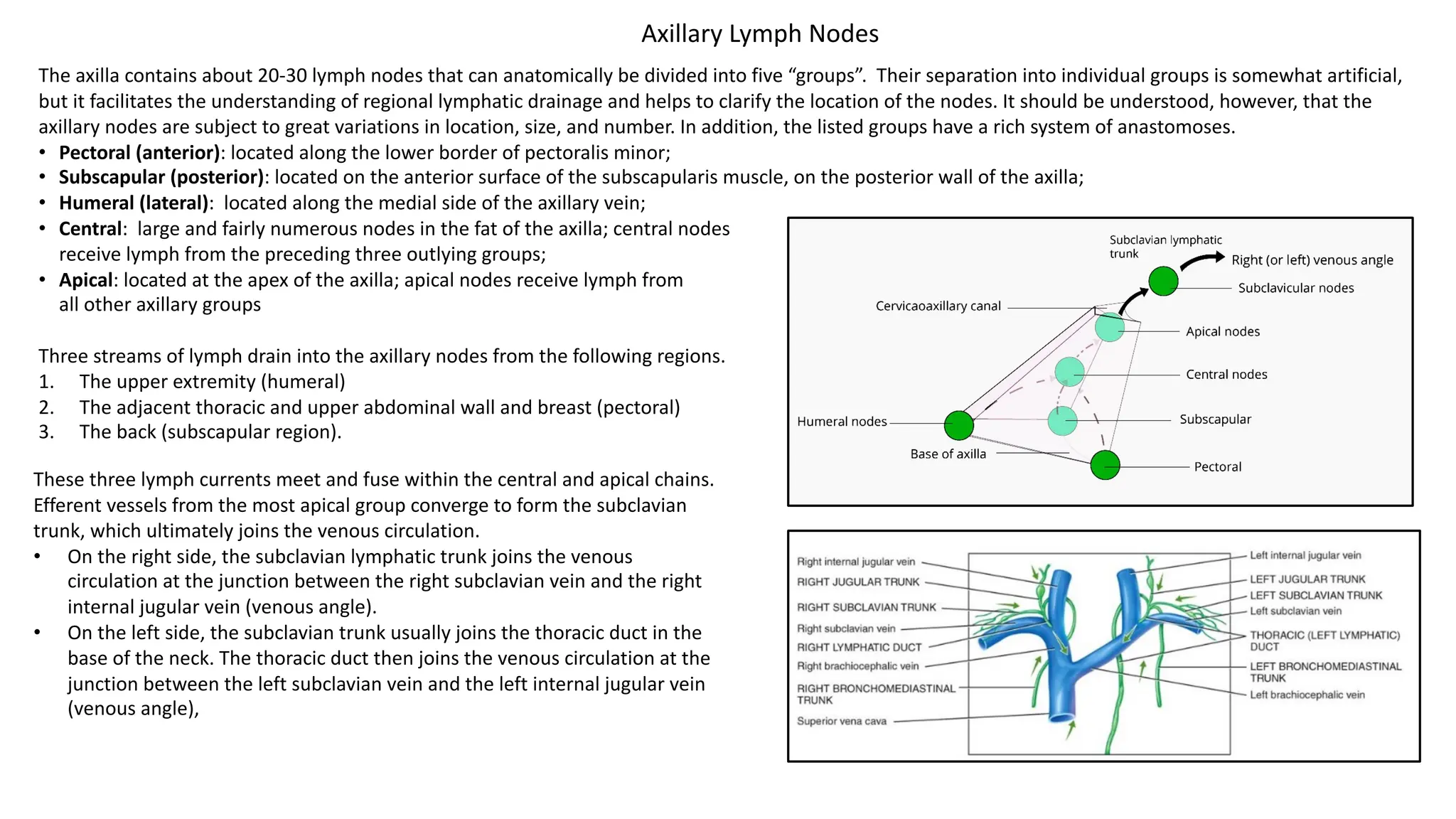
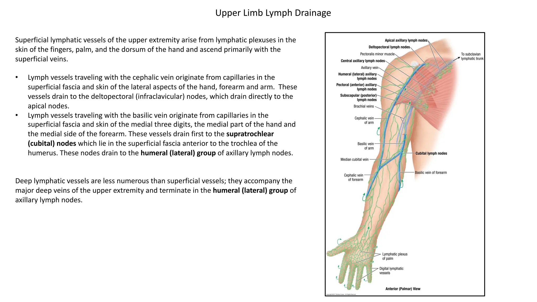
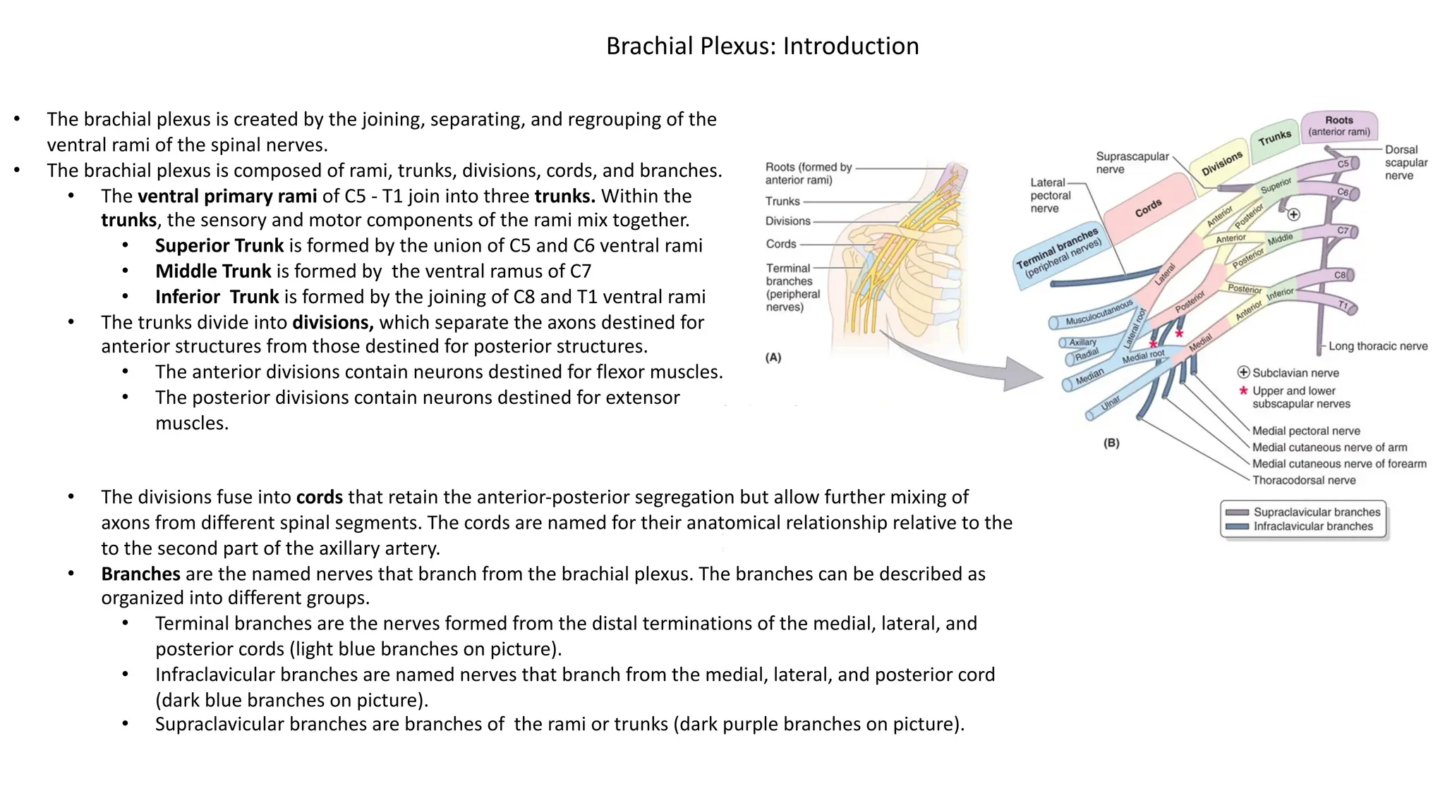
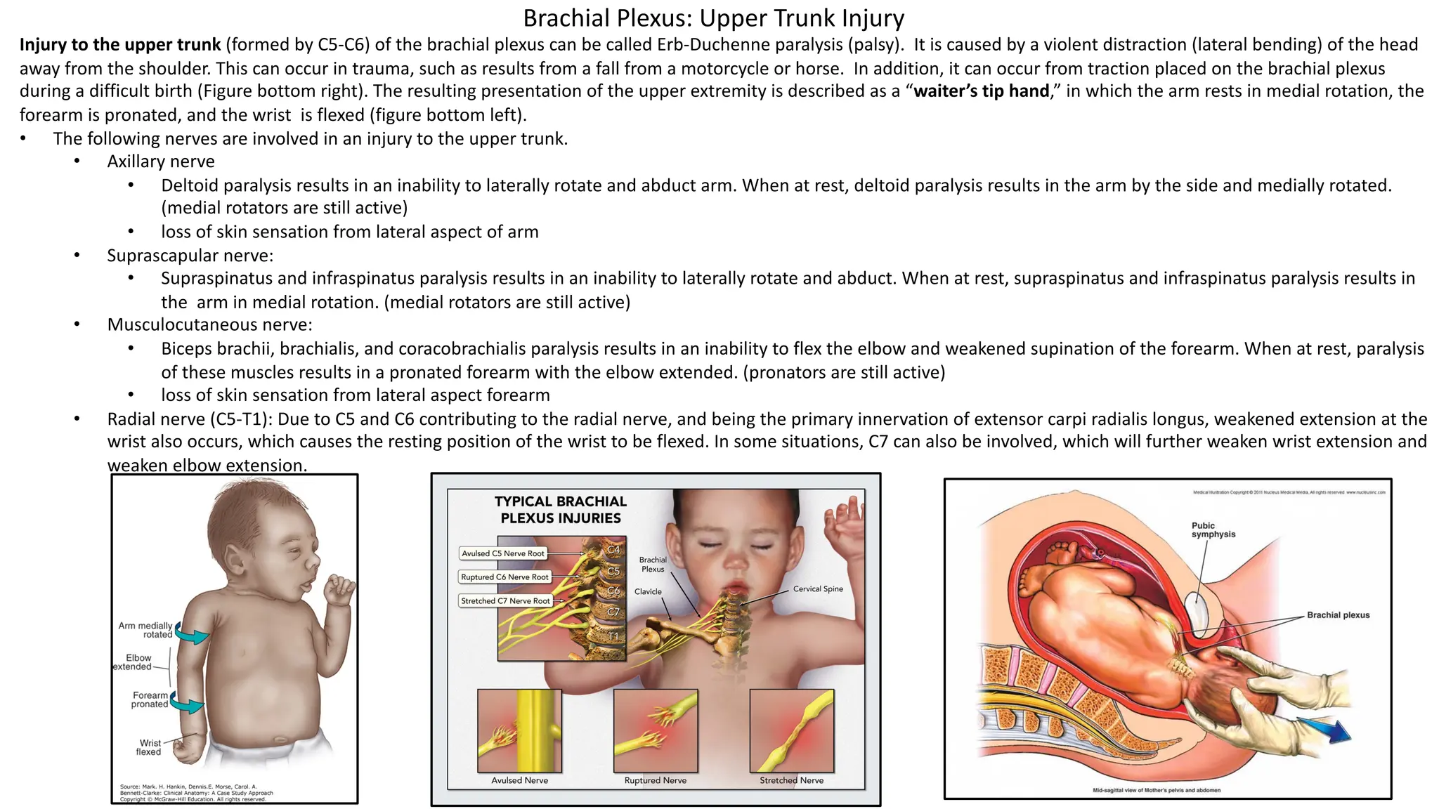
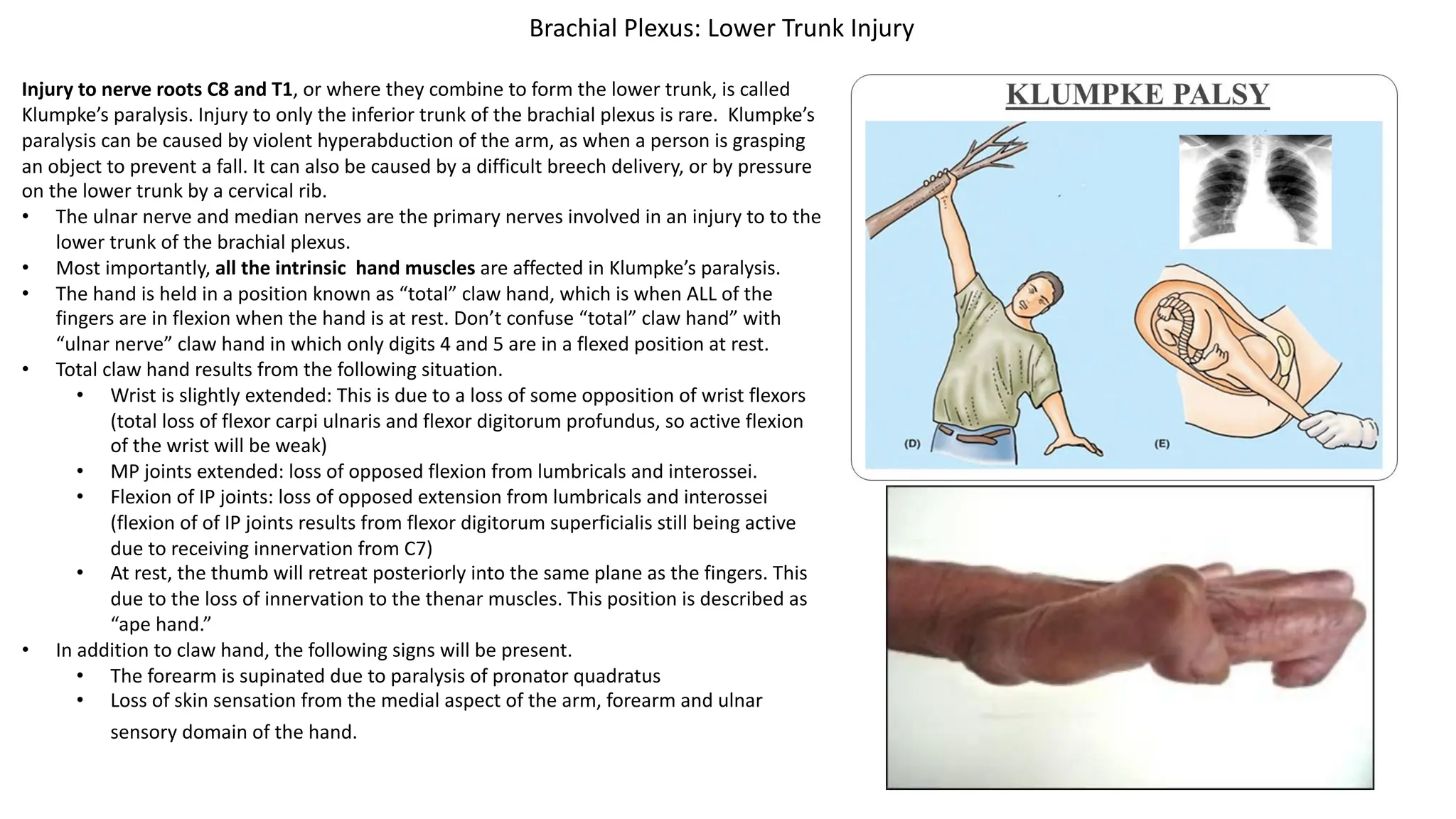
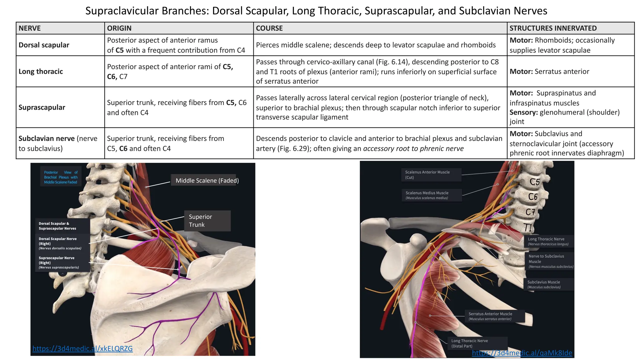
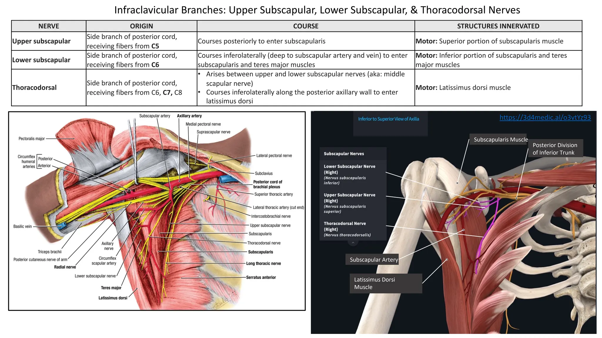
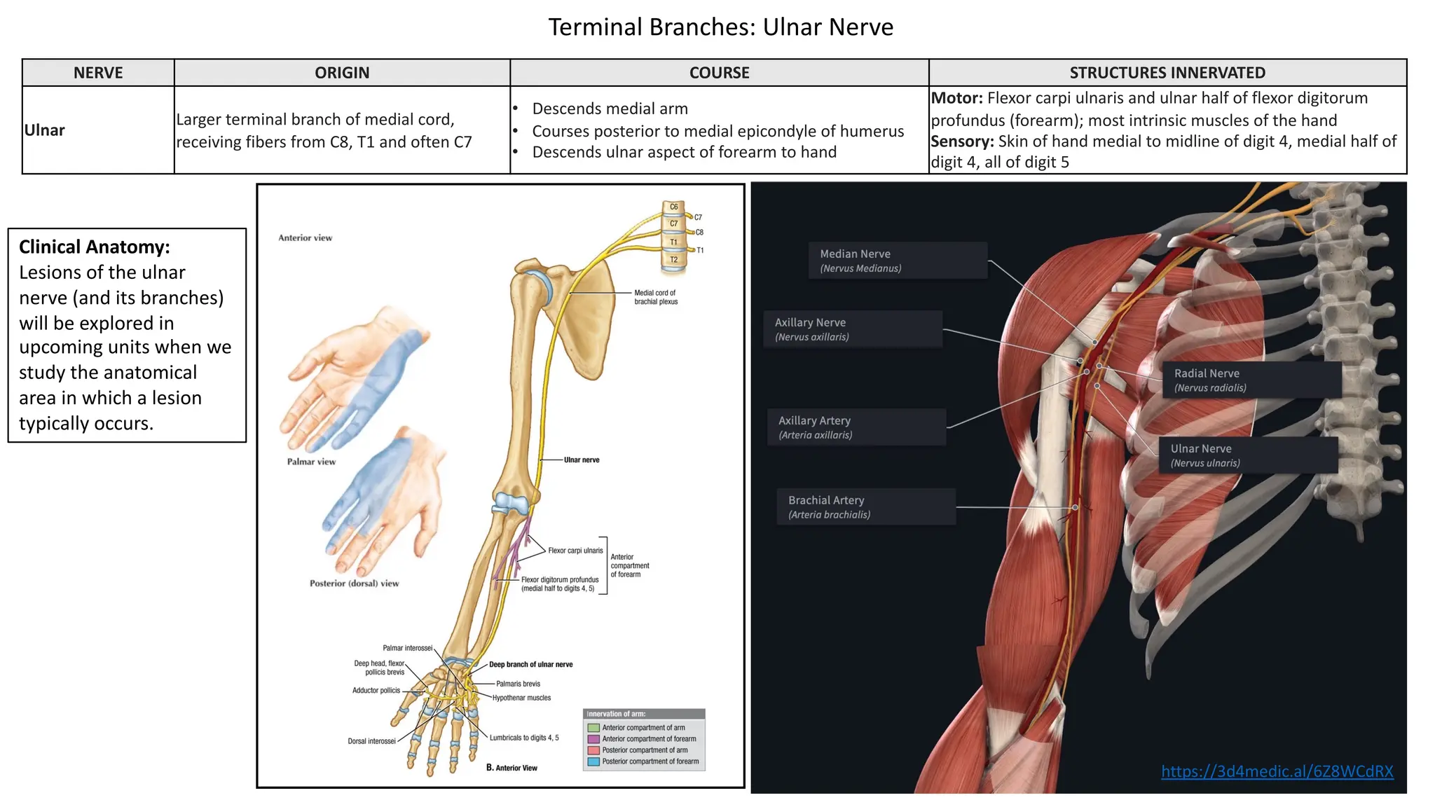
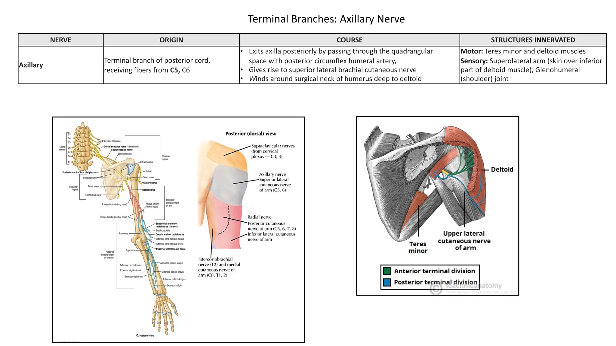
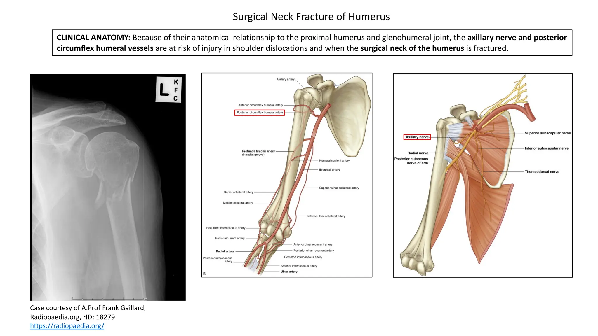
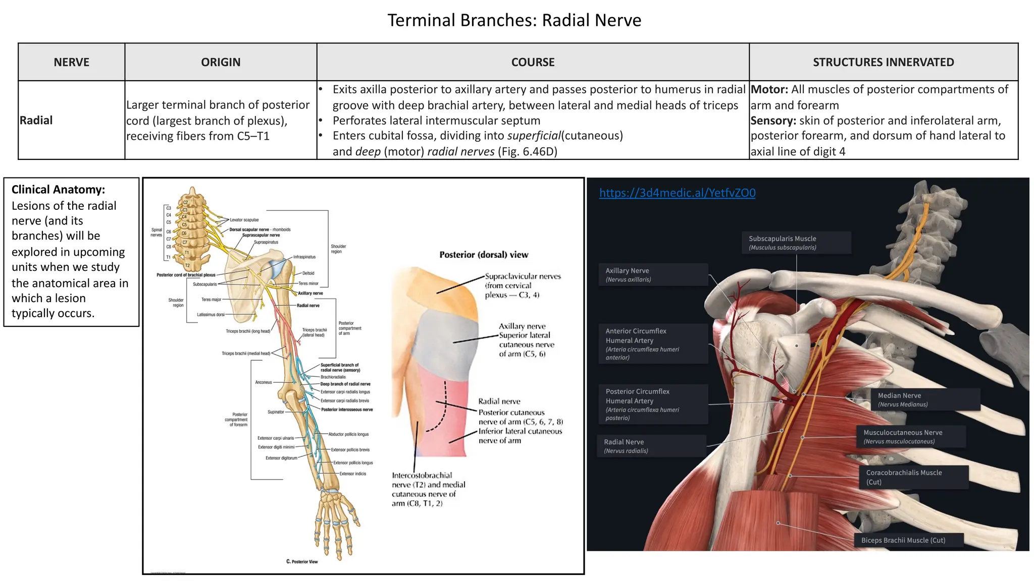
Slide Title Slide Number
Axilla: Introduction Slide 2
Axilla: Contents Slide 3
Axillary Artery: Review Slide 4
Axillary Vein Slide 5
Axillary Lymph Nodes Slide 6
Upper Limb Lymph Drainage Slide 7
Brachial Plexus: Introduction Slide 8
Brachial Plexus: Upper Trunk Injury Slide 9
Brachial Plexus: Lower Trunk Injury Slide 10
Supraclavicular Branches: Dorsal Scapular, Long
Thoracic, Suprascapular, and Subclavian Nerves
Slide 11
Scapular Winging Slide 12
Infraclavicular Branches Upper Subscapular, Lower
Subscapular, & Thoracodorsal Nerves
Slide 13
Slide Title Slide Number
Infraclavicular Branches: Medial & Lateral Pectoral &
Subclavian Nerve
Slide 14
Infraclavicular Branches: Medial Cutaneous Nerves
of Arm and Forearm
Slide 15
Terminal Branches: Musculocutaneous Nerve Slide 16
Terminal Branches: Median Nerve Slide 17
Terminal Branches: Ulnar Nerve Slide 18
Terminal Branches: Axillary Nerve Slide 19
Surgical Neck Fracture of Humerus Slide 20
Terminal Branches: Radial Nerve Slide 21
PowerPoint Handout: Lab 8, Axilla and Brachial Plexus
Axilla: Introduction
The axilla is a pyramidal space located between the proximal arm and the lateral
thoracic wall. The walls of the axilla are listed below.
• Anterior wall: pectoralis major, pectoralis minor, and subclavius muscle
• Posterior wall: latissimus dorsi, teres major, and subscapularis muscles
• Medial wall: serratus anterior
• Lateral wall: intertubercular groove on humerus
• Apex (passageway between the neck and axilla): lateral border 1st rib, superior
border scapula, posterior border clavicle
Axillary folds are visible surface features that demarcate the inferior parts of both
the anterior and posterior axillary boundaries..
• The posterior axillary fold is formed by the latissimus dorsi winding around the
lateral border of the teres major muscle. Latissimus dorsi forms much of the
muscle mass underlying the posterior axillary fold extending obliquely upward
from the trunk to the arm.
• The anterior axillary fold is formed by the inferior border of the pectoralis
major muscle.
Axilla: Contents
The following structures are located within the axilla
• Axillary artery
• Axillary vein
• Brachial Plexus cords and branches
• Lymphatic vessels and axillary lymph nodes
• Axillary fat
The prevertebral fascia that surrounds muscles associated with the vertebral column continues as the axillary sheath where the subclavian artery passes through
the interscalene triangle. The first part of the subclavian artery/axillary artery, axillary vein, and the brachial plexus, are all enclosed by the axillary sheath.
Prevertebral
Fascia
https://3d4medic.al/NLtfZEP8
Axillary Artery: Review
1. First part is proximal to the pectoralis minor and distal to the lateral edge
of the first rib. The first part gives off 1 branch.
• Superior thoracic artery (courses to anterior thoracic wall)
2. Second part is posterior to the pectoralis minor muscle and gives off 2
branches.
• The thoracoacromial artery forms four named branches that
ultimately supply to the pectoralis major and minor muscles, the
anterior part of the deltoid muscle, and skin overlying
the clavipectoral fascia.
• The lateral thoracic artery supplies the serratus anterior muscle
and the breast. It is accompanied by the long thoracic nerve.
3. Third Part is between the pectoralis minor muscle and the lateral border of
the teres major muscle and gives of 3 branches.
• The subscapular artery courses along the anterior aspect of the
subscapularis muscle and forms two branches.
• The thoracodorsal artery courses with the thoracodorsal
nerve to supply the latissimus dorsi muscle.
• The circumflex scapular artery passes through the triangular
space to form collateral circulation with the suprascapular and
dorsal scapular arteries.
• The posterior humeral circumflex artery passes through the
quadrangular space with the axillary nerve to course posteriorly
around the surgical neck of the humerus.
• The anterior humeral circumflex artery: Courses anterior around the
surgical neck of the humerus to anastomose with the posterior
circumflex artery.
The axillary artery is a continuation of the subclavian artery at the lateral edge of the first rib. Its distal boundary is at the inferior border of the teres major muscle where it
becomes the brachial artery. It is divided into three parts, with the pectoralis minor muscle defining the separation between the 3 parts. All of the branches are listed below,
but each of these branches will be explored in detail when we continue our anatomical study further into the axilla.
Axillary Vein
• The axillary vein is formed by the
union of the brachial veins (venae
comitantes) and the basilic vein at the
inferior border of teres major.
• The vein has 3 parts that correspond
to the 3 parts of the axillary artery.
The axilla contains about 20-30 lymph nodes that can anatomically be divided into five “groups”. Their separation into individual groups is somewhat artificial,
but it facilitates the understanding of regional lymphatic drainage and helps to clarify the location of the nodes. It should be understood, however, that the
axillary nodes are subject to great variations in location, size, and number. In addition, the listed groups have a rich system of anastomoses.
• Pectoral (anterior): located along the lower border of pectoralis minor;
• Subscapular (posterior): located on the anterior surface of the subscapularis muscle, on the posterior wall of the axilla;
• Humeral (lateral): located along the medial side of the axillary vein;
• Central: large and fairly numerous nodes in the fat of the axilla; central nodes
receive lymph from the preceding three outlying groups;
• Apical: located at the apex of the axilla; apical nodes receive lymph from
all other axillary groups
Three streams of lymph drain into the axillary nodes from the following regions.
1. The upper extremity (humeral)
2. The adjacent thoracic and upper abdominal wall and breast (pectoral)
3. The back (subscapular region).
Axillary Lymph Nodes
These three lymph currents meet and fuse within the central and apical chains.
Efferent vessels from the most apical group converge to form the subclavian
trunk, which ultimately joins the venous circulation.
• On the right side, the subclavian lymphatic trunk joins the venous
circulation at the junction between the right subclavian vein and the right
internal jugular vein (venous angle).
• On the left side, the subclavian trunk usually joins the thoracic duct in the
base of the neck. The thoracic duct then joins the venous circulation at the
junction between the left subclavian vein and the left internal jugular vein
(venous angle),
Superficial lymphatic vessels of the upper extremity arise from lymphatic plexuses in the
skin of the fingers, palm, and the dorsum of the hand and ascend primarily with the
superficial veins.
• Lymph vessels traveling with the cephalic vein originate from capillaries in the
superficial fascia and skin of the lateral aspects of the hand, forearm and arm. These
vessels drain to the deltopectoral (infraclavicular) nodes, which drain directly to the
apical nodes.
• Lymph vessels traveling with the basilic vein originate from capillaries in the
superficial fascia and skin of the medial three digits, the medial part of the hand and
the medial side of the forearm. These vessels drain first to the supratrochlear
(cubital) nodes which lie in the superficial fascia anterior to the trochlea of the
humerus. These nodes drain to the humeral (lateral) group of axillary lymph nodes.
Deep lymphatic vessels are less numerous than superficial vessels; they accompany the
major deep veins of the upper extremity and terminate in the humeral (lateral) group of
axillary lymph nodes.
Upper Limb Lymph Drainage
• The brachial plexus is created by the joining, separating, and regrouping of the
ventral rami of the spinal nerves.
• The brachial plexus is composed of rami, trunks, divisions, cords, and branches.
• The ventral primary rami of C5 - T1 join into three trunks. Within the
trunks, the sensory and motor components of the rami mix together.
• Superior Trunk is formed by the union of C5 and C6 ventral rami
• Middle Trunk is formed by the ventral ramus of C7
• Inferior Trunk is formed by the joining of C8 and T1 ventral rami
• The trunks divide into divisions, which separate the axons destined for
anterior structures from those destined for posterior structures.
• The anterior divisions contain neurons destined for flexor muscles.
• The posterior divisions contain neurons destined for extensor
muscles.
• The divisions fuse into cords that retain the anterior-posterior segregation but allow further mixing of
axons from different spinal segments. The cords are named for their anatomical relationship relative to the
to the second part of the axillary artery.
• Branches are the named nerves that branch from the brachial plexus. The branches can be described as
organized into different groups.
• Terminal branches are the nerves formed from the distal terminations of the medial, lateral, and
posterior cords (light blue branches on picture).
• Infraclavicular branches are named nerves that branch from the medial, lateral, and posterior cord
(dark blue branches on picture).
• Supraclavicular branches are branches of the rami or trunks (dark purple branches on picture).
Brachial Plexus: Introduction
Injury to the upper trunk (formed by C5-C6) of the brachial plexus can be called Erb-Duchenne paralysis (palsy). It is caused by a violent distraction (lateral bending) of the head
away from the shoulder. This can occur in trauma, such as results from a fall from a motorcycle or horse. In addition, it can occur from traction placed on the brachial plexus
during a difficult birth (Figure bottom right). The resulting presentation of the upper extremity is described as a “waiter’s tip hand,” in which the arm rests in medial rotation, the
forearm is pronated, and the wrist is flexed (figure bottom left).
• The following nerves are involved in an injury to the upper trunk.
• Axillary nerve
• Deltoid paralysis results in an inability to laterally rotate and abduct arm. When at rest, deltoid paralysis results in the arm by the side and medially rotated.
(medial rotators are still active)
• loss of skin sensation from lateral aspect of arm
• Suprascapular nerve:
• Supraspinatus and infraspinatus paralysis results in an inability to laterally rotate and abduct. When at rest, supraspinatus and infraspinatus paralysis results in
the arm in medial rotation. (medial rotators are still active)
• Musculocutaneous nerve:
• Biceps brachii, brachialis, and coracobrachialis paralysis results in an inability to flex the elbow and weakened supination of the forearm. When at rest, paralysis
of these muscles results in a pronated forearm with the elbow extended. (pronators are still active)
• loss of skin sensation from lateral aspect forearm
• Radial nerve (C5-T1): Due to C5 and C6 contributing to the radial nerve, and being the primary innervation of extensor carpi radialis longus, weakened extension at the
wrist also occurs, which causes the resting position of the wrist to be flexed. In some situations, C7 can also be involved, which will further weaken wrist extension and
weaken elbow extension.
Brachial Plexus: Upper Trunk Injury
Injury to nerve roots C8 and T1, or where they combine to form the lower trunk, is called
Klumpke’s paralysis. Injury to only the inferior trunk of the brachial plexus is rare. Klumpke’s
paralysis can be caused by violent hyperabduction of the arm, as when a person is grasping
an object to prevent a fall. It can also be caused by a difficult breech delivery, or by pressure
on the lower trunk by a cervical rib.
• The ulnar nerve and median nerves are the primary nerves involved in an injury to to the
lower trunk of the brachial plexus.
• Most importantly, all the intrinsic hand muscles are affected in Klumpke’s paralysis.
• The hand is held in a position known as “total” claw hand, which is when ALL of the
fingers are in flexion when the hand is at rest. Don’t confuse “total” claw hand” with
“ulnar nerve” claw hand in which only digits 4 and 5 are in a flexed position at rest.
• Total claw hand results from the following situation.
• Wrist is slightly extended: This is due to a loss of some opposition of wrist flexors
(total loss of flexor carpi ulnaris and flexor digitorum profundus, so active flexion
of the wrist will be weak)
• MP joints extended: loss of opposed flexion from lumbricals and interossei.
• Flexion of IP joints: loss of opposed extension from lumbricals and interossei
(flexion of of IP joints results from flexor digitorum superficialis still being active
due to receiving innervation from C7)
• At rest, the thumb will retreat posteriorly into the same plane as the fingers. This
due to the loss of innervation to the thenar muscles. This position is described as
“ape hand.”
• In addition to claw hand, the following signs will be present.
• The forearm is supinated due to paralysis of pronator quadratus
• Loss of skin sensation from the medial aspect of the arm, forearm and ulnar
sensory domain of the hand.
Brachial Plexus: Lower Trunk Injury
NERVE ORIGIN COURSE STRUCTURES INNERVATED
Dorsal scapular
Posterior aspect of anterior ramus
of C5 with a frequent contribution from C4
Pierces middle scalene; descends deep to levator scapulae and rhomboids
Motor: Rhomboids; occasionally
supplies levator scapulae
Long thoracic
Posterior aspect of anterior rami of C5,
C6, C7
Passes through cervico-axillary canal (Fig. 6.14), descending posterior to C8
and T1 roots of plexus (anterior rami); runs inferiorly on superficial surface
of serratus anterior
Motor: Serratus anterior
Suprascapular
Superior trunk, receiving fibers from C5, C6
and often C4
Passes laterally across lateral cervical region (posterior triangle of neck),
superior to brachial plexus; then through scapular notch inferior to superior
transverse scapular ligament
Motor: Supraspinatus and
infraspinatus muscles
Sensory: glenohumeral (shoulder)
joint
Subclavian nerve (nerve
to subclavius)
Superior trunk, receiving fibers from
C5, C6 and often C4
Descends posterior to clavicle and anterior to brachial plexus and subclavian
artery (Fig. 6.29); often giving an accessory root to phrenic nerve
Motor: Subclavius and
sternoclavicular joint (accessory
phrenic root innervates diaphragm)
Middle Scalene (Faded)
Superior
Trunk
Supraclavicular Branches: Dorsal Scapular, Long Thoracic, Suprascapular, and Subclavian Nerves
https://3d4medic.al/xkELQRZG
https://3d4medic.al/qaMk8Ide
Scapular Winging
CLINICAL ANATOMY: Medial winging of the scapula is the result of serratus anterior paralysis from
injury to the long thoracic nerve. The most common etiology is neuropraxia after blunt trauma or a
stretch injury. In addition, surgical procedures in the thoracic region such as radical mastectomy,
resection of the first rib, and transthoracic sympathectomy can expose the long thoracic nerve and
make it susceptible to damage.
To test for medial scapular winging, ask patient to push against a wall and observe the inferior angle
of scapula. If nerve is damaged, the inferior angle and medial border will project from the posterior
thoracic wall (medial scapular “winging”). In addition, a person with long thoracic nerve injury will
have difficulty abducting the arm past a horizonal position (90 degrees) due to an inability to
upwardly rotate the scapula.
Two mnemonics that might help remember medial scapular winging:
1. C5,6,7 wings to heaven: The serratus anterior is innervated by the long thoracic nerve (C5-C), so
damage to the nerve results in (medial) winging of the scapula.
2. SALT on the birds wings: SA=serratus anterior, LT=long thoracic nerve. Damage to the long
thoracic nerve results in (medial) winging of the scapula.
CLINICAL ANATOMY: Lateral Winging of the scapula results from a dysfunction in the
trapezius (spinal accessory nerve: CN XI) and/or rhomboid muscles (dorsal scapular nerve).
In lateral winging, the scapula is excessively protracted (abducted) on the thoracic wall.
NERVE ORIGIN COURSE STRUCTURES INNERVATED
Upper subscapular
Side branch of posterior cord,
receiving fibers from C5
Courses posteriorly to enter subscapularis Motor: Superior portion of subscapularis muscle
Lower subscapular
Side branch of posterior cord,
receiving fibers from C6
Courses inferolaterally (deep to subscapular artery and vein) to enter
subscapularis and teres major muscles
Motor: Inferior portion of subscapularis and teres
major muscles
Thoracodorsal
Side branch of posterior cord,
receiving fibers from C6, C7, C8
• Arises between upper and lower subscapular nerves (aka: middle
scapular nerve)
• Courses inferolaterally along the posterior axillary wall to enter
latissimus dorsi
Motor: Latissimus dorsi muscle
Subscapular Artery
Latissimus Dorsi
Muscle
Subscapularis Muscle
Posterior Division
of Inferior Trunk
Infraclavicular Branches: Upper Subscapular, Lower Subscapular, & Thoracodorsal Nerves
https://3d4medic.al/o3vtYz93
NERVE ORIGIN COURSE STRUCTURES INNERVATED
Lateral pectoral
Side branch of lateral
cord, receiving fibers
from C5, C6, C7
• Pierces costocoracoid membrane to reach deep surface of pectoral muscles
• Forms a communicating branch to the medial pectoral nerve
• Passes anterior to axillary artery and vein
Motor: Primarily pectoralis major; but some
lateral pectoral nerve fibers pass to pectoralis
minor via branch to medial pectoral nerve
Medial pectoral
Side branches of medial
cord, receiving fibers
from C8, T1
• Passes between axillary artery and vein
• Pierces pectoralis minor and enters deep surface of pectoralis major
• Although it is called medial for its origin from medial cord, its anatomical
position is lateral to lateral pectoral nerve
Motor: Pectoralis minor and sternocostal part of
pectoralis major
Subclavian nerve
(nerve to
subclavius)
Superior trunk, receiving
fibers from C5, C6 and
often C4
• Descends posterior to clavicle and anterior to brachial plexus and subclavian
artery
• Often contributes to accessory root to phrenic nerve
Motor: Subclavius and sternoclavicular joint
(accessory phrenic root innervates diaphragm)
Infraclavicular Branches: Medial & Lateral Pectoral & Subclavian Nerve
https://3d4medic.al/UkelIxUm
NERVE ORIGIN COURSE STRUCTURES INNERVATED
Medial cutaneous nerve of arm
Side branches of medial cord,
receiving fibers from C8, T1
• Smallest nerve of plexus
• Courses along medial side of axillary and brachial veins and
communicates with intercostobrachial nerve
Sensory: Skin of medial side of arm, as far
distal as medial epicondyle of humerus and
olecranon of ulna
Medial cutaneous nerve of forearm
• Initially runs with ulnar nerve
• Then pierces deep fascia with basilic vein and enters subcutaneous
tissue, dividing into anterior and posterior branches
• Often confused with ulnar nerve
Sensory: Skin of medial side of forearm, as far
distal as wrist
Infraclavicular Branches: Medial Cutaneous Nerves of Arm and Forearm
NERVE ORIGIN COURSE STRUCTURES INNERVATED
Musculocutaneous
Terminal branch of lateral cord, receiving fibers
from C5–C7
• Exits axilla by piercing coracobrachialis
• Descends through the arm between biceps brachii and brachialis
• Continues into forearm as lateral cutaneous nerve of forearm
• Motor: Muscles of anterior compartment
of the arm (coracobrachialis, biceps brachii
and brachialis)
• Sensory: Skin of lateral aspect of forearm
Terminal Branches: Musculocutaneous Nerve
https://3d4medic.al/ktFP2syE
CLINICAL ANATOMY: Lesions of the
musculocutaneous nerve can result
in both muscle weakness and
loss/reduction of sensation.
• Weakness of elbow flexion and
supination result. Supination
weakness occurs because the
biceps brachii is major
contributor to supination of the
forearm.
• Sensory loss occurs on the
lateral side of the forearm in the
domain of the lateral cutaneous
nerve of the forearm.
NERVE ORIGIN COURSE STRUCTURES INNERVATED
Median
Lateral root of median nerve is a
terminal branch of lateral cord (C6, C7)
Medial root of median nerve is a
terminal branch of medial cord (C8, T1)
• Lateral and medial roots merge to form median nerve
(lateral position relative to the axillary artery)
• Descends through arm adjacent to brachial artery, with
nerve gradually crossing anterior to artery to lie medial to
artery in cubital fossa
• Motor: Muscles of anterior forearm compartment (except flexor carpi
ulnaris and ulnar half of flexor digitorum profundus), lumbricals of digits
2 and 3, and thenar muscles (abductor pollicis brevis, opponens pollicis,
flexor pollicis brevis)
• Sensory: Skin of palm, palmar side of digits 1-3, lateral palmar side of
digit 4, and the distal half on the dorsal surface of digits 1-4.
Terminal Branches: Median Nerve
CLINICAL ANATOMY:
Lesions of the median
nerve (and its branches)
will be explored in
upcoming units when we
study the anatomical area
in which a lesion typically
occurs.
https://3d4medic.al/Jo1AzG38
NERVE ORIGIN COURSE STRUCTURES INNERVATED
Ulnar
Larger terminal branch of medial cord,
receiving fibers from C8, T1 and often C7
• Descends medial arm
• Courses posterior to medial epicondyle of humerus
• Descends ulnar aspect of forearm to hand
Motor: Flexor carpi ulnaris and ulnar half of flexor digitorum
profundus (forearm); most intrinsic muscles of the hand
Sensory: Skin of hand medial to midline of digit 4, medial half of
digit 4, all of digit 5
Terminal Branches: Ulnar Nerve
Clinical Anatomy:
Lesions of the ulnar
nerve (and its branches)
will be explored in
upcoming units when we
study the anatomical
area in which a lesion
typically occurs.
https://3d4medic.al/6Z8WCdRX
NERVE ORIGIN COURSE STRUCTURES INNERVATED
Axillary
Terminal branch of posterior cord,
receiving fibers from C5, C6
• Exits axilla posteriorly by passing through the quadrangular
space with posterior circumflex humeral artery,
• Gives rise to superior lateral brachial cutaneous nerve
• Winds around surgical neck of humerus deep to deltoid
Motor: Teres minor and deltoid muscles
Sensory: Superolateral arm (skin over inferior
part of deltoid muscle), Glenohumeral
(shoulder) joint
Terminal Branches: Axillary Nerve
CLINICAL ANATOMY: Because of their anatomical relationship to the proximal humerus and glenohumeral joint, the axillary nerve and posterior
circumflex humeral vessels are at risk of injury in shoulder dislocations and when the surgical neck of the humerus is fractured.
Surgical Neck Fracture of Humerus
Case courtesy of A.Prof Frank Gaillard,
Radiopaedia.org, rID: 18279
https://radiopaedia.org/
NERVE ORIGIN COURSE STRUCTURES INNERVATED
Radial
Larger terminal branch of posterior
cord (largest branch of plexus),
receiving fibers from C5–T1
• Exits axilla posterior to axillary artery and passes posterior to humerus in radial
groove with deep brachial artery, between lateral and medial heads of triceps
• Perforates lateral intermuscular septum
• Enters cubital fossa, dividing into superficial(cutaneous)
and deep (motor) radial nerves (Fig. 6.46D)
Motor: All muscles of posterior compartments of
arm and forearm
Sensory: skin of posterior and inferolateral arm,
posterior forearm, and dorsum of hand lateral to
axial line of digit 4
Terminal Branches: Radial Nerve
Clinical Anatomy:
Lesions of the radial
nerve (and its
branches) will be
explored in upcoming
units when we study
the anatomical area in
which a lesion
typically occurs.
https://3d4medic.al/YetfvZO0

PowerPointHandout_AxillaBrachialPlexus.pdf

  • 1.
    Slide Title SlideNumber Axilla: Introduction Slide 2 Axilla: Contents Slide 3 Axillary Artery: Review Slide 4 Axillary Vein Slide 5 Axillary Lymph Nodes Slide 6 Upper Limb Lymph Drainage Slide 7 Brachial Plexus: Introduction Slide 8 Brachial Plexus: Upper Trunk Injury Slide 9 Brachial Plexus: Lower Trunk Injury Slide 10 Supraclavicular Branches: Dorsal Scapular, Long Thoracic, Suprascapular, and Subclavian Nerves Slide 11 Scapular Winging Slide 12 Infraclavicular Branches Upper Subscapular, Lower Subscapular, & Thoracodorsal Nerves Slide 13 Slide Title Slide Number Infraclavicular Branches: Medial & Lateral Pectoral & Subclavian Nerve Slide 14 Infraclavicular Branches: Medial Cutaneous Nerves of Arm and Forearm Slide 15 Terminal Branches: Musculocutaneous Nerve Slide 16 Terminal Branches: Median Nerve Slide 17 Terminal Branches: Ulnar Nerve Slide 18 Terminal Branches: Axillary Nerve Slide 19 Surgical Neck Fracture of Humerus Slide 20 Terminal Branches: Radial Nerve Slide 21 PowerPoint Handout: Lab 8, Axilla and Brachial Plexus
  • 2.
    Axilla: Introduction The axillais a pyramidal space located between the proximal arm and the lateral thoracic wall. The walls of the axilla are listed below. • Anterior wall: pectoralis major, pectoralis minor, and subclavius muscle • Posterior wall: latissimus dorsi, teres major, and subscapularis muscles • Medial wall: serratus anterior • Lateral wall: intertubercular groove on humerus • Apex (passageway between the neck and axilla): lateral border 1st rib, superior border scapula, posterior border clavicle Axillary folds are visible surface features that demarcate the inferior parts of both the anterior and posterior axillary boundaries.. • The posterior axillary fold is formed by the latissimus dorsi winding around the lateral border of the teres major muscle. Latissimus dorsi forms much of the muscle mass underlying the posterior axillary fold extending obliquely upward from the trunk to the arm. • The anterior axillary fold is formed by the inferior border of the pectoralis major muscle.
  • 3.
    Axilla: Contents The followingstructures are located within the axilla • Axillary artery • Axillary vein • Brachial Plexus cords and branches • Lymphatic vessels and axillary lymph nodes • Axillary fat The prevertebral fascia that surrounds muscles associated with the vertebral column continues as the axillary sheath where the subclavian artery passes through the interscalene triangle. The first part of the subclavian artery/axillary artery, axillary vein, and the brachial plexus, are all enclosed by the axillary sheath. Prevertebral Fascia
  • 4.
    https://3d4medic.al/NLtfZEP8 Axillary Artery: Review 1.First part is proximal to the pectoralis minor and distal to the lateral edge of the first rib. The first part gives off 1 branch. • Superior thoracic artery (courses to anterior thoracic wall) 2. Second part is posterior to the pectoralis minor muscle and gives off 2 branches. • The thoracoacromial artery forms four named branches that ultimately supply to the pectoralis major and minor muscles, the anterior part of the deltoid muscle, and skin overlying the clavipectoral fascia. • The lateral thoracic artery supplies the serratus anterior muscle and the breast. It is accompanied by the long thoracic nerve. 3. Third Part is between the pectoralis minor muscle and the lateral border of the teres major muscle and gives of 3 branches. • The subscapular artery courses along the anterior aspect of the subscapularis muscle and forms two branches. • The thoracodorsal artery courses with the thoracodorsal nerve to supply the latissimus dorsi muscle. • The circumflex scapular artery passes through the triangular space to form collateral circulation with the suprascapular and dorsal scapular arteries. • The posterior humeral circumflex artery passes through the quadrangular space with the axillary nerve to course posteriorly around the surgical neck of the humerus. • The anterior humeral circumflex artery: Courses anterior around the surgical neck of the humerus to anastomose with the posterior circumflex artery. The axillary artery is a continuation of the subclavian artery at the lateral edge of the first rib. Its distal boundary is at the inferior border of the teres major muscle where it becomes the brachial artery. It is divided into three parts, with the pectoralis minor muscle defining the separation between the 3 parts. All of the branches are listed below, but each of these branches will be explored in detail when we continue our anatomical study further into the axilla.
  • 5.
    Axillary Vein • Theaxillary vein is formed by the union of the brachial veins (venae comitantes) and the basilic vein at the inferior border of teres major. • The vein has 3 parts that correspond to the 3 parts of the axillary artery.
  • 6.
    The axilla containsabout 20-30 lymph nodes that can anatomically be divided into five “groups”. Their separation into individual groups is somewhat artificial, but it facilitates the understanding of regional lymphatic drainage and helps to clarify the location of the nodes. It should be understood, however, that the axillary nodes are subject to great variations in location, size, and number. In addition, the listed groups have a rich system of anastomoses. • Pectoral (anterior): located along the lower border of pectoralis minor; • Subscapular (posterior): located on the anterior surface of the subscapularis muscle, on the posterior wall of the axilla; • Humeral (lateral): located along the medial side of the axillary vein; • Central: large and fairly numerous nodes in the fat of the axilla; central nodes receive lymph from the preceding three outlying groups; • Apical: located at the apex of the axilla; apical nodes receive lymph from all other axillary groups Three streams of lymph drain into the axillary nodes from the following regions. 1. The upper extremity (humeral) 2. The adjacent thoracic and upper abdominal wall and breast (pectoral) 3. The back (subscapular region). Axillary Lymph Nodes These three lymph currents meet and fuse within the central and apical chains. Efferent vessels from the most apical group converge to form the subclavian trunk, which ultimately joins the venous circulation. • On the right side, the subclavian lymphatic trunk joins the venous circulation at the junction between the right subclavian vein and the right internal jugular vein (venous angle). • On the left side, the subclavian trunk usually joins the thoracic duct in the base of the neck. The thoracic duct then joins the venous circulation at the junction between the left subclavian vein and the left internal jugular vein (venous angle),
  • 7.
    Superficial lymphatic vesselsof the upper extremity arise from lymphatic plexuses in the skin of the fingers, palm, and the dorsum of the hand and ascend primarily with the superficial veins. • Lymph vessels traveling with the cephalic vein originate from capillaries in the superficial fascia and skin of the lateral aspects of the hand, forearm and arm. These vessels drain to the deltopectoral (infraclavicular) nodes, which drain directly to the apical nodes. • Lymph vessels traveling with the basilic vein originate from capillaries in the superficial fascia and skin of the medial three digits, the medial part of the hand and the medial side of the forearm. These vessels drain first to the supratrochlear (cubital) nodes which lie in the superficial fascia anterior to the trochlea of the humerus. These nodes drain to the humeral (lateral) group of axillary lymph nodes. Deep lymphatic vessels are less numerous than superficial vessels; they accompany the major deep veins of the upper extremity and terminate in the humeral (lateral) group of axillary lymph nodes. Upper Limb Lymph Drainage
  • 8.
    • The brachialplexus is created by the joining, separating, and regrouping of the ventral rami of the spinal nerves. • The brachial plexus is composed of rami, trunks, divisions, cords, and branches. • The ventral primary rami of C5 - T1 join into three trunks. Within the trunks, the sensory and motor components of the rami mix together. • Superior Trunk is formed by the union of C5 and C6 ventral rami • Middle Trunk is formed by the ventral ramus of C7 • Inferior Trunk is formed by the joining of C8 and T1 ventral rami • The trunks divide into divisions, which separate the axons destined for anterior structures from those destined for posterior structures. • The anterior divisions contain neurons destined for flexor muscles. • The posterior divisions contain neurons destined for extensor muscles. • The divisions fuse into cords that retain the anterior-posterior segregation but allow further mixing of axons from different spinal segments. The cords are named for their anatomical relationship relative to the to the second part of the axillary artery. • Branches are the named nerves that branch from the brachial plexus. The branches can be described as organized into different groups. • Terminal branches are the nerves formed from the distal terminations of the medial, lateral, and posterior cords (light blue branches on picture). • Infraclavicular branches are named nerves that branch from the medial, lateral, and posterior cord (dark blue branches on picture). • Supraclavicular branches are branches of the rami or trunks (dark purple branches on picture). Brachial Plexus: Introduction
  • 9.
    Injury to theupper trunk (formed by C5-C6) of the brachial plexus can be called Erb-Duchenne paralysis (palsy). It is caused by a violent distraction (lateral bending) of the head away from the shoulder. This can occur in trauma, such as results from a fall from a motorcycle or horse. In addition, it can occur from traction placed on the brachial plexus during a difficult birth (Figure bottom right). The resulting presentation of the upper extremity is described as a “waiter’s tip hand,” in which the arm rests in medial rotation, the forearm is pronated, and the wrist is flexed (figure bottom left). • The following nerves are involved in an injury to the upper trunk. • Axillary nerve • Deltoid paralysis results in an inability to laterally rotate and abduct arm. When at rest, deltoid paralysis results in the arm by the side and medially rotated. (medial rotators are still active) • loss of skin sensation from lateral aspect of arm • Suprascapular nerve: • Supraspinatus and infraspinatus paralysis results in an inability to laterally rotate and abduct. When at rest, supraspinatus and infraspinatus paralysis results in the arm in medial rotation. (medial rotators are still active) • Musculocutaneous nerve: • Biceps brachii, brachialis, and coracobrachialis paralysis results in an inability to flex the elbow and weakened supination of the forearm. When at rest, paralysis of these muscles results in a pronated forearm with the elbow extended. (pronators are still active) • loss of skin sensation from lateral aspect forearm • Radial nerve (C5-T1): Due to C5 and C6 contributing to the radial nerve, and being the primary innervation of extensor carpi radialis longus, weakened extension at the wrist also occurs, which causes the resting position of the wrist to be flexed. In some situations, C7 can also be involved, which will further weaken wrist extension and weaken elbow extension. Brachial Plexus: Upper Trunk Injury
  • 10.
    Injury to nerveroots C8 and T1, or where they combine to form the lower trunk, is called Klumpke’s paralysis. Injury to only the inferior trunk of the brachial plexus is rare. Klumpke’s paralysis can be caused by violent hyperabduction of the arm, as when a person is grasping an object to prevent a fall. It can also be caused by a difficult breech delivery, or by pressure on the lower trunk by a cervical rib. • The ulnar nerve and median nerves are the primary nerves involved in an injury to to the lower trunk of the brachial plexus. • Most importantly, all the intrinsic hand muscles are affected in Klumpke’s paralysis. • The hand is held in a position known as “total” claw hand, which is when ALL of the fingers are in flexion when the hand is at rest. Don’t confuse “total” claw hand” with “ulnar nerve” claw hand in which only digits 4 and 5 are in a flexed position at rest. • Total claw hand results from the following situation. • Wrist is slightly extended: This is due to a loss of some opposition of wrist flexors (total loss of flexor carpi ulnaris and flexor digitorum profundus, so active flexion of the wrist will be weak) • MP joints extended: loss of opposed flexion from lumbricals and interossei. • Flexion of IP joints: loss of opposed extension from lumbricals and interossei (flexion of of IP joints results from flexor digitorum superficialis still being active due to receiving innervation from C7) • At rest, the thumb will retreat posteriorly into the same plane as the fingers. This due to the loss of innervation to the thenar muscles. This position is described as “ape hand.” • In addition to claw hand, the following signs will be present. • The forearm is supinated due to paralysis of pronator quadratus • Loss of skin sensation from the medial aspect of the arm, forearm and ulnar sensory domain of the hand. Brachial Plexus: Lower Trunk Injury
  • 11.
    NERVE ORIGIN COURSESTRUCTURES INNERVATED Dorsal scapular Posterior aspect of anterior ramus of C5 with a frequent contribution from C4 Pierces middle scalene; descends deep to levator scapulae and rhomboids Motor: Rhomboids; occasionally supplies levator scapulae Long thoracic Posterior aspect of anterior rami of C5, C6, C7 Passes through cervico-axillary canal (Fig. 6.14), descending posterior to C8 and T1 roots of plexus (anterior rami); runs inferiorly on superficial surface of serratus anterior Motor: Serratus anterior Suprascapular Superior trunk, receiving fibers from C5, C6 and often C4 Passes laterally across lateral cervical region (posterior triangle of neck), superior to brachial plexus; then through scapular notch inferior to superior transverse scapular ligament Motor: Supraspinatus and infraspinatus muscles Sensory: glenohumeral (shoulder) joint Subclavian nerve (nerve to subclavius) Superior trunk, receiving fibers from C5, C6 and often C4 Descends posterior to clavicle and anterior to brachial plexus and subclavian artery (Fig. 6.29); often giving an accessory root to phrenic nerve Motor: Subclavius and sternoclavicular joint (accessory phrenic root innervates diaphragm) Middle Scalene (Faded) Superior Trunk Supraclavicular Branches: Dorsal Scapular, Long Thoracic, Suprascapular, and Subclavian Nerves https://3d4medic.al/xkELQRZG https://3d4medic.al/qaMk8Ide
  • 12.
    Scapular Winging CLINICAL ANATOMY:Medial winging of the scapula is the result of serratus anterior paralysis from injury to the long thoracic nerve. The most common etiology is neuropraxia after blunt trauma or a stretch injury. In addition, surgical procedures in the thoracic region such as radical mastectomy, resection of the first rib, and transthoracic sympathectomy can expose the long thoracic nerve and make it susceptible to damage. To test for medial scapular winging, ask patient to push against a wall and observe the inferior angle of scapula. If nerve is damaged, the inferior angle and medial border will project from the posterior thoracic wall (medial scapular “winging”). In addition, a person with long thoracic nerve injury will have difficulty abducting the arm past a horizonal position (90 degrees) due to an inability to upwardly rotate the scapula. Two mnemonics that might help remember medial scapular winging: 1. C5,6,7 wings to heaven: The serratus anterior is innervated by the long thoracic nerve (C5-C), so damage to the nerve results in (medial) winging of the scapula. 2. SALT on the birds wings: SA=serratus anterior, LT=long thoracic nerve. Damage to the long thoracic nerve results in (medial) winging of the scapula. CLINICAL ANATOMY: Lateral Winging of the scapula results from a dysfunction in the trapezius (spinal accessory nerve: CN XI) and/or rhomboid muscles (dorsal scapular nerve). In lateral winging, the scapula is excessively protracted (abducted) on the thoracic wall.
  • 13.
    NERVE ORIGIN COURSESTRUCTURES INNERVATED Upper subscapular Side branch of posterior cord, receiving fibers from C5 Courses posteriorly to enter subscapularis Motor: Superior portion of subscapularis muscle Lower subscapular Side branch of posterior cord, receiving fibers from C6 Courses inferolaterally (deep to subscapular artery and vein) to enter subscapularis and teres major muscles Motor: Inferior portion of subscapularis and teres major muscles Thoracodorsal Side branch of posterior cord, receiving fibers from C6, C7, C8 • Arises between upper and lower subscapular nerves (aka: middle scapular nerve) • Courses inferolaterally along the posterior axillary wall to enter latissimus dorsi Motor: Latissimus dorsi muscle Subscapular Artery Latissimus Dorsi Muscle Subscapularis Muscle Posterior Division of Inferior Trunk Infraclavicular Branches: Upper Subscapular, Lower Subscapular, & Thoracodorsal Nerves https://3d4medic.al/o3vtYz93
  • 14.
    NERVE ORIGIN COURSESTRUCTURES INNERVATED Lateral pectoral Side branch of lateral cord, receiving fibers from C5, C6, C7 • Pierces costocoracoid membrane to reach deep surface of pectoral muscles • Forms a communicating branch to the medial pectoral nerve • Passes anterior to axillary artery and vein Motor: Primarily pectoralis major; but some lateral pectoral nerve fibers pass to pectoralis minor via branch to medial pectoral nerve Medial pectoral Side branches of medial cord, receiving fibers from C8, T1 • Passes between axillary artery and vein • Pierces pectoralis minor and enters deep surface of pectoralis major • Although it is called medial for its origin from medial cord, its anatomical position is lateral to lateral pectoral nerve Motor: Pectoralis minor and sternocostal part of pectoralis major Subclavian nerve (nerve to subclavius) Superior trunk, receiving fibers from C5, C6 and often C4 • Descends posterior to clavicle and anterior to brachial plexus and subclavian artery • Often contributes to accessory root to phrenic nerve Motor: Subclavius and sternoclavicular joint (accessory phrenic root innervates diaphragm) Infraclavicular Branches: Medial & Lateral Pectoral & Subclavian Nerve https://3d4medic.al/UkelIxUm
  • 15.
    NERVE ORIGIN COURSESTRUCTURES INNERVATED Medial cutaneous nerve of arm Side branches of medial cord, receiving fibers from C8, T1 • Smallest nerve of plexus • Courses along medial side of axillary and brachial veins and communicates with intercostobrachial nerve Sensory: Skin of medial side of arm, as far distal as medial epicondyle of humerus and olecranon of ulna Medial cutaneous nerve of forearm • Initially runs with ulnar nerve • Then pierces deep fascia with basilic vein and enters subcutaneous tissue, dividing into anterior and posterior branches • Often confused with ulnar nerve Sensory: Skin of medial side of forearm, as far distal as wrist Infraclavicular Branches: Medial Cutaneous Nerves of Arm and Forearm
  • 16.
    NERVE ORIGIN COURSESTRUCTURES INNERVATED Musculocutaneous Terminal branch of lateral cord, receiving fibers from C5–C7 • Exits axilla by piercing coracobrachialis • Descends through the arm between biceps brachii and brachialis • Continues into forearm as lateral cutaneous nerve of forearm • Motor: Muscles of anterior compartment of the arm (coracobrachialis, biceps brachii and brachialis) • Sensory: Skin of lateral aspect of forearm Terminal Branches: Musculocutaneous Nerve https://3d4medic.al/ktFP2syE CLINICAL ANATOMY: Lesions of the musculocutaneous nerve can result in both muscle weakness and loss/reduction of sensation. • Weakness of elbow flexion and supination result. Supination weakness occurs because the biceps brachii is major contributor to supination of the forearm. • Sensory loss occurs on the lateral side of the forearm in the domain of the lateral cutaneous nerve of the forearm.
  • 17.
    NERVE ORIGIN COURSESTRUCTURES INNERVATED Median Lateral root of median nerve is a terminal branch of lateral cord (C6, C7) Medial root of median nerve is a terminal branch of medial cord (C8, T1) • Lateral and medial roots merge to form median nerve (lateral position relative to the axillary artery) • Descends through arm adjacent to brachial artery, with nerve gradually crossing anterior to artery to lie medial to artery in cubital fossa • Motor: Muscles of anterior forearm compartment (except flexor carpi ulnaris and ulnar half of flexor digitorum profundus), lumbricals of digits 2 and 3, and thenar muscles (abductor pollicis brevis, opponens pollicis, flexor pollicis brevis) • Sensory: Skin of palm, palmar side of digits 1-3, lateral palmar side of digit 4, and the distal half on the dorsal surface of digits 1-4. Terminal Branches: Median Nerve CLINICAL ANATOMY: Lesions of the median nerve (and its branches) will be explored in upcoming units when we study the anatomical area in which a lesion typically occurs. https://3d4medic.al/Jo1AzG38
  • 18.
    NERVE ORIGIN COURSESTRUCTURES INNERVATED Ulnar Larger terminal branch of medial cord, receiving fibers from C8, T1 and often C7 • Descends medial arm • Courses posterior to medial epicondyle of humerus • Descends ulnar aspect of forearm to hand Motor: Flexor carpi ulnaris and ulnar half of flexor digitorum profundus (forearm); most intrinsic muscles of the hand Sensory: Skin of hand medial to midline of digit 4, medial half of digit 4, all of digit 5 Terminal Branches: Ulnar Nerve Clinical Anatomy: Lesions of the ulnar nerve (and its branches) will be explored in upcoming units when we study the anatomical area in which a lesion typically occurs. https://3d4medic.al/6Z8WCdRX
  • 19.
    NERVE ORIGIN COURSESTRUCTURES INNERVATED Axillary Terminal branch of posterior cord, receiving fibers from C5, C6 • Exits axilla posteriorly by passing through the quadrangular space with posterior circumflex humeral artery, • Gives rise to superior lateral brachial cutaneous nerve • Winds around surgical neck of humerus deep to deltoid Motor: Teres minor and deltoid muscles Sensory: Superolateral arm (skin over inferior part of deltoid muscle), Glenohumeral (shoulder) joint Terminal Branches: Axillary Nerve
  • 20.
    CLINICAL ANATOMY: Becauseof their anatomical relationship to the proximal humerus and glenohumeral joint, the axillary nerve and posterior circumflex humeral vessels are at risk of injury in shoulder dislocations and when the surgical neck of the humerus is fractured. Surgical Neck Fracture of Humerus Case courtesy of A.Prof Frank Gaillard, Radiopaedia.org, rID: 18279 https://radiopaedia.org/
  • 21.
    NERVE ORIGIN COURSESTRUCTURES INNERVATED Radial Larger terminal branch of posterior cord (largest branch of plexus), receiving fibers from C5–T1 • Exits axilla posterior to axillary artery and passes posterior to humerus in radial groove with deep brachial artery, between lateral and medial heads of triceps • Perforates lateral intermuscular septum • Enters cubital fossa, dividing into superficial(cutaneous) and deep (motor) radial nerves (Fig. 6.46D) Motor: All muscles of posterior compartments of arm and forearm Sensory: skin of posterior and inferolateral arm, posterior forearm, and dorsum of hand lateral to axial line of digit 4 Terminal Branches: Radial Nerve Clinical Anatomy: Lesions of the radial nerve (and its branches) will be explored in upcoming units when we study the anatomical area in which a lesion typically occurs. https://3d4medic.al/YetfvZO0