Downloaded 613 times
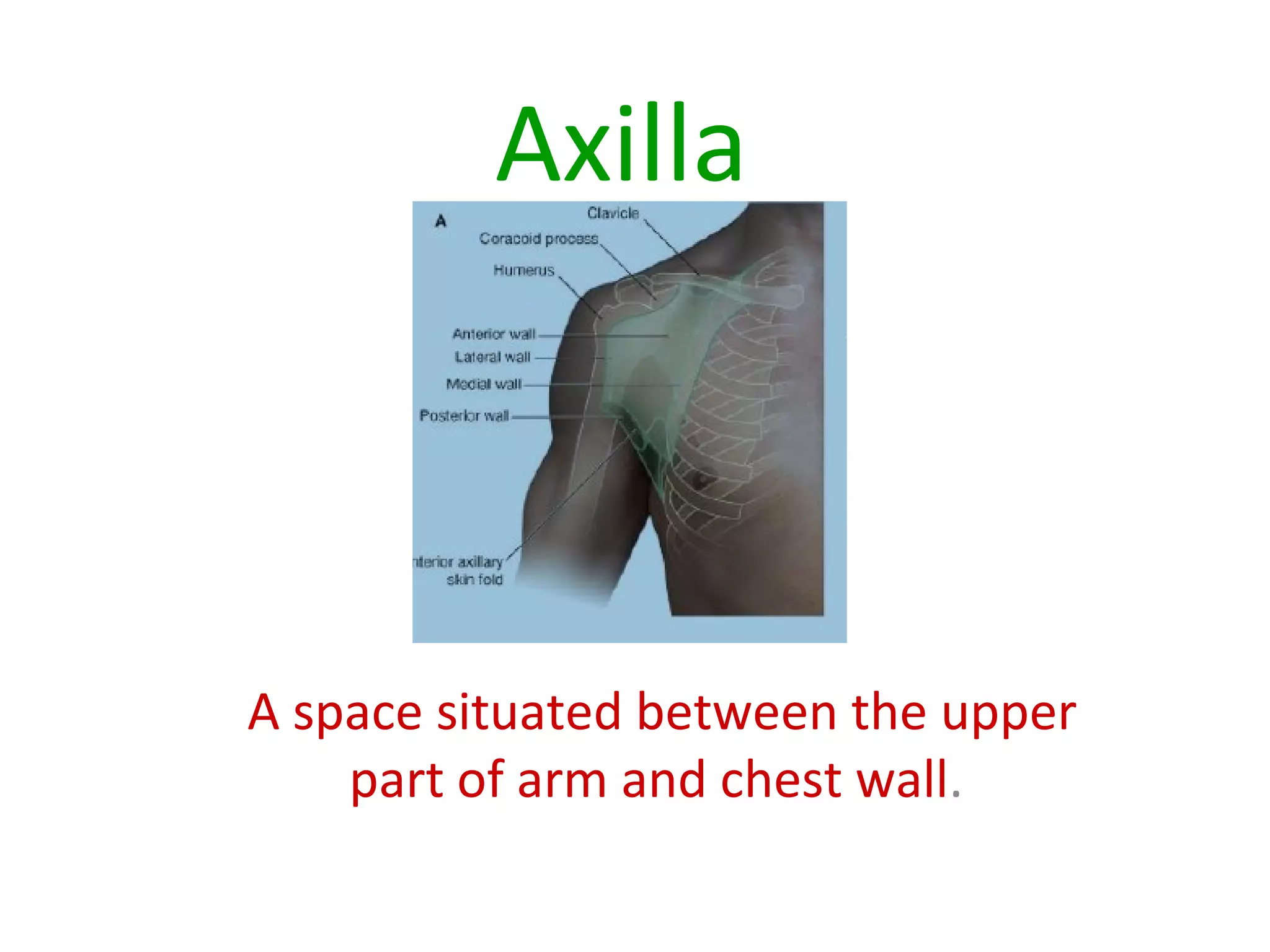
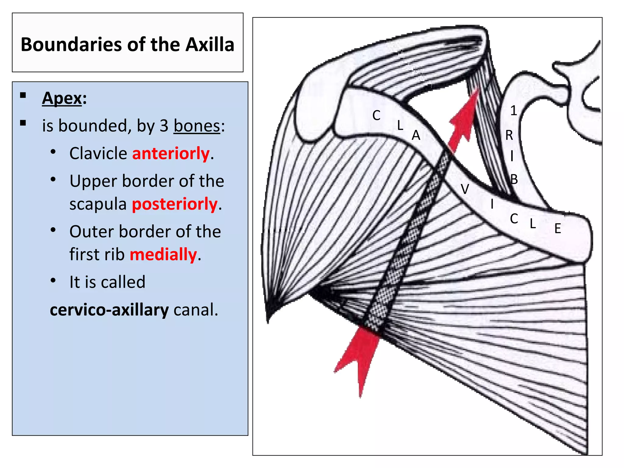
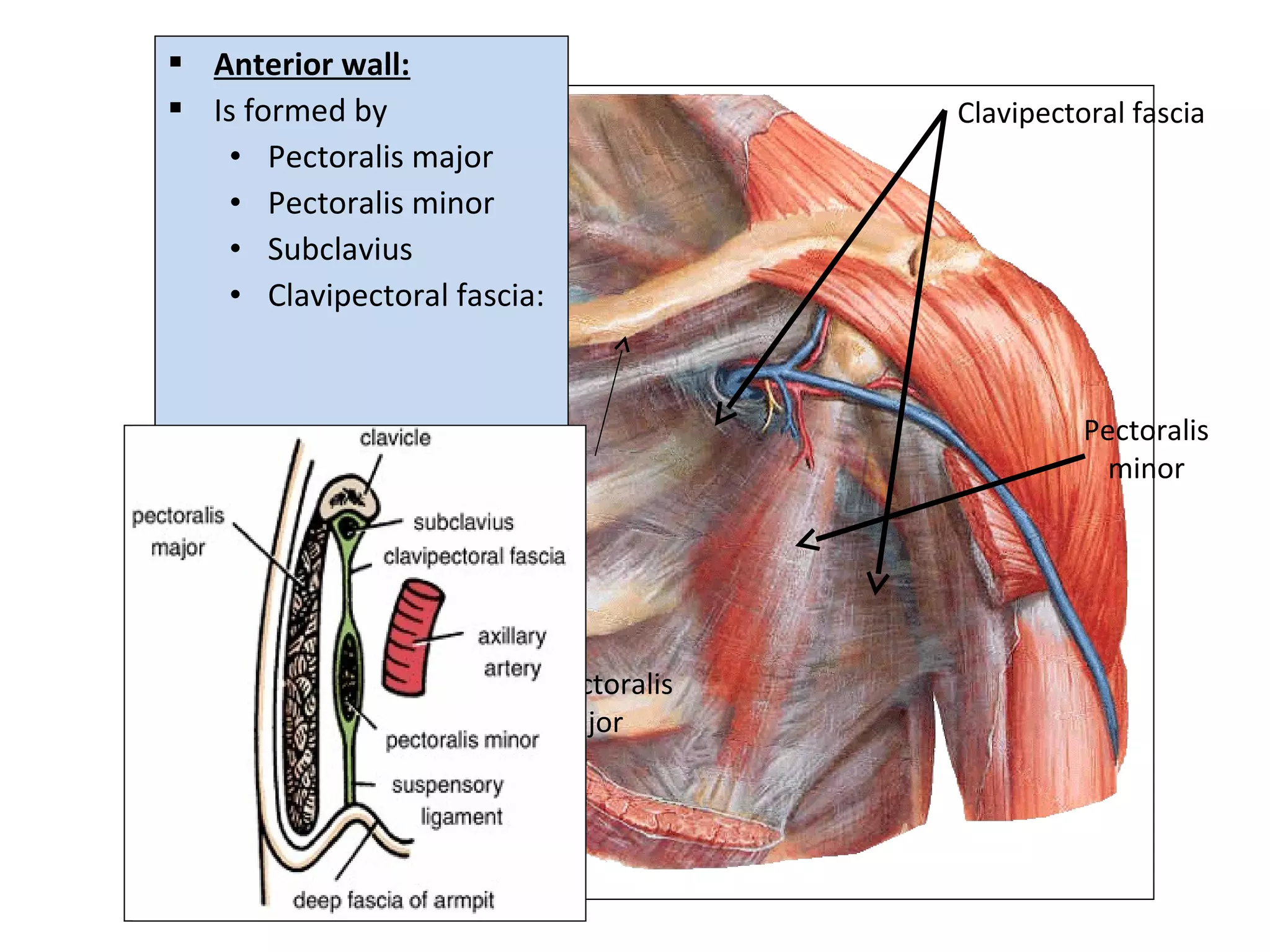
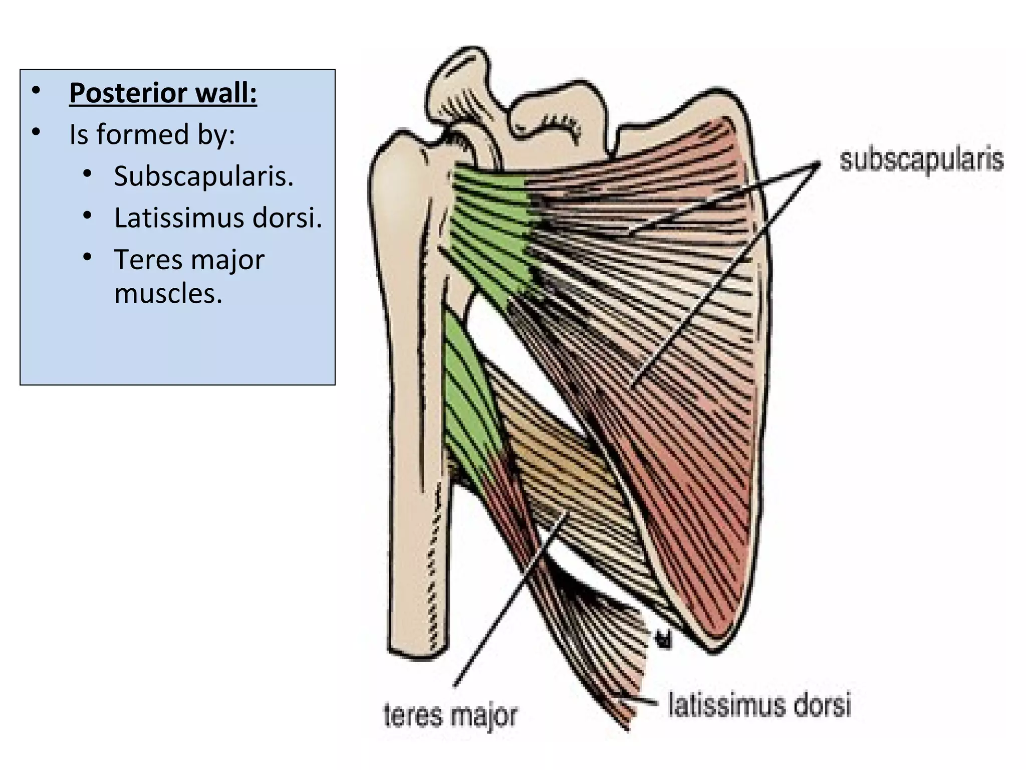
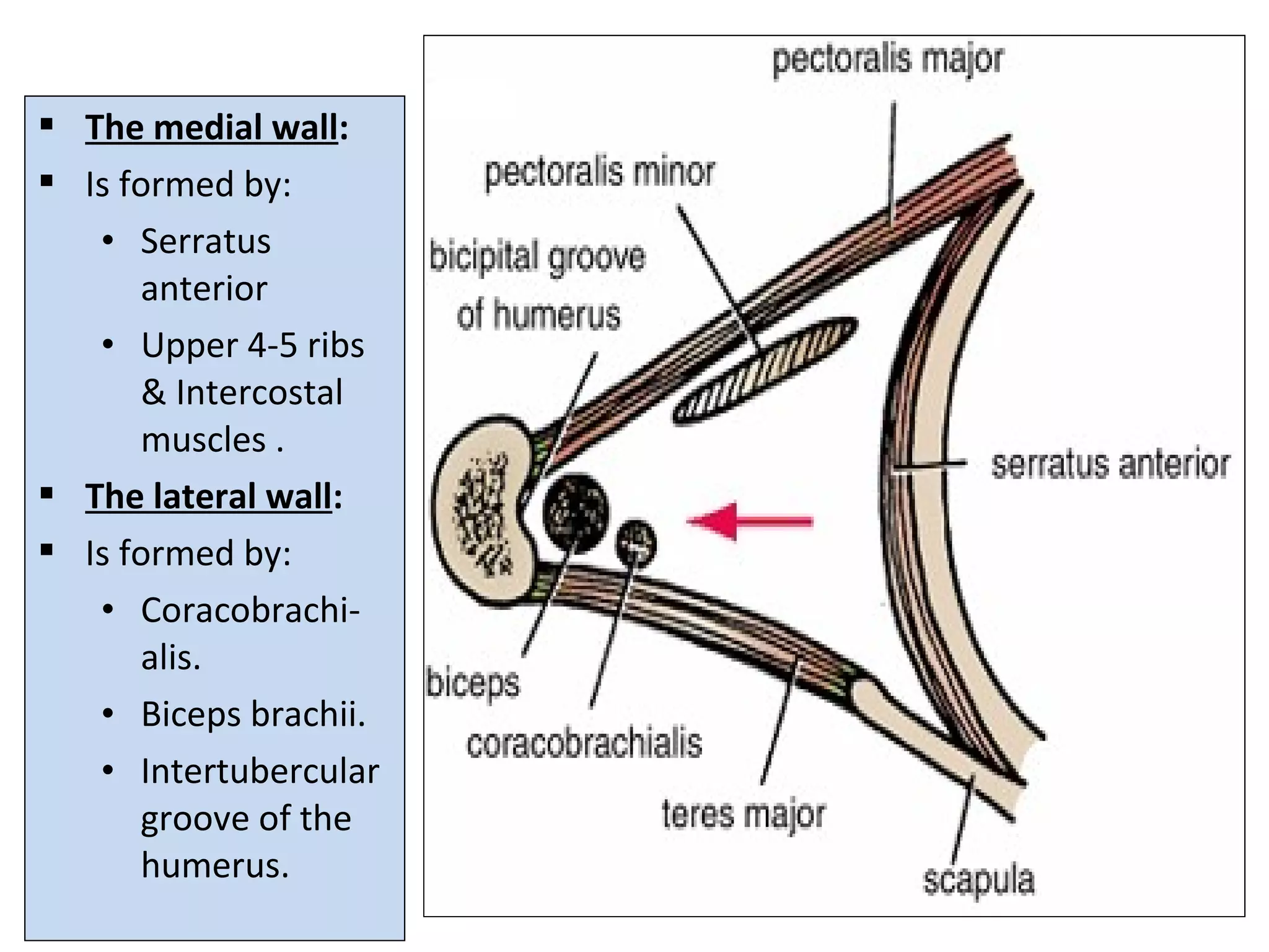
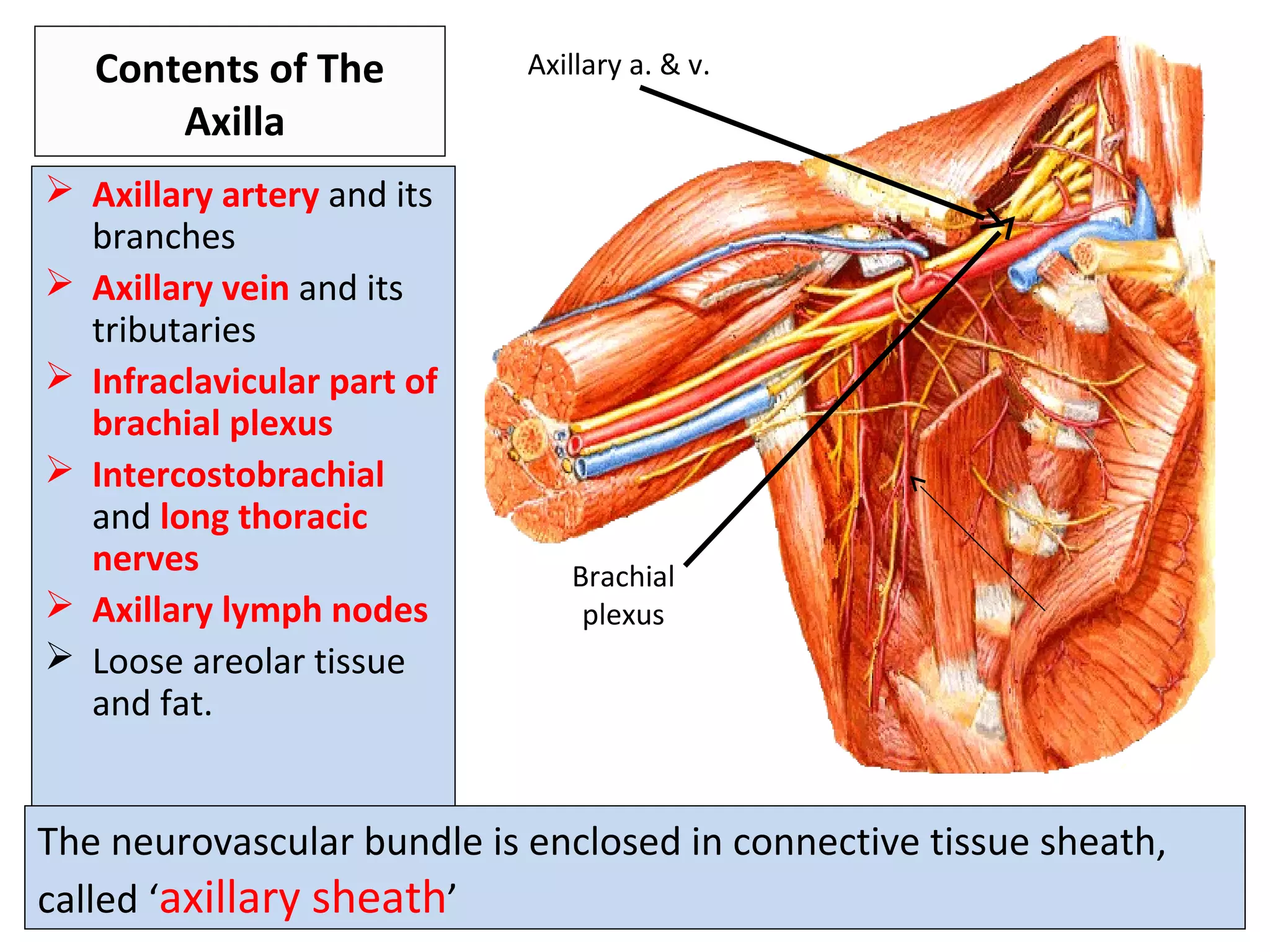
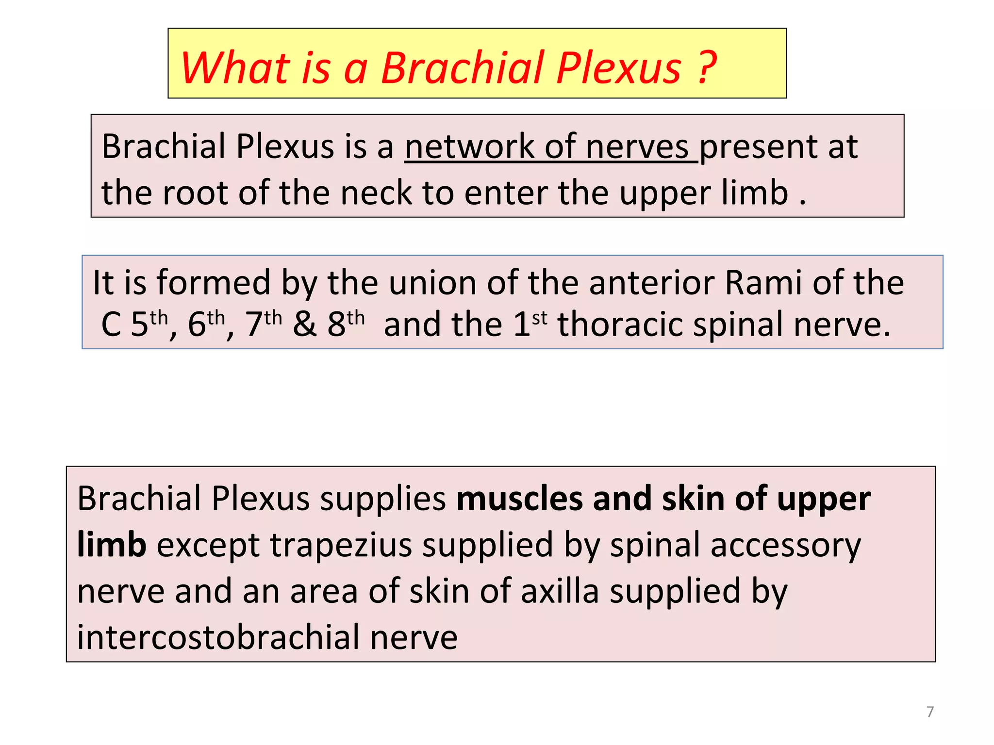
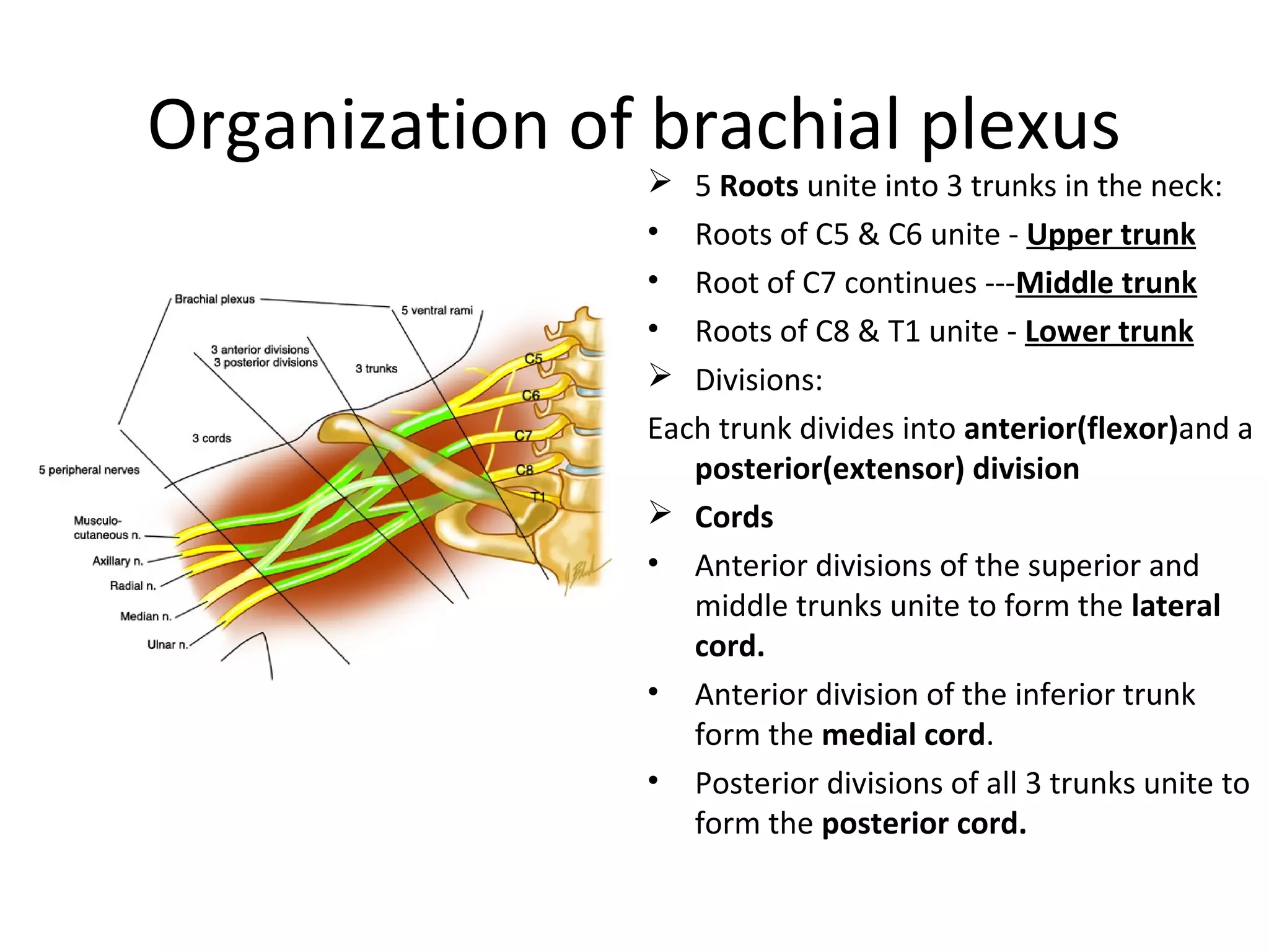

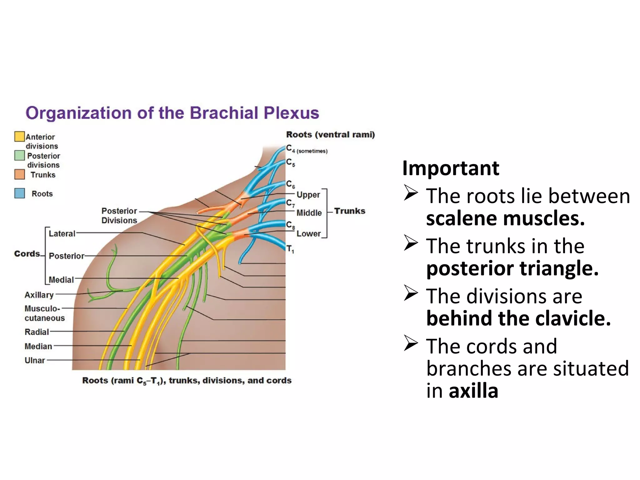
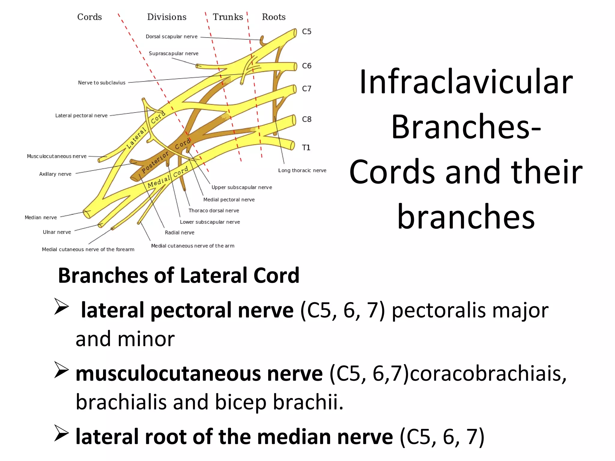
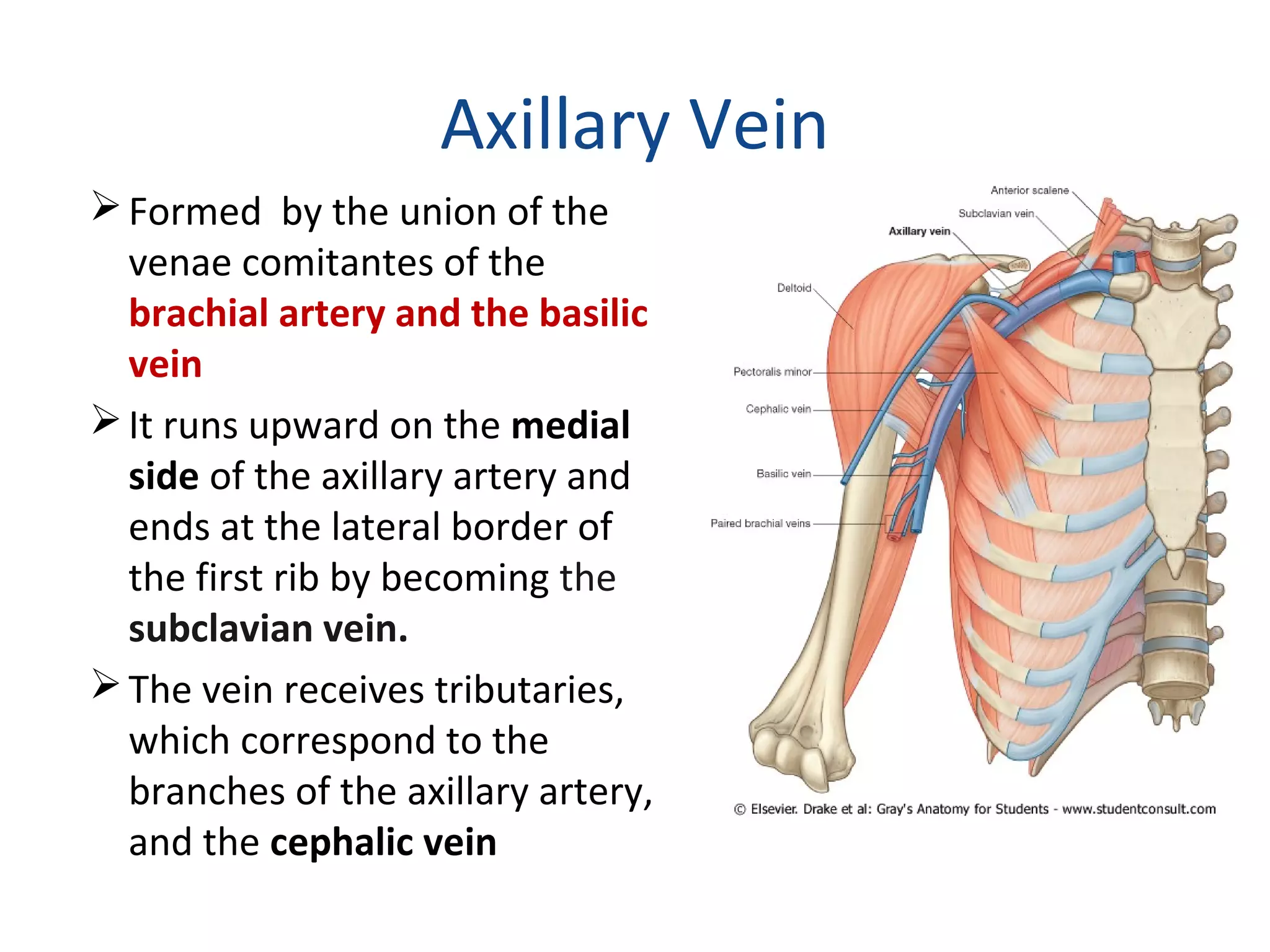
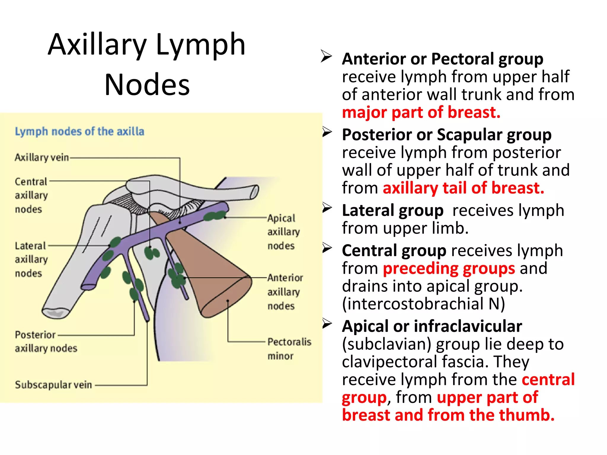

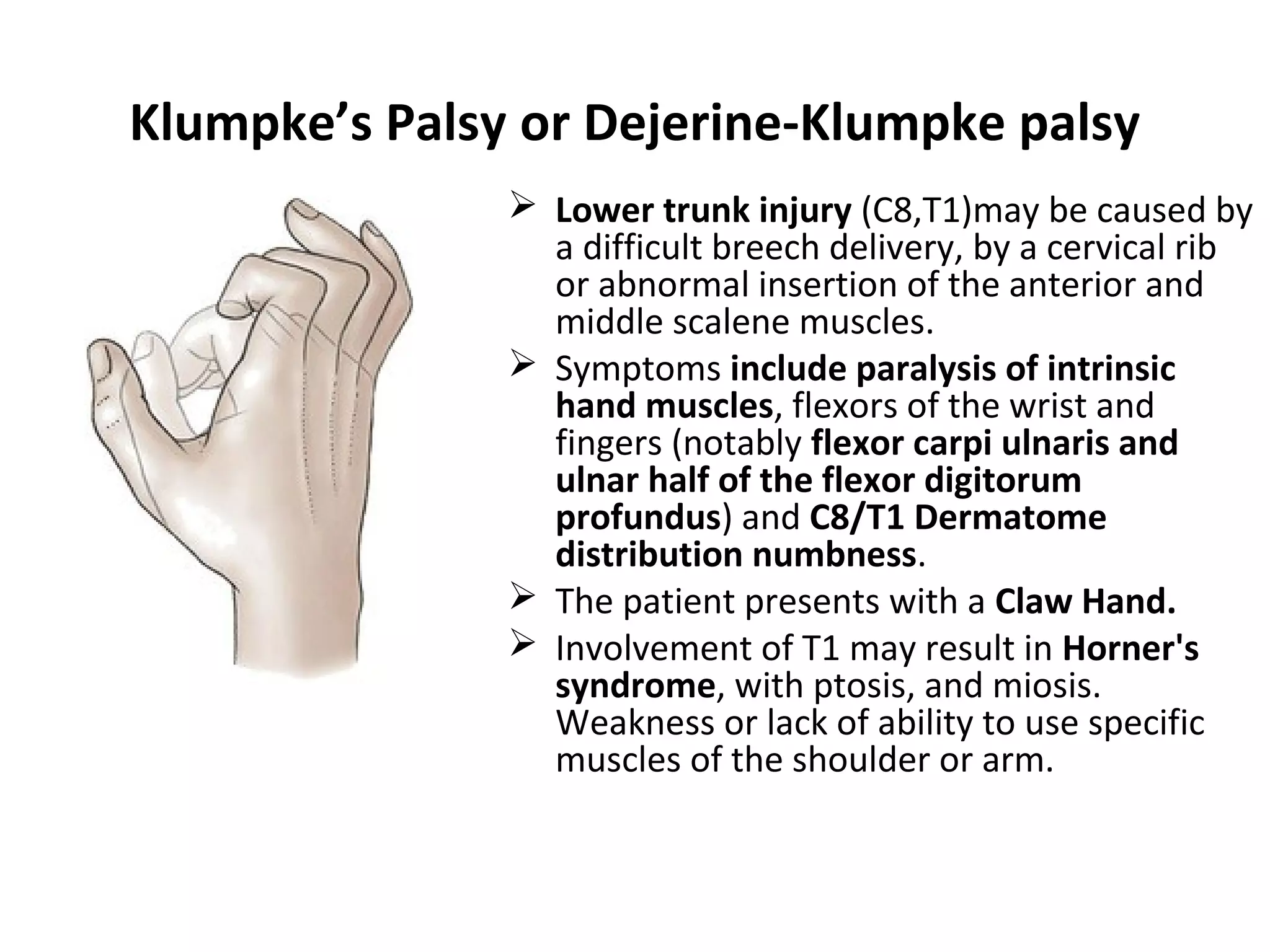

The axilla is the space between the upper arm and chest wall. It is bounded by the clavicle, first rib, and scapula. The axilla contains the axillary artery and vein, brachial plexus nerves, lymph nodes, and loose connective tissue. The brachial plexus is formed by the union of cervical and upper thoracic spinal nerves and provides motor and sensory innervation to the upper limb. Injuries to different parts of the brachial plexus can result in specific neuropathies like Erb's palsy or Klumpke's palsy, characterized by weakness or paralysis of certain muscles.


