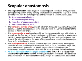
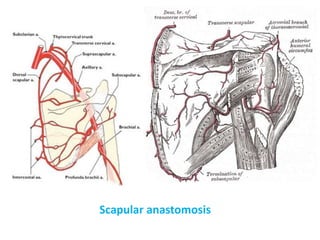
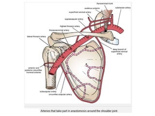
Downloaded 541 times


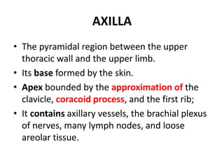
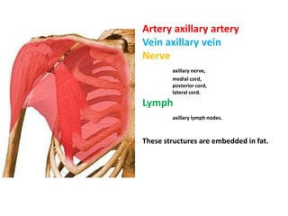
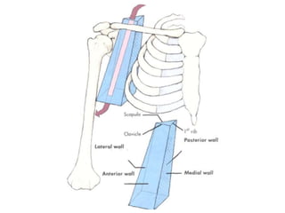
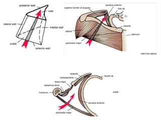
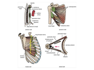
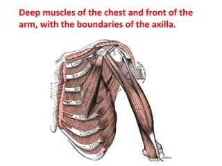
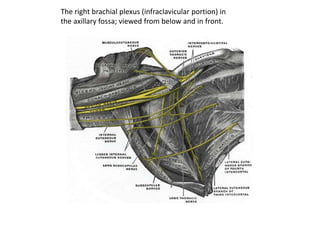

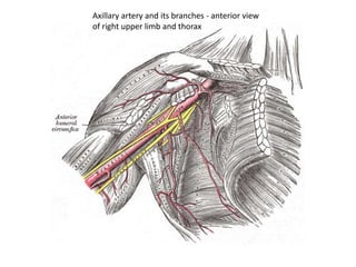
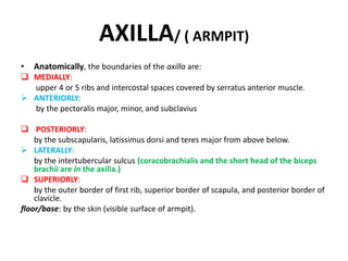

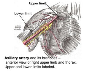
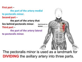
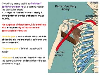
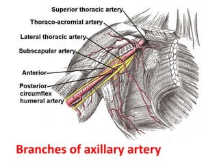
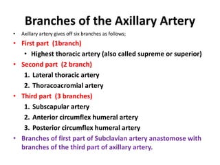
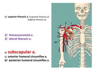
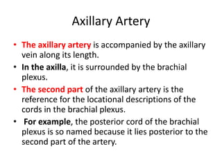
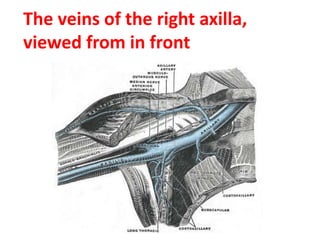
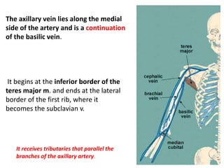
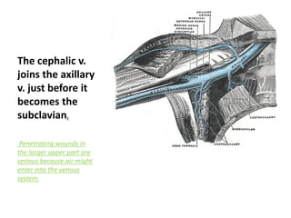
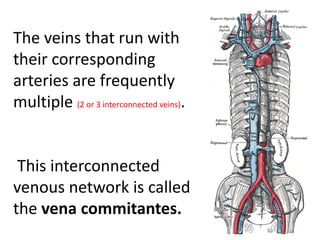
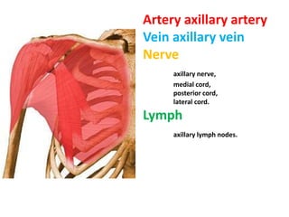
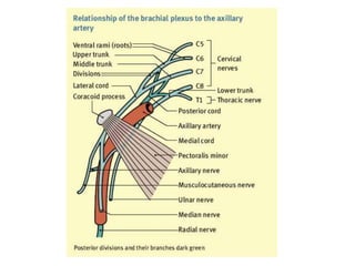
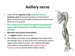
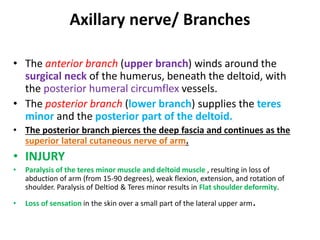
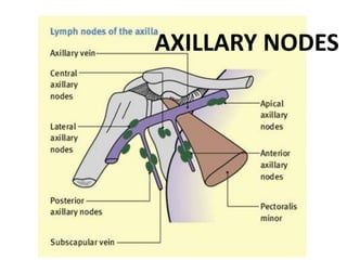
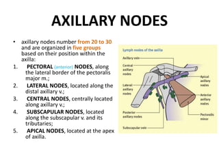

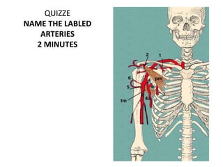
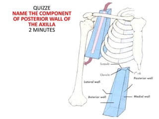

The document provides detailed information about the anatomy of the axilla region. It discusses the boundaries, contents, neurovasculature and lymph nodes of the axilla. The key points are: The axilla is bounded superiorly by the clavicle, first rib and scapula. It contains the axillary vessels (artery and vein), brachial plexus nerves, lymph nodes and loose connective tissue. The axillary artery divides into three parts based on its relationship to the pectoralis minor muscle and gives off six branches. The axillary vein receives tributaries that parallel the arterial branches. The axillary nerve originates from the brachial plexus and innervates the deltoid


![Lecture 25 Intermuscular sapces and axilla [Autosaved].pptx](https://cdn.slidesharecdn.com/ss_thumbnails/lecture25intermuscularsapcesandaxillaautosaved-251110002658-47b36c78-thumbnail.jpg?width=640&height=640&fit=bounds)


