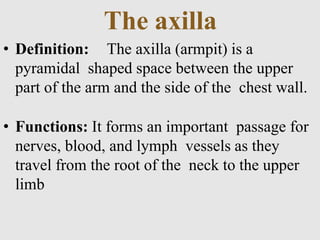
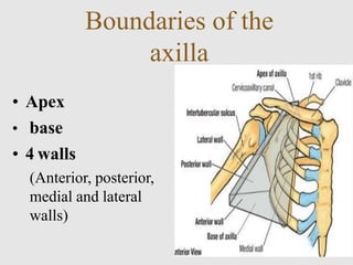
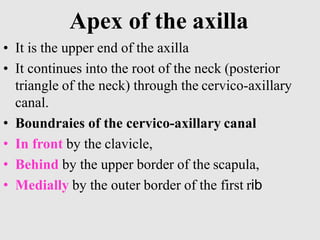
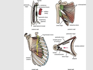
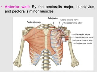
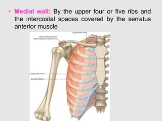
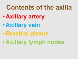
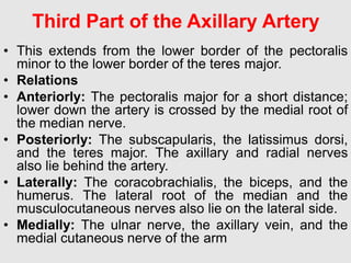
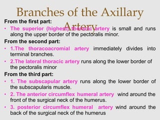
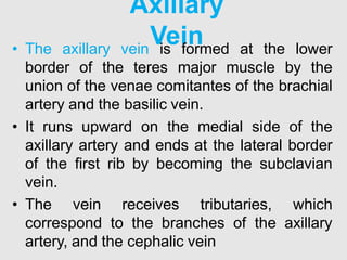
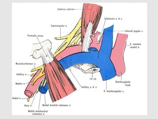
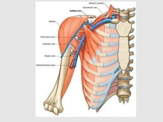
Dr. Heta Patel(PT)
Assistant Professor
ARIP
CHARUSAT
The axilla
• Definition: The axilla (armpit) is a
pyramidal shaped space between the upper
part of the arm and the side of the chest wall.
• Functions: It forms an important passage for
nerves, blood, and lymph vessels as they
travel from the root of the neck to the upper
limb
Boundaries of the
axilla
• Apex
• base
• 4 walls
(Anterior, posterior,
medial and lateral
walls)
Apex of the axilla
• It is the upper end of the axilla
• It continues into the root of the neck (posterior
triangle of the neck) through the cervico-axillary
canal.
• Boundraies of the cervico-axillary canal
• In front by the clavicle,
• Behind by the upper border of the scapula,
• Medially by the outer border of the first rib
Base of the
axilla• It is the lower end of the axilla.
• The base is formed by the skin stretching
between the anterior and posterior walls.
• Boundaries
• In front by the anterior axillary fold (formed
by the lower border of the pectoralis major
muscle),
• behind by the posterior axillary fold (formed
by the tendon of latissimus dorsi and the
teres major muscle),
• medially by the chest wall
 The walls of the axilla are made up as follows:
• Anterior wall: By the pectoralis major, subclavius,
and pectoralis minor muscles
• Posterior wall: By the subscapularis, latissimus dorsi,
and teres major muscles
• Medial wall: By the upper four or five ribs and the
intercostal spaces covered by the serratus anterior
muscle
• Lateral wall: By the coracobrachialis and biceps
muscles in the bicipital groove of the humerus
Walls of the
Axilla
• Anterior wall: By the pectoralis major, subclavius,
and pectoralis minor muscles
• Posterior wall: By the subscapularis, latissimus
dorsi, and teres major muscles
• Medial wall: By the upper four or five ribs and
the intercostal spaces covered by the serratus
anterior muscle
Contents of the axilla
•Axillary artery
•Axillary vein
•Brachial plexus
•Axillary lymph nodes
Axillary
Artery• Beginning: The axillary artery begins at the lateral
border of the first rib as a continuation of the
subclavian artery
• Termination: at the lower border of the teres major
muscle, where it continues as the brachial artery.
• The artery is closely related to the cords of the
brachial plexus and their branches and is enclosed
with them in a connective tissue sheath called the
axillary sheath. If this sheath is traced upward into
the root of the neck, it is seen to be continuous with
the prevertebral fascia
Parts of the axillary
artery
• The pectoralis minor muscle crosses
in front of the axillary artery and
divides it into three parts:
• First part above the pectoralis
minor muscle
• Second part behind the pectoralis
minor muscle
• Third part below the pectoralis
minor muscle
 This extends from the lateral border of the first rib to
the upper border of the pectoralis minor.
• Relations
• Anteriorly: The pectoralis major and the skin.
• Posteriorly: The long thoracic nerve (nerve to the
serratus anterior)
• Laterally: The three cords of the brachial plexus
• Medially: The axillary vein
First Part of the Axillary
Artery
Second Part of the Axillary Artery
• This lies behind the pectoralis minor muscle.
• Relations
• Anteriorly: The pectoralis minor, the pectoralis
major, and the skin
• Posteriorly: The posterior cord of the brachial
plexus, the subscapularis muscle, and the shoulder
joint
• Laterally: The lateral cord of the brachial plexus
• Medially: The medial cord of the brachial plexus and
the axillary vein
Third Part of the Axillary Artery
• This extends from the lower border of the pectoralis
minor to the lower border of the teres major.
• Relations
• Anteriorly: The pectoralis major for a short distance;
lower down the artery is crossed by the medial root of
the median nerve.
• Posteriorly: The subscapularis, the latissimus dorsi,
and the teres major. The axillary and radial nerves
also lie behind the artery.
• Laterally: The coracobrachialis, the biceps, and the
humerus. The lateral root of the median and the
musculocutaneous nerves also lie on the lateral side.
• Medially: The ulnar nerve, the axillary vein, and the
medial cutaneous nerve of the arm
Branches of the Axillary
Artery
From the first part:
• The superior (highest) thoracic artery is small and runs
along the upper border of the pectoralis minor.
From the second part:
• 1.The thoracoacromial artery immediately divides into
terminal branches.
• 2.The lateral thoracic artery runs along the lower border of
the pectoralis minor
From the third part:
• 1. The subscapular artery runs along the lower border of
the subscapularis muscle.
• 2. The anterior circumflex humeral artery wind around the
front of the surgical neck of the humerus.
• 3. posterior circumflex humeral artery wind around the
back of the surgical neck of the humerus
Axillary
Vein• The axillary vein is formed at the lower
border of the teres major muscle by the
union of the venae comitantes of the brachial
artery and the basilic vein.
• It runs upward on the medial side of the
axillary artery and ends at the lateral border
of the first rib by becoming the subclavian
vein.
• The vein receives tributaries, which
correspond to the branches of the axillary
artery, and the cephalic vein
Axilla
Axilla
Axilla

Axilla

  • 1.
    Dr. Heta Patel(PT) AssistantProfessor ARIP CHARUSAT
  • 2.
    The axilla • Definition:The axilla (armpit) is a pyramidal shaped space between the upper part of the arm and the side of the chest wall. • Functions: It forms an important passage for nerves, blood, and lymph vessels as they travel from the root of the neck to the upper limb
  • 3.
    Boundaries of the axilla •Apex • base • 4 walls (Anterior, posterior, medial and lateral walls)
  • 4.
    Apex of theaxilla • It is the upper end of the axilla • It continues into the root of the neck (posterior triangle of the neck) through the cervico-axillary canal. • Boundraies of the cervico-axillary canal • In front by the clavicle, • Behind by the upper border of the scapula, • Medially by the outer border of the first rib
  • 6.
    Base of the axilla•It is the lower end of the axilla. • The base is formed by the skin stretching between the anterior and posterior walls. • Boundaries • In front by the anterior axillary fold (formed by the lower border of the pectoralis major muscle), • behind by the posterior axillary fold (formed by the tendon of latissimus dorsi and the teres major muscle), • medially by the chest wall
  • 8.
     The wallsof the axilla are made up as follows: • Anterior wall: By the pectoralis major, subclavius, and pectoralis minor muscles • Posterior wall: By the subscapularis, latissimus dorsi, and teres major muscles • Medial wall: By the upper four or five ribs and the intercostal spaces covered by the serratus anterior muscle • Lateral wall: By the coracobrachialis and biceps muscles in the bicipital groove of the humerus Walls of the Axilla
  • 10.
    • Anterior wall:By the pectoralis major, subclavius, and pectoralis minor muscles
  • 11.
    • Posterior wall:By the subscapularis, latissimus dorsi, and teres major muscles
  • 13.
    • Medial wall:By the upper four or five ribs and the intercostal spaces covered by the serratus anterior muscle
  • 14.
    Contents of theaxilla •Axillary artery •Axillary vein •Brachial plexus •Axillary lymph nodes
  • 15.
    Axillary Artery• Beginning: Theaxillary artery begins at the lateral border of the first rib as a continuation of the subclavian artery • Termination: at the lower border of the teres major muscle, where it continues as the brachial artery. • The artery is closely related to the cords of the brachial plexus and their branches and is enclosed with them in a connective tissue sheath called the axillary sheath. If this sheath is traced upward into the root of the neck, it is seen to be continuous with the prevertebral fascia
  • 16.
    Parts of theaxillary artery • The pectoralis minor muscle crosses in front of the axillary artery and divides it into three parts: • First part above the pectoralis minor muscle • Second part behind the pectoralis minor muscle • Third part below the pectoralis minor muscle
  • 17.
     This extendsfrom the lateral border of the first rib to the upper border of the pectoralis minor. • Relations • Anteriorly: The pectoralis major and the skin. • Posteriorly: The long thoracic nerve (nerve to the serratus anterior) • Laterally: The three cords of the brachial plexus • Medially: The axillary vein First Part of the Axillary Artery
  • 18.
    Second Part ofthe Axillary Artery • This lies behind the pectoralis minor muscle. • Relations • Anteriorly: The pectoralis minor, the pectoralis major, and the skin • Posteriorly: The posterior cord of the brachial plexus, the subscapularis muscle, and the shoulder joint • Laterally: The lateral cord of the brachial plexus • Medially: The medial cord of the brachial plexus and the axillary vein
  • 19.
    Third Part ofthe Axillary Artery • This extends from the lower border of the pectoralis minor to the lower border of the teres major. • Relations • Anteriorly: The pectoralis major for a short distance; lower down the artery is crossed by the medial root of the median nerve. • Posteriorly: The subscapularis, the latissimus dorsi, and the teres major. The axillary and radial nerves also lie behind the artery. • Laterally: The coracobrachialis, the biceps, and the humerus. The lateral root of the median and the musculocutaneous nerves also lie on the lateral side. • Medially: The ulnar nerve, the axillary vein, and the medial cutaneous nerve of the arm
  • 20.
    Branches of theAxillary Artery From the first part: • The superior (highest) thoracic artery is small and runs along the upper border of the pectoralis minor. From the second part: • 1.The thoracoacromial artery immediately divides into terminal branches. • 2.The lateral thoracic artery runs along the lower border of the pectoralis minor From the third part: • 1. The subscapular artery runs along the lower border of the subscapularis muscle. • 2. The anterior circumflex humeral artery wind around the front of the surgical neck of the humerus. • 3. posterior circumflex humeral artery wind around the back of the surgical neck of the humerus
  • 22.
    Axillary Vein• The axillaryvein is formed at the lower border of the teres major muscle by the union of the venae comitantes of the brachial artery and the basilic vein. • It runs upward on the medial side of the axillary artery and ends at the lateral border of the first rib by becoming the subclavian vein. • The vein receives tributaries, which correspond to the branches of the axillary artery, and the cephalic vein