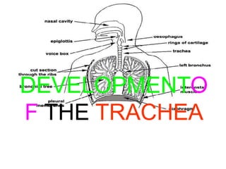
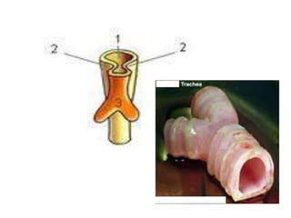
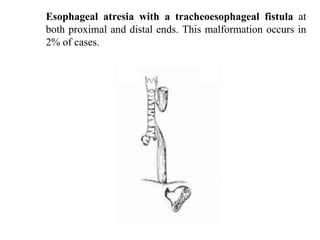
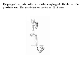
ANA 206
SYSTEMIC EMBRYOLOGY
DEVELOPMENTO
F THE TRACHEA
Brief insight into the
Anatomical & Physiological
Characteristics of the
Trachea
The trachea is a flexible pipe which travels from
the throat down into the thorax. It is supported
by rings of a flexible but strong material called

cartilage.

It

prevents

the

trachea

from

collapsing as inspired and expired air move

through it. The cartilage rings also allow for
continue breathing when the neck is bent. The

inside of the trachea is covered by millions of
microscopic hairs called cilia.
The cilia are covered with a layer of liquid called
mucus. Very fine dust particles and microbes
get trapped in the mucus and cilia. Although the
cilia are attached at one end, each one is able to
move (beat). As the cilia beat they tend to push
the dust-containing mucus toward the nose and
the mouth. In this way the lungs are kept free of
most of the dust and microbes inhaled.
Development of the
Trachea
The trachea develops caudal to the larynx.
The endodermal lining of the laryngotracheal
tube differentiates into the epithelium and
glands of the trachea and the pulmonary

epihelium.
The epithelium develops from the endoderm
and the tracheal cartilage and muscles develop
from splanchnic mesoderm.

Early in development, the trachea bifurcates
into the left and right bronchi.
CLINICAL CORRELATES
Tracheoesophageal fistula is an abnormal communication
between the trachea and esophagus that results from improper
division of foregut by the tracheoesophageal septum. It is
generally

associated

polyhydramnios.

with

esophageal

atresia

and
Clinical features include excessive accumulation of saliva or
mucus in the nose and mouth; episodes of gagging and
cyanosis after swallowing milk; abdominal distention after
crying; and reflux of gastric contents into lungs, causing
pneumonitis.
Diagnostic features include inability to pass a catheter into the

stomach and radiographs demonstrating air in the infant's
stomach. There are five different anatomical types of

esophagus and trachea malformations as follows:
Esophageal atresia with a tracheoesophageal fistula at the
distal one-third end of the trachea. This is the most common

type, occurring in 82% of cases. The anteroposterior (AP)
radiograph (the image on the next slide) of this malformation
shows an enteric tube (arrow) coiled in the upper esophageal
pouch.

The

air

in

tracheoesophageal fistula.

the

bowel

indicates

a

distal
Esophageal atresia with a tracheoesophageal fistula at the distal one-third end of the
trachea
Esophageal atresia only. This malformation occurs in
9% of cases.
H-type tracheoesophageal fistula only. This malformation
occurs in 6% of cases. The barium swallow radiograph (in
the image below) shows a normal esophagus (E), but dye
has spilled into the trachea (T) through the fistula and
outlines the upper trachea and larynx.
Esophageal atresia with a tracheoesophageal fistula at
both proximal and distal ends. This malformation occurs in
2% of cases.
Esophageal atresia with a tracheoesophageal fistula at the
proximal end. This malformation occurs in 1% of cases
COMPLIMENT OF
THE SEASON
&
Have a
pleasant day
Ch33r5!!!!!!!!!!!!!!!!!!!!!!!!!!

Trachea

  • 1.
  • 2.
  • 3.
    Brief insight intothe Anatomical & Physiological Characteristics of the Trachea
  • 4.
    The trachea isa flexible pipe which travels from the throat down into the thorax. It is supported by rings of a flexible but strong material called cartilage. It prevents the trachea from collapsing as inspired and expired air move through it. The cartilage rings also allow for continue breathing when the neck is bent. The inside of the trachea is covered by millions of microscopic hairs called cilia.
  • 5.
    The cilia arecovered with a layer of liquid called mucus. Very fine dust particles and microbes get trapped in the mucus and cilia. Although the cilia are attached at one end, each one is able to move (beat). As the cilia beat they tend to push the dust-containing mucus toward the nose and the mouth. In this way the lungs are kept free of most of the dust and microbes inhaled.
  • 6.
  • 7.
    The trachea developscaudal to the larynx. The endodermal lining of the laryngotracheal tube differentiates into the epithelium and glands of the trachea and the pulmonary epihelium.
  • 8.
    The epithelium developsfrom the endoderm and the tracheal cartilage and muscles develop from splanchnic mesoderm. Early in development, the trachea bifurcates into the left and right bronchi.
  • 10.
    CLINICAL CORRELATES Tracheoesophageal fistulais an abnormal communication between the trachea and esophagus that results from improper division of foregut by the tracheoesophageal septum. It is generally associated polyhydramnios. with esophageal atresia and
  • 11.
    Clinical features includeexcessive accumulation of saliva or mucus in the nose and mouth; episodes of gagging and cyanosis after swallowing milk; abdominal distention after crying; and reflux of gastric contents into lungs, causing pneumonitis. Diagnostic features include inability to pass a catheter into the stomach and radiographs demonstrating air in the infant's stomach. There are five different anatomical types of esophagus and trachea malformations as follows:
  • 12.
    Esophageal atresia witha tracheoesophageal fistula at the distal one-third end of the trachea. This is the most common type, occurring in 82% of cases. The anteroposterior (AP) radiograph (the image on the next slide) of this malformation shows an enteric tube (arrow) coiled in the upper esophageal pouch. The air in tracheoesophageal fistula. the bowel indicates a distal
  • 13.
    Esophageal atresia witha tracheoesophageal fistula at the distal one-third end of the trachea
  • 14.
    Esophageal atresia only.This malformation occurs in 9% of cases.
  • 15.
    H-type tracheoesophageal fistulaonly. This malformation occurs in 6% of cases. The barium swallow radiograph (in the image below) shows a normal esophagus (E), but dye has spilled into the trachea (T) through the fistula and outlines the upper trachea and larynx.
  • 16.
    Esophageal atresia witha tracheoesophageal fistula at both proximal and distal ends. This malformation occurs in 2% of cases.
  • 17.
    Esophageal atresia witha tracheoesophageal fistula at the proximal end. This malformation occurs in 1% of cases
  • 18.
    COMPLIMENT OF THE SEASON & Havea pleasant day Ch33r5!!!!!!!!!!!!!!!!!!!!!!!!!!

Editor's Notes

  • #12 Cyanosis, bluish skin colour
  • #15 Fistula – openin, opp. of atresia