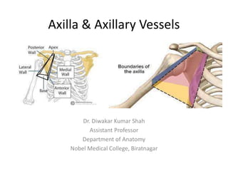
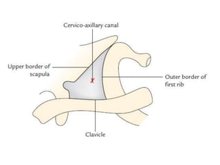
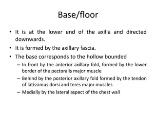
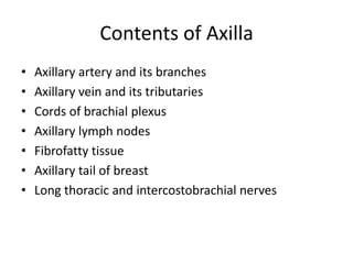
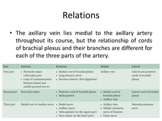
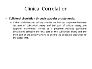
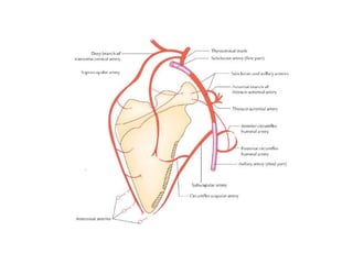
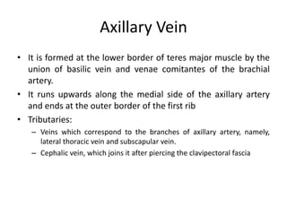
The axilla contains the brachial plexus, axillary vessels, and lymph nodes. It has boundaries formed by muscles and acts as a passage from the neck to the upper limb. The axillary artery passes through the axilla in three parts, giving off branches. The axillary vein lies medial to the artery, draining the upper limb. Lymph nodes in the axilla drain the arm and breast. The scapular anastomosis connects arterial branches to ensure circulation if main vessels are blocked.
Anatomy of axilla with Dr- Ameera Al-Humidi .pptxAmeera Al-Humidi
The axilla is the anatomical region under the shoulder joint where the arm connects to the shoulder.
The axilla has five anatomic borders: superior, anterior, posterior, lateral, and medial walls.
The borders of the axilla are composed of muscles, including the serratus anterior, coracobrachialis, and short head of the biceps
The axillary walls are used as landmarks by surgeons to prevent damage to the neurovascular structures within the axilla during surgery
The contents of the axilla include muscles, nerves, vessels, and lymphatics
The axillary artery and vein, brachial plexus, and axillary lymph nodes are some of the neurovascular structures found in the axilla
Seminar presentation on arterial supply of human head & neck - carotid artery, maxillary artery, ophthalmic artery
post-graduate level
MDS- oral & maxillofacial surgery
The arterial supply to the upper limb is delivered via five main vessels (proximal to distal):
Subclavian artery
Axillary artery
Brachial artery
Radial artery
Ulnar artery
In this article, we shall look at the anatomy of the arteries of the upper limb – their anatomical course, branches and clinical correlations.
Best Ayurvedic medicine for Gas and IndigestionSwastikAyurveda
Here is the updated list of Top Best Ayurvedic medicine for Gas and Indigestion and those are Gas-O-Go Syp for Dyspepsia | Lavizyme Syrup for Acidity | Yumzyme Hepatoprotective Capsules etc
micro teaching on communication m.sc nursing.pdfAnurag Sharma
Microteaching is a unique model of practice teaching. It is a viable instrument for the. desired change in the teaching behavior or the behavior potential which, in specified types of real. classroom situations, tends to facilitate the achievement of specified types of objectives.
These lecture slides, by Dr Sidra Arshad, offer a quick overview of the physiological basis of a normal electrocardiogram.
Learning objectives:
1. Define an electrocardiogram (ECG) and electrocardiography
2. Describe how dipoles generated by the heart produce the waveforms of the ECG
3. Describe the components of a normal electrocardiogram of a typical bipolar lead (limb II)
4. Differentiate between intervals and segments
5. Enlist some common indications for obtaining an ECG
6. Describe the flow of current around the heart during the cardiac cycle
7. Discuss the placement and polarity of the leads of electrocardiograph
8. Describe the normal electrocardiograms recorded from the limb leads and explain the physiological basis of the different records that are obtained
9. Define mean electrical vector (axis) of the heart and give the normal range
10. Define the mean QRS vector
11. Describe the axes of leads (hexagonal reference system)
12. Comprehend the vectorial analysis of the normal ECG
13. Determine the mean electrical axis of the ventricular QRS and appreciate the mean axis deviation
14. Explain the concepts of current of injury, J point, and their significance
Study Resources:
1. Chapter 11, Guyton and Hall Textbook of Medical Physiology, 14th edition
2. Chapter 9, Human Physiology - From Cells to Systems, Lauralee Sherwood, 9th edition
3. Chapter 29, Ganong’s Review of Medical Physiology, 26th edition
4. Electrocardiogram, StatPearls - https://www.ncbi.nlm.nih.gov/books/NBK549803/
5. ECG in Medical Practice by ABM Abdullah, 4th edition
6. Chapter 3, Cardiology Explained, https://www.ncbi.nlm.nih.gov/books/NBK2214/
7. ECG Basics, http://www.nataliescasebook.com/tag/e-c-g-basics
Basavarajeeyam is an important text for ayurvedic physician belonging to andhra pradehs. It is a popular compendium in various parts of our country as well as in andhra pradesh. The content of the text was presented in sanskrit and telugu language (Bilingual). One of the most famous book in ayurvedic pharmaceutics and therapeutics. This book contains 25 chapters called as prakaranas. Many rasaoushadis were explained, pioneer of dhatu druti, nadi pareeksha, mutra pareeksha etc. Belongs to the period of 15-16 century. New diseases like upadamsha, phiranga rogas are explained.
Adv. biopharm. APPLICATION OF PHARMACOKINETICS : TARGETED DRUG DELIVERY SYSTEMSAkankshaAshtankar
MIP 201T & MPH 202T
ADVANCED BIOPHARMACEUTICS & PHARMACOKINETICS : UNIT 5
APPLICATION OF PHARMACOKINETICS : TARGETED DRUG DELIVERY SYSTEMS By - AKANKSHA ASHTANKAR
Muktapishti is a traditional Ayurvedic preparation made from Shoditha Mukta (Purified Pearl), is believed to help regulate thyroid function and reduce symptoms of hyperthyroidism due to its cooling and balancing properties. Clinical evidence on its efficacy remains limited, necessitating further research to validate its therapeutic benefits.
- Video recording of this lecture in English language: https://youtu.be/kqbnxVAZs-0
- Video recording of this lecture in Arabic language: https://youtu.be/SINlygW1Mpc
- Link to download the book free: https://nephrotube.blogspot.com/p/nephrotube-nephrology-books.html
- Link to NephroTube website: www.NephroTube.com
- Link to NephroTube social media accounts: https://nephrotube.blogspot.com/p/join-nephrotube-on-social-media.html
Title: Sense of Taste
Presenter: Dr. Faiza, Assistant Professor of Physiology
Qualifications:
MBBS (Best Graduate, AIMC Lahore)
FCPS Physiology
ICMT, CHPE, DHPE (STMU)
MPH (GC University, Faisalabad)
MBA (Virtual University of Pakistan)
Learning Objectives:
Describe the structure and function of taste buds.
Describe the relationship between the taste threshold and taste index of common substances.
Explain the chemical basis and signal transduction of taste perception for each type of primary taste sensation.
Recognize different abnormalities of taste perception and their causes.
Key Topics:
Significance of Taste Sensation:
Differentiation between pleasant and harmful food
Influence on behavior
Selection of food based on metabolic needs
Receptors of Taste:
Taste buds on the tongue
Influence of sense of smell, texture of food, and pain stimulation (e.g., by pepper)
Primary and Secondary Taste Sensations:
Primary taste sensations: Sweet, Sour, Salty, Bitter, Umami
Chemical basis and signal transduction mechanisms for each taste
Taste Threshold and Index:
Taste threshold values for Sweet (sucrose), Salty (NaCl), Sour (HCl), and Bitter (Quinine)
Taste index relationship: Inversely proportional to taste threshold
Taste Blindness:
Inability to taste certain substances, particularly thiourea compounds
Example: Phenylthiocarbamide
Structure and Function of Taste Buds:
Composition: Epithelial cells, Sustentacular/Supporting cells, Taste cells, Basal cells
Features: Taste pores, Taste hairs/microvilli, and Taste nerve fibers
Location of Taste Buds:
Found in papillae of the tongue (Fungiform, Circumvallate, Foliate)
Also present on the palate, tonsillar pillars, epiglottis, and proximal esophagus
Mechanism of Taste Stimulation:
Interaction of taste substances with receptors on microvilli
Signal transduction pathways for Umami, Sweet, Bitter, Sour, and Salty tastes
Taste Sensitivity and Adaptation:
Decrease in sensitivity with age
Rapid adaptation of taste sensation
Role of Saliva in Taste:
Dissolution of tastants to reach receptors
Washing away the stimulus
Taste Preferences and Aversions:
Mechanisms behind taste preference and aversion
Influence of receptors and neural pathways
Impact of Sensory Nerve Damage:
Degeneration of taste buds if the sensory nerve fiber is cut
Abnormalities of Taste Detection:
Conditions: Ageusia, Hypogeusia, Dysgeusia (parageusia)
Causes: Nerve damage, neurological disorders, infections, poor oral hygiene, adverse drug effects, deficiencies, aging, tobacco use, altered neurotransmitter levels
Neurotransmitters and Taste Threshold:
Effects of serotonin (5-HT) and norepinephrine (NE) on taste sensitivity
Supertasters:
25% of the population with heightened sensitivity to taste, especially bitterness
Increased number of fungiform papillae
Ozempic: Preoperative Management of Patients on GLP-1 Receptor Agonists Saeid Safari
Preoperative Management of Patients on GLP-1 Receptor Agonists like Ozempic and Semiglutide
ASA GUIDELINE
NYSORA Guideline
2 Case Reports of Gastric Ultrasound
Knee anatomy and clinical tests 2024.pdfvimalpl1234
This includes all relevant anatomy and clinical tests compiled from standard textbooks, Campbell,netter etc..It is comprehensive and best suited for orthopaedicians and orthopaedic residents.
1. Axilla & Axillary Vessels
Dr. Diwakar Kumar Shah
Assistant Professor
Department of Anatomy
Nobel Medical College, Biratnagar
2.
3. Introduction
• The axilla or armpit is a fat-filled pyramid-shaped space, between the
upper part of the arm and the side of the chest wall.
• It contains the brachial plexus, axillary vessels, and lymph nodes.
• It also acts as a funnel shaped tunnel for neurovascular structures to pass
from the root of the neck to the upper limb and vice versa.
• Groups of lymph nodes within it drain the upper limb and the breast.
• Axillary lymph nodes are often enlarged and hence routinely palpated
during physical examination of the patient.
• Abscess in this region is also common.
4. BOUNDARIES
• The axilla resembles a truncated four-sided pyramid and presents an apex,
a base and four walls (anterior, posterior, medial, and lateral).
Apex/cervico-axillary canal:
• It is a passageway between the neck and axilla.
• It is directed upwards and medially into the root of the neck and
corresponds to the triangular space bounded in front by the clavicle,
behind by the upper border of the scapula and medially by the outer
border of the first rib.
• The axillary artery and brachial plexus enter the axilla from neck through
this gap, hence it is also termed cervico-axillary canal.
• The axillary vein enters the neck from axilla into the neck through this
canal.
5.
6.
7.
8. Base/floor
• It is at the lower end of the axilla and directed
downwards.
• It is formed by the axillary fascia.
• The base corresponds to the hollow bounded
– In front by the anterior axillary fold, formed by the lower
border of the pectoralis major muscle
– Behind by the posterior axillary fold formed by the tendon
of latissimus dorsi and teres major muscles
– Medially by the lateral aspect of the chest wall
9. • Anterior wall:
– It is formed by the pectoralis major, subclavius, and pectoralis minor
muscle.
• Posterior wall:
– It is formed by the subscapular muscle above and latissimus dorsi and
teres major muscle below.
• Medial wall:
– It is formed by the upper four or five ribs, and corresponding
intercostal spaces covered by the serratus anterior muscle
• Lateral wall:
– It is formed by tendon of biceps brachii in the bicipital groove of
humerus, coracobrachialis and short head of biceps brachii.
– The lateral wall is extremely narrow because anterior and posterior
walls of the axilla converge at this site.
10. Contents of Axilla
• Axillary artery and its branches
• Axillary vein and its tributaries
• Cords of brachial plexus
• Axillary lymph nodes
• Fibrofatty tissue
• Axillary tail of breast
• Long thoracic and intercostobrachial nerves
11. Axillary artery
• It is the main artery of the upper limb.
• It begins at the outer border of the first rib as the
continuation of subclavian artery and ends by becoming
brachial artery at the lower border of teres major.
• In axilla, it runs from its apex to the base along the lateral wall
nearer to the anterior wall than the posterior wall.
• During its course through axilla, it is crossed on its superficial
aspect by the pectoralis minor muscle, which divides it into
three parts.
• Axillary vein is medial to the artery and the cords of brachial
plexus are arranged around the second part of the artery (i.e.,
part deep to the pectoralis minor); the lateral cord being
lateral, the medial cord medial, and posterior cord behind.
12. Parts
• First part, superior (or proximal) to the muscle
• Second part, posterior (or deep) to the muscle
• Third part, inferior (or distal) to the muscle
13. Relations
• The axillary vein lies medial to the axillary artery
throughout its course, but the relationship of cords
of brachial plexus and their branches are different for
each of the three parts of the artery.
14.
15. Branches of Axillary Artery
• From First Part
– Superior thoracic artery, a very small branch, arises near the
subclavius, passes between the pectoralis major and minor muscle,
and supplies these muscles and medial wall of the axilla
16. From Second part
• Thoraco-acromial artery (acromiothoracic artery), emerges at the upper
border of pectoralis minor, pierces clavipectoral fascia and soon breaks up
into four branches:
• Pectoral branch
• Deltoid branch
• Acromial branch
• Clavicular branch.
– These branches radiate at right angle to each other.
– The pectoral branch supplies pectoral muscles, deltoid branch, ends by
joining anastomosis over the acromion, clavicular branch supplied
sternoclavicular joint.
• Lateral thoracic artery, emerges at and runs along the inferior border of
pectoralis minor, supplying the branches to pectoralis major and minor
and serratus anterior muscles.
– In the females, the lateral thoracic artery is large and provides
important supply to the breast through its lateral mammary branches.
17.
18. From Third part
• Subscapular artery, the largest branch of axillary
artery, runs along the lower border of the
subscapularis and ends near the inferior angle of the
scapula.
– It gives a large branch, the circumflex scapular artery,
which passes through upper triangular intermuscular
space, winds round the lateral border of scapula to enter
infraspinous fossa.
– In addition, it gives numerous small branches.
19. • Anterior circumflex humeral artery, a small branch, passes in
front of surgical neck of humerus and anastomoses with the
posterior circumflex humeral artery to form an arterial circle
around the surgical neck of humerus.
– It gives an ascending branch, which runs upwards into the
intertubercular sulcus of humerus to supply the head of humerus and
shoulder joint.
• Posterior circumflex humeral artery, larger than the anterior
circumflex humeral artery, passes backwards, along with
axillary nerve through the quadrangular intermuscular space,
crosses the posterior aspect of surgical neck of humerus to
anastomose with the anterior circumflex humeral artery.
– It supplies the deltoid muscle and shoulder joint.
20. Arterial Anastomosis Around Scapula
• The arterial anastomosis around scapula is principally formed between the
branches of the first part of the subclavian and the third part of the
axillary arteries.
• The scapular anastomosis takes place at two sites: around the body of
scapula and over the acromion process of the scapula.
• Around the body of scapula: it occurs between the
– Suprascapular artery, a branch of the thyrocervical trunk from the first part of
the subclavian artery
– Circumflex scapular artery, a branch of the subscapular artery from the third
part of the axillary artery
– Deep branch of the transverse cervical artery, a branch of the thyrocervical
trunk
• Over the acromion process: it occurs between the
– Acromial branch of the thoraco-acromial artery
– Acromial branch of the suprascapular artery
– Acromial branch of the posterior circumflex humeral artery
21. Clinical Correlation
• Collateral circulation through scapular anastomosis:
– If the subclavian and axillary arteries are blocked anywhere between
1st part of subclavian artery and 3rd part of axillary artery, the
scapular anastomosis serves as a potential pathway (collateral
circulation) between the first part of the subclavian artery and the
third part of the axillary artery, to ensure the adequate circulation to
the upper limb.
22.
23. Axillary Vein
• It is formed at the lower border of teres major muscle by the
union of basilic vein and venae comitantes of the brachial
artery.
• It runs upwards along the medial side of the axillary artery
and ends at the outer border of the first rib
• Tributaries:
– Veins which correspond to the branches of axillary artery, namely,
lateral thoracic vein and subscapular vein.
– Cephalic vein, which joins it after piercing the clavipectoral fascia
24. Clinical Correlation
• Spontaneous thrombosis of the axillary vein:
– Occasionally, a muscular band- the axillary arch, overlies
the vein.
– It may compress the vein, following excessive and
unaccustomed movements of the arm at the shoulder
joint and cause spontaneous thrombosis of the axillary
vein