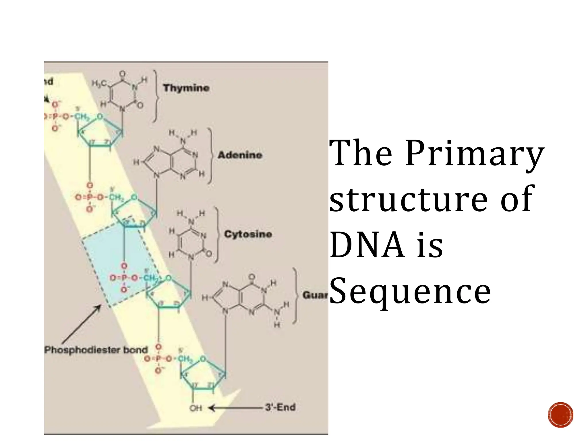
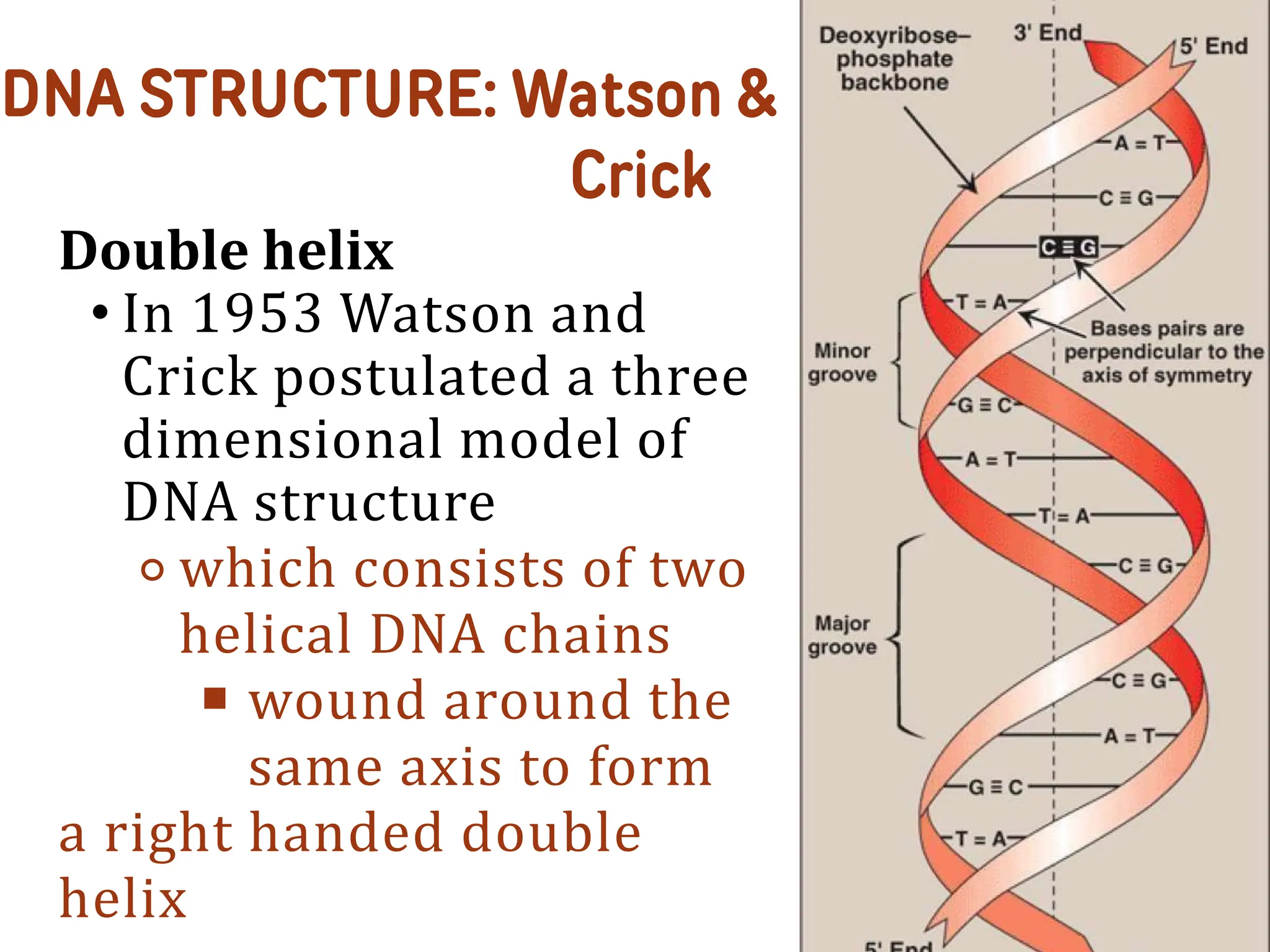
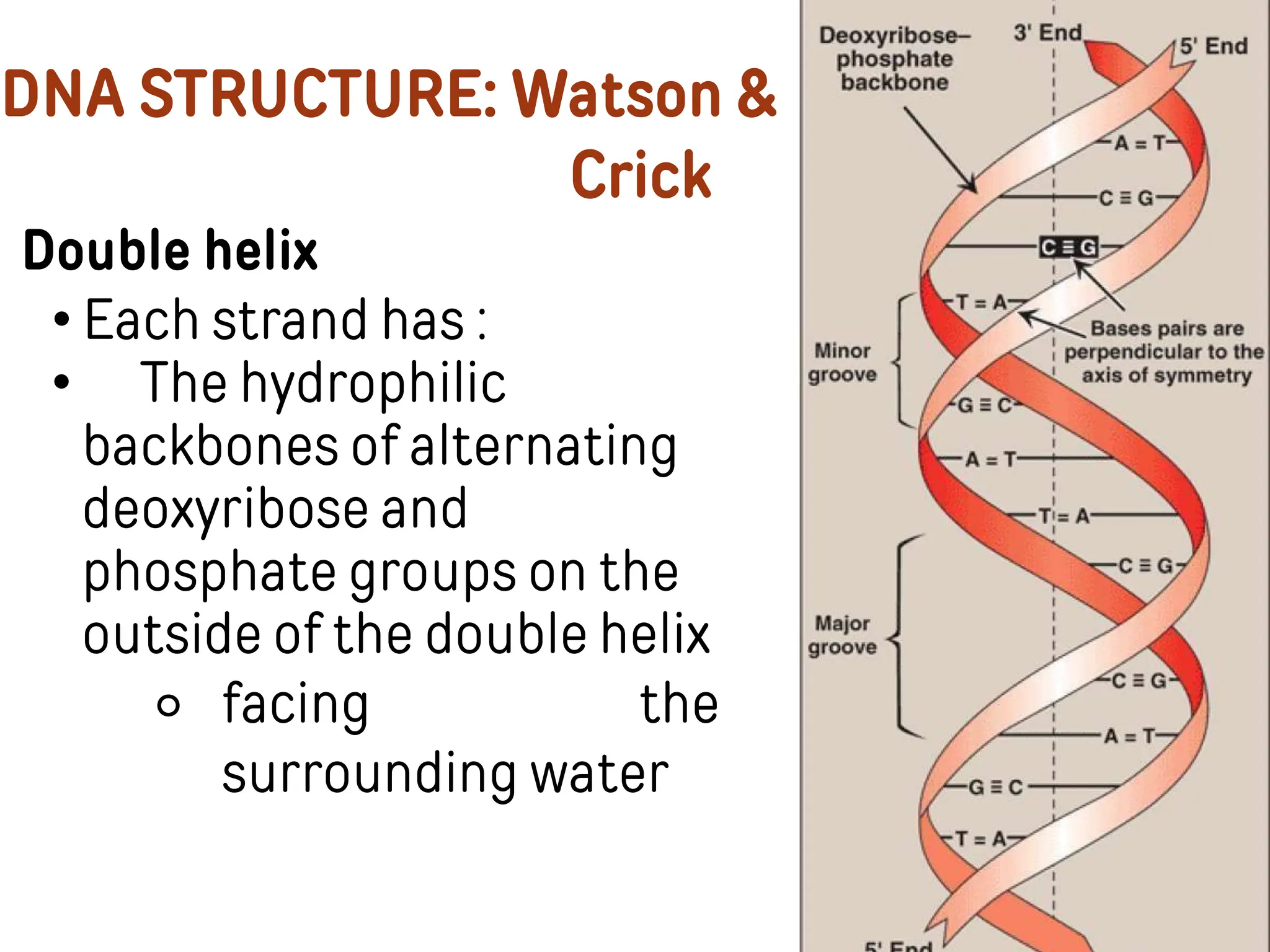
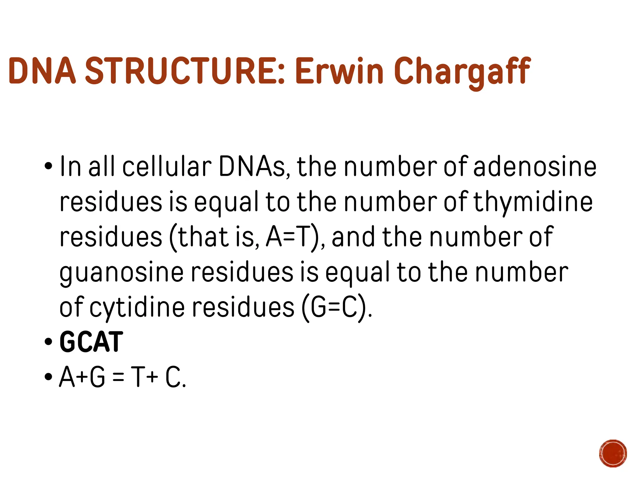
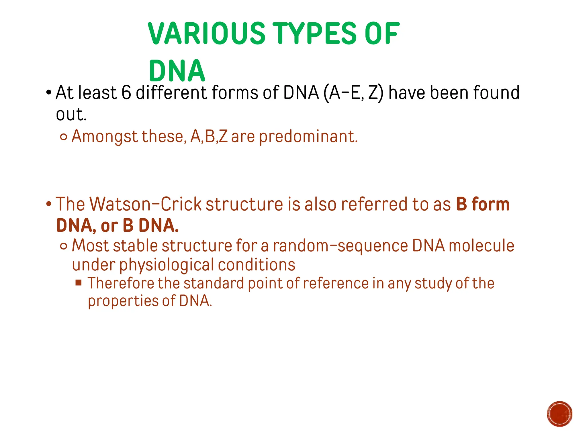
Past Inquiries
1.Why do children sometimes
resemble their parents?
Past Inquiries
2.What process do people use to
test if they're the biological
parents?
Dad + Mom = ?
Based on the pictures of
the parents, describe what
their child will look like.
DNA
STRUCTURE
LESSON OBJECTIVES
1.Conduct DNA denaturation using
tomatoes; and
2.Understand the structure of DNA.
DNA
Denaturation
Conduct an experiment
entitled DNA denaturation by
following the procedure
shown in the video.
• History
• Introduction
• Complementary Base
pairing
TIMELINE
Friedrich Miescher : identified the “nuclein” by isolating a
molecule from a cell nucleus that would later become known as DNA
Albrecht Kossel, who is credited with naming DNA, identified nuclein as a nucleic acid. He also
isolated those five nitrogen bases : adenine (A), cytosine (C), guanine (G), thymine
(T) and uracil (U)
Avery, McCleod & McCarty- Transforming principle is DNA. i.e carrier of genetic
information.
Erwin Chargaff :Chargaff ’s Rules, proved that guanine = cytosine , as well as adenine= thymine .
Sum of Purine is equal to sum of Pyrimidine
Roslind Franklin’s work in X-ray crystallography, started taking X-ray diffraction photographs
of DNA. Images showed the helical form, which was confirmed by Watson and Crick nearly
two years later.
J Watson and F Crick :published on DNA’s double helix structure that twists to form
the ladder-like structure
1869
1881
1944
1950
1951
1953
• DNA is a polymer of
deoxyribonucleoside
monophosphates covalently
linked by 3′→5′– phosphodiester
bonds
• Found in chromosomes,
mitochondria and
chloroplasts
DNA: Deoxyribonucleic
Acid
• Genetic Information (Genetic Blue
Print):
DNA contains the genetic instructions
necessary for the development, growth,
functioning, and reproduction of all
known living organisms.
SAMPLE FUNCTIONS OF DNA
2. Protein Synthesis:
DNA provides the instructions for
protein synthesis through a process
called gene expression. Genes,
segments of DNA, are transcribed into
RNA, which is then translated into
proteins.
SAMPLE FUNCTIONS OF DNA
The Primary
structure of
DNA is
Sequence
The secondary structure of DNA is the double
helix
• DNA double helical structure coils round Histones.
• DNA bound to histones forms NUCLEOSOMES
(10nm FIBRES)
• Nucleosomes contain 146 nucleotides
DNA STRUCTURE: Watson &
Crick
Double helix
• In 1953 Watson and
Crick postulated a three
dimensional model of
DNA structure
⚬ which consists of two
helical DNA chains
■ wound around the
same axis to form
a right handed double
helix
Double helix
• Each strand has :
• The hydrophilic
backbones of alternating
deoxyribose and
phosphate groups on the
outside of the double helix
⚬ facing the
surrounding water
DNA STRUCTURE: Watson &
Crick
DNA STRUCTURE: Erwin Chargaff
• Most important clue to the structure of DNA
came from the work of Erwin Chargaff and
his colleagues in the late 1940s.
• The base composition of DNA generally
varies from one species to another.
DNA STRUCTURE: Erwin Chargaff
• In all cellular DNAs, the number of adenosine
residues is equal to the number of thymidine
residues (that is, A=T), and the number of
guanosine residues is equal to the number
of cytidine residues (G=C).
• GCAT
• A+G = T+ C.
BASE PAIRING
• Purines -
adenine (A)
and guanine (G)
• Pyrimidines -
cytosine
(C) and thymine (T)
• The two strands
are
complementary
• DNA double helix is
held
together by 2 forces
• H-bonding between
complementary base
pairs
• van
der
interactions
stacked
bases
Waals
between
Major groove :
• Sequence-specific interactions
⚬ provide access for the binding of regulatory proteins
■ Maintained by hydrogen bonds, ionic interactions and van der Waals
forces.
Minor groove
• Offers the highest sequence specificity binding between a small molecule and DNA
⚬ To disrupt the specific gene expression
■ Certain anticancer drugs, such adactinomycin (actinomycin D), exert their
cytotoxic effect by intercalating into the narrow groove of the DNA double
helix, thus interfering with DNA synthesis
• Compared to minor
groove
binding,
interactions between small molecules and
the DNA major groove have not been
extensively explored.
DIFFERENT 3-DIMENSIONAL FORMS OF
DNA
• DNA is a remarkably flexible.
⚬ Considerable rotation is possible around several types of bonds
⚬ Thermal fluctuation can produce bending, stretching and unpairing
(melting) of the strands.
The structural variations reflect 3 things:
⚬ Different possible conformations of deoxyribose
⚬ Rotation about the contiguous bonds that make up
the phospho- deoxyribose backbone
⚬ Free rotation about C-1’-N glycosyl bond
VARIOUS TYPES OF
DNA
• At least 6 different forms of DNA (A-E, Z) have been found
out.
⚬ Amongst these, A,B,Z are predominant.
• The Watson-Crick structure is also referred to as B form
DNA, or B DNA.
⚬ Most stable structure for a random-sequence DNA molecule
under physiological conditions
■ Therefore the standard point of reference in any study of the
properties of DNA.
A-
DNA
• X-ray diffraction studies of less-hydrated DNA fibers revealed a A
form DNA
• When the relative humidity of B-form DNA falls to less than
75%,
o the B-form undergoes a reversible transition into the A-form of DNA
• It is also a right-handed helix
⚬ Helix is wider ,
⚬ Number of base pairs per helical turn is 11
⚬ The plane of the base pairs is tilted about 20° with respect to the
helix axis.
• In A-DNA, C-3’ lies out of the plane (a conformation referred
to as C-3’endo) formed by the
other four atoms of the ring
Z-
DNA
• Z-DNA is a left handed helix containing 12 base pairs per turn.
• To form the left-handed helix in Z-DNA
o the purine residues flip to the synconformation, alternating with pyrimidines in
the anti conformation.
owhenthe sequence of
nucleotides purine/pyrimidine
stretches- form, Z-DNA
• is also favored at high ionic concentrations
consists of
alternating
• DNA with a zigzag configuration along the sugar phosphate
backbone.
o hence named Z-DNA
• The major groove is barely apparent in Z-DNA, and the minor groove
is narrow and deep.
• The Z-DNA tracts may play a role (as yet undefined) in regulating
the
expression of some genes or in genetic
recombination.
• Blue: sugar phosphate
backbone
• Yellow: for pyrimidines
(thymine and cytosine)
• brown for
purines (adenine and
guanine)
COMPARISON OF DIFFERENT FORMS OF DNA
Ref: Lehninger Principles of Biochemistry
• Observed at some conditions such as
relatively low humidity and the presence of
certain ions, such as Li+ or Mg2+
• Not very stable and Not very common.
• Right handed helice
• Has 9 base pairs per turn of spiral
• Has diameter of 19A°,smaller than that
of A-&B- DNA.
• The tilt of base is 7.8°
• Extremely rare variant with only 8base pairs per helical
turn .
• This forms of DNA found in some DNA molecules devoid
of guanine.
• Axial rise of 3.03A°per base pairs .
• Tilt of 16.7° from axis of helix
• E-DNA - extended &eccentric double helix
⚬ Cytosine methylation of or bromination of DNA
sequence d(GGCGCC)₂
• E-DNA has a long helical axis rise and base
perpendicular to the helical axis.
• Deep major groove and shallow minor groove.
• E-DNA allowed to crystallize for a period time
longer, the methylated sequence forms
standard A-DNA.
• E-DNA is the intermediate in the transition to A-
DNA.
• E-DNA is the intermediate in the
crystallographic pathway from B-DNA to A-DNA
• Mitochondria also have a small amount of their own DNA.
⚬ mitochondrial DNA
• In humans, mitochondrial DNA spans about 16,569 DNA
building blocks (base pairs)
• Mitochondrial DNA contains 37 genes,
⚬ Thirteen of these genes
■ Provide instructions for making enzymes involved in oxidative
phosphorylation.
⚬ The remaining genes
■ Provide instructions for making molecules called transfer RNA
(tRNA) and ribosomal RNA (rRNA), which are chemical cousins of
DNA.
Mitochondrial DNA Nuclear DNA
Location Mitochondria Cell Nucleus
Copies per somatic cell 100-1,000 2
Structure Circular and closed Linear and open ended
Membrane enclosure
Not enveloped by a
membrane
Enclosed by a nuclear
membrane
Genome size
1 chromosome with 16,569
base pairs
46 chromosomes with 3.3
billion base pairs
Number of genes 37 genes 20,000-25,000 genes
Method of inheritance Maternal Maternal and Paternal
Method of translation
Some codons do not follow
universal codon pattern
Follows universal codon
pattern
Method of transcription Polycistronic Monocistronic
SATELLITE DNA
• Originally identified as a subfraction of DNA with a
buoyant density slightly lower than that of genomic DNA
⚬ because of its higher content of AT base pairs.
• Consists of clusters of short, species-specific, nearly
identical sequences that are tandemly repeated hundreds
of thousands of times.
• These clusters lack protein-coding genes
⚬ Found principally near the centromeres of chromosomes.
• Two types
⚬ Microsatellite sequences
■ 1-6 bp repeat units flanked by conserved sequences
⚬ Minisatellite sequences
■ 11-60 bp flanked by conserved restriction sites.
• Used for DNA matching or finger printing as first found
out by Jeffreys et al (1985).
• Viral DNA (and in some, RNA)
⚬ Some circular, some linear
⚬ Some double stranded, some single stranded
⚬ Very small amount, packed very tightly
⚬ Small size is an advantage
⚬ Viruses use host cell enzymes, need few genes
• Bacterial DNA
⚬ Usually single copy of double stranded
⚬ Usually circular
• Eukaryotic DNA
⚬ Linear, in several pieces
UNUSUAL SEQUENCES
/ STRUCTURES OF DNA
• Important for molecular recognition of DNA
by proteins and enzymes
• Bent DNA:
• Bends occur in the DNA helix
wherever four or more adenosine residues appear sequentially
in one strand.
• Cause a collapse of the helix into the
minor groove.
• Six adenosines in a row produce a bend of about 18°.
• Drugs, photochemical damage, mispairing of bases can also
produce
• DNA duplex possesses areas where sequence of nucleotides is the
same but opposite in the two strands.
• These sequences are recognized by restriction endonucleases and
are used in genetic engineering.
• It is similar to palindrome words having same words in both
forward and backward direction, e.g., NITIN, MALAYALAM
• When the inverted repeat occurs within each individual strand
of the DNA, the sequence is called a mirror repeat.
• Palindromic DNA sequences can form alternative
structures with intrastrand base pairing.
⚬ (a) When only a single DNA (or RNA) strand is involved, the
structure is called a hairpin.
⚬ (b) When both strands of a duplex DNA are involved, it is called a
cruciform.
TRIPLEX DNA/
TRIPLE
STRANDED DNA
• Due to additional H-bond between bases particularly
with functional groups arrayed in the major groove.
• The bonds are called as Hoogsteen H-bonds
• The non-Watson-Crick pairing is called Hoogsteen
pairing, after Karst Hoogsteen, who in 1963 first
recognized the potential for these unusual pairings
• Triple helical structure is less stable than double
helix - increased electrostatic repulsion.
G-TETRAPLEX / FOUR STRANDED
DNA
• High content of Guanine – form tetrameric structure called G-quartets.
• These structures are planar & are connected by Hoogsteen hydrogen
bonds.
• Eukaryotic chromosomes - Telomeres are rich in guanine - forms G-
tetraplexes.
Ref: Lehninger Principles of Biochemistry
• Type of deoxyribonucleic acid, discovered in cell
nuclei in 2018,
⚬ that is found in the nuclei of human cells.
• I-motifs are four-stranded quadruplex
structures formed by cytosine-rich DNA.
⚬ C-rich DNA regions are common in gene regulation
portions of the genome.
(Zeraati et al., Nat Chem, 2018)
• Postulated to play a role in gene regulation and
expression in the cell
• I-motifs have potential applications in nano-
technology
and nano-medicine,
⚬ because size is more than 1nm and less than 100nm due to
their unique pH sensitivity
• Have been used as biosensors, nanomachines, and
molecular switches.
Nucleic Acids Research, Volume 30, Issue 21, 1 November 2002, Pages 4618–4625, https://doi.org/10.1093/nar/gkf597
The content of this slide may be subject to copyright: please see the slide notes for details.
FIGURE 6. SOME POSSIBLE MODELS OF DUPLEX, I‐MOTIF AND G‐QUADRUPLEX
ASSOCIATION FOR THE HUMAN TELOMERE ...
NUCLEIC ACID
CHEMISTRY
• Thechemical transformations that do occur are
generally very low in the absence of an enzyme catalyst.
• Understanding the nucleic acid chemistry gives us:
⚬ Powerful array of technologies that have applications in molecular
biology, medicine & forensic medicine.
• DENATURATION
• Disruption of the hydrogen bonds between paired bases
and of base stacking
⚬ causes unwinding of the double helix to
form two single
strands,
■ completely separate from each other along the entire length
or part of length
DENATURATION
• Causes:
⚬ Extreme pH,
⚬ Temperature above 80 ̊C,
⚬ Chemicals such as formamide and urea
• Hyperchromic effect
⚬ Denaturation of a double stranded nucleic acid produces an increase
in absorption of UV light
■ Relative to that of a solution with the same concentration of paired
complementary nucleic acid strands
⚬ The transition from double stranded DNA to the single stranded
denatured form can be detected by monitoring UV absorption at
260nm
DENATURATION
43
Each species of DNA has a characteristic
denaturation temperature or melting poin
Melting temperatures (Tm) of DNA
molecules with different nucleotide
compositions
ANNEALIN
G
• Process by whichseparated complementary
strands form a double helix
• Occurs when the temperature and pH is returned to the
range in which most organisms live
• Rapid one step process
⚬ If double helical segment of a dozen or more residues still
unites the two strands
ANNEALING
• If the two strands are completely separated, renaturation
occurs in two steps
• First step:
⚬ Slow, the two strands find each other by random collisons
■ Form a short segment of complementary double helix
• Second step:
⚬ Faster, the remaining unpaired bases successively
come into register as base pair
■ The two strands unite to form double helix
Fig :Reversible denaturation and
annealing (renaturation) of DNA.
Ref: Lehninger Principles of Biochemistry
ORGANIZATION OF DNA
• A segment of DNA molecule that contains the
information required for the synthesis of a functional
biological product (RNA or protein) is called gene
• A cell has many thousands of genes and so the DNA
molecules are very large
• Large DNA molecules must be packaged in such a way
that they can fit inside the cell and still be functional
ORGANIZATION OF DNA
• Nuclear DNA in eukaryotes is found in chromatin associated with
histones and non histone proteins
ORGANIZATION OF DNA
• DNA (negatively charged) loops twice around the histone octamer to
form nucleosomes (series of nucleosomes: beads on a string)
• Histone H1 is associated with the linker DNA
found between nucleosomes to help package them into a
solenoid like structure
• Further condensation occurs to eventually form the chromosome.
ORGANIZATION OF DNA
Polynucleosomes(nucleofilaments)
REFERENCE
S
• Lehninger Principle of Biochemistry 4th edition
• Robert k. Murray, D.K.Granner ,P.A.Mayes & Victor W.Rodwell Harpers
illustrated biochemistry 26th edition
• Lubert Stryer, Jeremy M. Berg , John L. Tymoczko Biochemistry, 4th
edition
• Lippincott’s Illustrated Reviews: Biochemistry, 4th Edition
• https://www.lunadna.com/blog/history-of-dna/
• http://www.biologydiscussion.com/dna/dna-types-structure-and- function-
of-dna/1301
• http://www.differencebetween.net/science/difference-between-
mitochondrial-dna-and-nuclear-dna/
• https://bio.libretexts.org/Bookshelves/Genetics/Book%3A_Working_with
_Molecular_Genetics_(Hardison)/Unit_I%3A_Genes%2C_Nucleic_Acids
%2C_Genomes_and_Chromosomes/2%3A_Structures_of_Nucleic_Acids/
2.5%3A_B-Form%2C_A-Form%2C_and_Z-Form_of_DNA
• https://www.ncbi.nlm.nih.gov/pubmed/10966645
• https://academic.oup.com/nar/article/30/21/4618/1105212
DNA STRUCTURE AND INTRODUCTION TO THE REPLICATION.pptx

DNA STRUCTURE AND INTRODUCTION TO THE REPLICATION.pptx

  • 1.
    Past Inquiries 1.Why dochildren sometimes resemble their parents?
  • 2.
    Past Inquiries 2.What processdo people use to test if they're the biological parents?
  • 3.
  • 4.
    Based on thepictures of the parents, describe what their child will look like.
  • 9.
  • 10.
    LESSON OBJECTIVES 1.Conduct DNAdenaturation using tomatoes; and 2.Understand the structure of DNA.
  • 11.
  • 12.
    Conduct an experiment entitledDNA denaturation by following the procedure shown in the video.
  • 13.
    • History • Introduction •Complementary Base pairing
  • 14.
    TIMELINE Friedrich Miescher :identified the “nuclein” by isolating a molecule from a cell nucleus that would later become known as DNA Albrecht Kossel, who is credited with naming DNA, identified nuclein as a nucleic acid. He also isolated those five nitrogen bases : adenine (A), cytosine (C), guanine (G), thymine (T) and uracil (U) Avery, McCleod & McCarty- Transforming principle is DNA. i.e carrier of genetic information. Erwin Chargaff :Chargaff ’s Rules, proved that guanine = cytosine , as well as adenine= thymine . Sum of Purine is equal to sum of Pyrimidine Roslind Franklin’s work in X-ray crystallography, started taking X-ray diffraction photographs of DNA. Images showed the helical form, which was confirmed by Watson and Crick nearly two years later. J Watson and F Crick :published on DNA’s double helix structure that twists to form the ladder-like structure 1869 1881 1944 1950 1951 1953
  • 15.
    • DNA isa polymer of deoxyribonucleoside monophosphates covalently linked by 3′→5′– phosphodiester bonds • Found in chromosomes, mitochondria and chloroplasts DNA: Deoxyribonucleic Acid
  • 16.
    • Genetic Information(Genetic Blue Print): DNA contains the genetic instructions necessary for the development, growth, functioning, and reproduction of all known living organisms. SAMPLE FUNCTIONS OF DNA
  • 17.
    2. Protein Synthesis: DNAprovides the instructions for protein synthesis through a process called gene expression. Genes, segments of DNA, are transcribed into RNA, which is then translated into proteins. SAMPLE FUNCTIONS OF DNA
  • 18.
  • 19.
    The secondary structureof DNA is the double helix
  • 20.
    • DNA doublehelical structure coils round Histones. • DNA bound to histones forms NUCLEOSOMES (10nm FIBRES) • Nucleosomes contain 146 nucleotides
  • 22.
    DNA STRUCTURE: Watson& Crick Double helix • In 1953 Watson and Crick postulated a three dimensional model of DNA structure ⚬ which consists of two helical DNA chains ■ wound around the same axis to form a right handed double helix
  • 23.
    Double helix • Eachstrand has : • The hydrophilic backbones of alternating deoxyribose and phosphate groups on the outside of the double helix ⚬ facing the surrounding water DNA STRUCTURE: Watson & Crick
  • 24.
    DNA STRUCTURE: ErwinChargaff • Most important clue to the structure of DNA came from the work of Erwin Chargaff and his colleagues in the late 1940s. • The base composition of DNA generally varies from one species to another.
  • 25.
    DNA STRUCTURE: ErwinChargaff • In all cellular DNAs, the number of adenosine residues is equal to the number of thymidine residues (that is, A=T), and the number of guanosine residues is equal to the number of cytidine residues (G=C). • GCAT • A+G = T+ C.
  • 26.
    BASE PAIRING • Purines- adenine (A) and guanine (G) • Pyrimidines - cytosine (C) and thymine (T) • The two strands are complementary • DNA double helix is held together by 2 forces • H-bonding between complementary base pairs • van der interactions stacked bases Waals between
  • 27.
    Major groove : •Sequence-specific interactions ⚬ provide access for the binding of regulatory proteins ■ Maintained by hydrogen bonds, ionic interactions and van der Waals forces. Minor groove • Offers the highest sequence specificity binding between a small molecule and DNA ⚬ To disrupt the specific gene expression ■ Certain anticancer drugs, such adactinomycin (actinomycin D), exert their cytotoxic effect by intercalating into the narrow groove of the DNA double helix, thus interfering with DNA synthesis • Compared to minor groove binding, interactions between small molecules and the DNA major groove have not been extensively explored.
  • 28.
    DIFFERENT 3-DIMENSIONAL FORMSOF DNA • DNA is a remarkably flexible. ⚬ Considerable rotation is possible around several types of bonds ⚬ Thermal fluctuation can produce bending, stretching and unpairing (melting) of the strands. The structural variations reflect 3 things: ⚬ Different possible conformations of deoxyribose ⚬ Rotation about the contiguous bonds that make up the phospho- deoxyribose backbone ⚬ Free rotation about C-1’-N glycosyl bond
  • 29.
    VARIOUS TYPES OF DNA •At least 6 different forms of DNA (A-E, Z) have been found out. ⚬ Amongst these, A,B,Z are predominant. • The Watson-Crick structure is also referred to as B form DNA, or B DNA. ⚬ Most stable structure for a random-sequence DNA molecule under physiological conditions ■ Therefore the standard point of reference in any study of the properties of DNA.
  • 30.
    A- DNA • X-ray diffractionstudies of less-hydrated DNA fibers revealed a A form DNA • When the relative humidity of B-form DNA falls to less than 75%, o the B-form undergoes a reversible transition into the A-form of DNA • It is also a right-handed helix ⚬ Helix is wider , ⚬ Number of base pairs per helical turn is 11 ⚬ The plane of the base pairs is tilted about 20° with respect to the helix axis. • In A-DNA, C-3’ lies out of the plane (a conformation referred to as C-3’endo) formed by the other four atoms of the ring
  • 31.
    Z- DNA • Z-DNA isa left handed helix containing 12 base pairs per turn. • To form the left-handed helix in Z-DNA o the purine residues flip to the synconformation, alternating with pyrimidines in the anti conformation. owhenthe sequence of nucleotides purine/pyrimidine stretches- form, Z-DNA • is also favored at high ionic concentrations consists of alternating • DNA with a zigzag configuration along the sugar phosphate backbone. o hence named Z-DNA • The major groove is barely apparent in Z-DNA, and the minor groove is narrow and deep. • The Z-DNA tracts may play a role (as yet undefined) in regulating the expression of some genes or in genetic recombination.
  • 32.
    • Blue: sugarphosphate backbone • Yellow: for pyrimidines (thymine and cytosine) • brown for purines (adenine and guanine)
  • 33.
    COMPARISON OF DIFFERENTFORMS OF DNA Ref: Lehninger Principles of Biochemistry
  • 35.
    • Observed atsome conditions such as relatively low humidity and the presence of certain ions, such as Li+ or Mg2+ • Not very stable and Not very common. • Right handed helice • Has 9 base pairs per turn of spiral • Has diameter of 19A°,smaller than that of A-&B- DNA. • The tilt of base is 7.8°
  • 36.
    • Extremely rarevariant with only 8base pairs per helical turn . • This forms of DNA found in some DNA molecules devoid of guanine. • Axial rise of 3.03A°per base pairs . • Tilt of 16.7° from axis of helix
  • 37.
    • E-DNA -extended &eccentric double helix ⚬ Cytosine methylation of or bromination of DNA sequence d(GGCGCC)₂ • E-DNA has a long helical axis rise and base perpendicular to the helical axis. • Deep major groove and shallow minor groove. • E-DNA allowed to crystallize for a period time longer, the methylated sequence forms standard A-DNA.
  • 38.
    • E-DNA isthe intermediate in the transition to A- DNA. • E-DNA is the intermediate in the crystallographic pathway from B-DNA to A-DNA
  • 40.
    • Mitochondria alsohave a small amount of their own DNA. ⚬ mitochondrial DNA • In humans, mitochondrial DNA spans about 16,569 DNA building blocks (base pairs) • Mitochondrial DNA contains 37 genes, ⚬ Thirteen of these genes ■ Provide instructions for making enzymes involved in oxidative phosphorylation. ⚬ The remaining genes ■ Provide instructions for making molecules called transfer RNA (tRNA) and ribosomal RNA (rRNA), which are chemical cousins of DNA.
  • 41.
    Mitochondrial DNA NuclearDNA Location Mitochondria Cell Nucleus Copies per somatic cell 100-1,000 2 Structure Circular and closed Linear and open ended Membrane enclosure Not enveloped by a membrane Enclosed by a nuclear membrane Genome size 1 chromosome with 16,569 base pairs 46 chromosomes with 3.3 billion base pairs Number of genes 37 genes 20,000-25,000 genes Method of inheritance Maternal Maternal and Paternal Method of translation Some codons do not follow universal codon pattern Follows universal codon pattern Method of transcription Polycistronic Monocistronic
  • 42.
    SATELLITE DNA • Originallyidentified as a subfraction of DNA with a buoyant density slightly lower than that of genomic DNA ⚬ because of its higher content of AT base pairs. • Consists of clusters of short, species-specific, nearly identical sequences that are tandemly repeated hundreds of thousands of times. • These clusters lack protein-coding genes ⚬ Found principally near the centromeres of chromosomes. • Two types ⚬ Microsatellite sequences ■ 1-6 bp repeat units flanked by conserved sequences ⚬ Minisatellite sequences ■ 11-60 bp flanked by conserved restriction sites. • Used for DNA matching or finger printing as first found out by Jeffreys et al (1985).
  • 43.
    • Viral DNA(and in some, RNA) ⚬ Some circular, some linear ⚬ Some double stranded, some single stranded ⚬ Very small amount, packed very tightly ⚬ Small size is an advantage ⚬ Viruses use host cell enzymes, need few genes • Bacterial DNA ⚬ Usually single copy of double stranded ⚬ Usually circular • Eukaryotic DNA ⚬ Linear, in several pieces
  • 44.
    UNUSUAL SEQUENCES / STRUCTURESOF DNA • Important for molecular recognition of DNA by proteins and enzymes • Bent DNA: • Bends occur in the DNA helix wherever four or more adenosine residues appear sequentially in one strand. • Cause a collapse of the helix into the minor groove. • Six adenosines in a row produce a bend of about 18°. • Drugs, photochemical damage, mispairing of bases can also produce
  • 45.
    • DNA duplexpossesses areas where sequence of nucleotides is the same but opposite in the two strands. • These sequences are recognized by restriction endonucleases and are used in genetic engineering. • It is similar to palindrome words having same words in both forward and backward direction, e.g., NITIN, MALAYALAM • When the inverted repeat occurs within each individual strand of the DNA, the sequence is called a mirror repeat.
  • 46.
    • Palindromic DNAsequences can form alternative structures with intrastrand base pairing. ⚬ (a) When only a single DNA (or RNA) strand is involved, the structure is called a hairpin. ⚬ (b) When both strands of a duplex DNA are involved, it is called a cruciform.
  • 47.
    TRIPLEX DNA/ TRIPLE STRANDED DNA •Due to additional H-bond between bases particularly with functional groups arrayed in the major groove. • The bonds are called as Hoogsteen H-bonds • The non-Watson-Crick pairing is called Hoogsteen pairing, after Karst Hoogsteen, who in 1963 first recognized the potential for these unusual pairings • Triple helical structure is less stable than double helix - increased electrostatic repulsion.
  • 48.
    G-TETRAPLEX / FOURSTRANDED DNA • High content of Guanine – form tetrameric structure called G-quartets. • These structures are planar & are connected by Hoogsteen hydrogen bonds. • Eukaryotic chromosomes - Telomeres are rich in guanine - forms G- tetraplexes. Ref: Lehninger Principles of Biochemistry
  • 50.
    • Type ofdeoxyribonucleic acid, discovered in cell nuclei in 2018, ⚬ that is found in the nuclei of human cells. • I-motifs are four-stranded quadruplex structures formed by cytosine-rich DNA. ⚬ C-rich DNA regions are common in gene regulation portions of the genome. (Zeraati et al., Nat Chem, 2018)
  • 51.
    • Postulated toplay a role in gene regulation and expression in the cell • I-motifs have potential applications in nano- technology and nano-medicine, ⚬ because size is more than 1nm and less than 100nm due to their unique pH sensitivity • Have been used as biosensors, nanomachines, and molecular switches.
  • 52.
    Nucleic Acids Research,Volume 30, Issue 21, 1 November 2002, Pages 4618–4625, https://doi.org/10.1093/nar/gkf597 The content of this slide may be subject to copyright: please see the slide notes for details. FIGURE 6. SOME POSSIBLE MODELS OF DUPLEX, I‐MOTIF AND G‐QUADRUPLEX ASSOCIATION FOR THE HUMAN TELOMERE ...
  • 53.
    NUCLEIC ACID CHEMISTRY • Thechemicaltransformations that do occur are generally very low in the absence of an enzyme catalyst. • Understanding the nucleic acid chemistry gives us: ⚬ Powerful array of technologies that have applications in molecular biology, medicine & forensic medicine. • DENATURATION • Disruption of the hydrogen bonds between paired bases and of base stacking ⚬ causes unwinding of the double helix to form two single strands, ■ completely separate from each other along the entire length or part of length
  • 54.
    DENATURATION • Causes: ⚬ ExtremepH, ⚬ Temperature above 80 ̊C, ⚬ Chemicals such as formamide and urea • Hyperchromic effect ⚬ Denaturation of a double stranded nucleic acid produces an increase in absorption of UV light ■ Relative to that of a solution with the same concentration of paired complementary nucleic acid strands ⚬ The transition from double stranded DNA to the single stranded denatured form can be detected by monitoring UV absorption at 260nm
  • 55.
    DENATURATION 43 Each species ofDNA has a characteristic denaturation temperature or melting poin Melting temperatures (Tm) of DNA molecules with different nucleotide compositions
  • 56.
    ANNEALIN G • Process bywhichseparated complementary strands form a double helix • Occurs when the temperature and pH is returned to the range in which most organisms live • Rapid one step process ⚬ If double helical segment of a dozen or more residues still unites the two strands
  • 57.
    ANNEALING • If thetwo strands are completely separated, renaturation occurs in two steps • First step: ⚬ Slow, the two strands find each other by random collisons ■ Form a short segment of complementary double helix • Second step: ⚬ Faster, the remaining unpaired bases successively come into register as base pair ■ The two strands unite to form double helix
  • 58.
    Fig :Reversible denaturationand annealing (renaturation) of DNA. Ref: Lehninger Principles of Biochemistry
  • 59.
    ORGANIZATION OF DNA •A segment of DNA molecule that contains the information required for the synthesis of a functional biological product (RNA or protein) is called gene • A cell has many thousands of genes and so the DNA molecules are very large • Large DNA molecules must be packaged in such a way that they can fit inside the cell and still be functional
  • 60.
    ORGANIZATION OF DNA •Nuclear DNA in eukaryotes is found in chromatin associated with histones and non histone proteins
  • 61.
    ORGANIZATION OF DNA •DNA (negatively charged) loops twice around the histone octamer to form nucleosomes (series of nucleosomes: beads on a string) • Histone H1 is associated with the linker DNA found between nucleosomes to help package them into a solenoid like structure • Further condensation occurs to eventually form the chromosome.
  • 62.
  • 63.
    REFERENCE S • Lehninger Principleof Biochemistry 4th edition • Robert k. Murray, D.K.Granner ,P.A.Mayes & Victor W.Rodwell Harpers illustrated biochemistry 26th edition • Lubert Stryer, Jeremy M. Berg , John L. Tymoczko Biochemistry, 4th edition • Lippincott’s Illustrated Reviews: Biochemistry, 4th Edition • https://www.lunadna.com/blog/history-of-dna/ • http://www.biologydiscussion.com/dna/dna-types-structure-and- function- of-dna/1301 • http://www.differencebetween.net/science/difference-between- mitochondrial-dna-and-nuclear-dna/ • https://bio.libretexts.org/Bookshelves/Genetics/Book%3A_Working_with _Molecular_Genetics_(Hardison)/Unit_I%3A_Genes%2C_Nucleic_Acids %2C_Genomes_and_Chromosomes/2%3A_Structures_of_Nucleic_Acids/ 2.5%3A_B-Form%2C_A-Form%2C_and_Z-Form_of_DNA • https://www.ncbi.nlm.nih.gov/pubmed/10966645 • https://academic.oup.com/nar/article/30/21/4618/1105212