Download to read offline


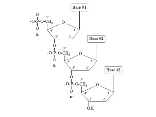
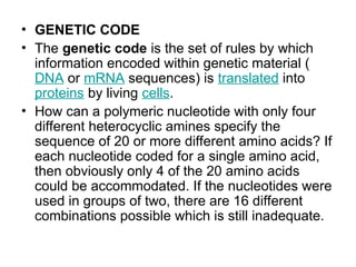
![Deoxyribose versus Ribose sugars
• Ribose is a single-ring pentose [5-C]
sugar. The numbering of the carbon atoms
runs clockwise, following organic
chemistry rules. Note the absence of the
hydroxyl (-OH) group on the 2’ carbon in
the deoxy-ribose sugar in DNA as
compared with the ribose sugar in RNA.](https://image.slidesharecdn.com/nitrogenbases-241107184450-59a6ed41/85/Nitrogenous-bases-and-genetic-code-types-7-320.jpg)


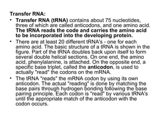


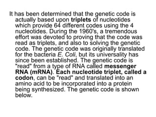
![Standard genetic code
1st
base
2nd base 3rd
base
U C A G
U
UUU (Phe/F)
Phenylalanine
UCU
(Ser/S) Serine
UAU
(Tyr/Y) Tyrosine
UGU
(Cys/C) Cysteine
U
UUC UCC UAC UGC C
UUA
(Leu/L)
Leucine
UCA UAA Stop (Ochre) UGA Stop (Opal) A
UUG UCG UAG Stop (Amber) UGG
(Trp/W)
Tryptophan
G
C
CUU CCU
(Pro/P) Proline
CAU
(His/H) Histidine
CGU
(Arg/R) Arginine
U
CUC CCC CAC CGC C
CUA CCA CAA
(Gln/Q) Glutamine
CGA A
CUG CCG CAG CGG G
A
AUU
(Ile/I)
Isoleucine
ACU
(Thr/T)
Threonine
AAU
(Asn/N) Asparagine
AGU
(Ser/S) Serine
U
AUC ACC AAC AGC C
AUA ACA AAA
(Lys/K) Lysine
AGA
(Arg/R) Arginine
A
AUG[A
]
(Met/M)
Methionine
ACG AAG AGG G
G
GUU
(Val/V)
Valine
GCU
(Ala/A) Alanine
GAU (Asp/D) Aspartic
acid
GGU
(Gly/G) Glycine
U
GUC GCC GAC GGC C
GUA GCA GAA (Glu/E) Glutamic
acid
GGA A
GUG GCG GAG GGG G](https://image.slidesharecdn.com/nitrogenbases-241107184450-59a6ed41/85/Nitrogenous-bases-and-genetic-code-types-25-320.jpg)

The document discusses molecular biology, emphasizing the roles of nitrogen bases in genetic coding, specifically purines and pyrimidines. It outlines the structure and function of nucleic acids (DNA and RNA), detailing components like nucleosides, nucleotides, and the specific types of RNA involved in protein synthesis. Additionally, it explains how the genetic code is based on nucleotide triplets that correspond to amino acids, enabling the translation of genetic information into proteins.