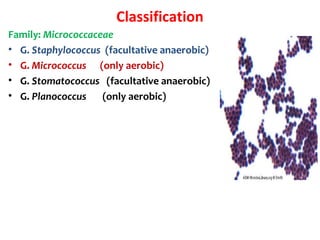
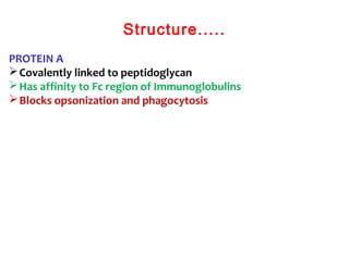
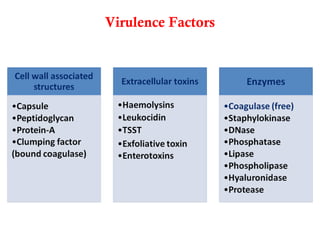
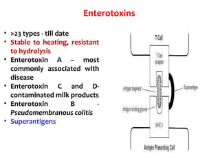
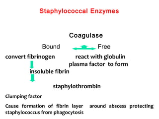
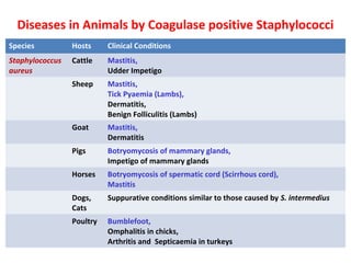
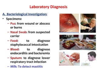
This document provides information on the genus Staphylococcus. It discusses the morphology, classification, virulence factors, and diseases caused by Staphylococcus species. Some key points include:
- Staphylococcus is a genus of gram-positive bacteria that forms grapelike clusters and includes major human pathogens.
- Important virulence factors include toxins like alpha toxin, enterotoxins, and exfoliative toxins. Enzymes and surface proteins also contribute to pathogenesis.
- Major diseases caused by coagulase-positive Staphylococcus include mastitis, tick pyemia, exudative dermatitis, and botryomycosis in various animal
The genus Shigella exclusively infects human intestine.
Shigella dysenteriae is the causative agent of bacillary dysentery or shigellosis in humans.
It is a diarrheal illness which is characterized by frequent passage of blood stained mucopurulent stools.
The four important species of the genus Shigella are:
Shigella dysenteriae
Shigella flexneri
Shigella sonnei
Shigella boydii.
Clostridium are anerobic gram positive rod shaped spore forming organisms responsible to cause various life threatening diseases in humans like Gas gangrene, Tetanus, Botulism, etc
Staphylococcus aureus is a Gram-positive, round-shaped bacterium, a member of the Firmicutes, and is a usual member of the microbiota of the body, frequently found in the upper respiratory tract and on the skin. It is the leading cause of skin and soft tissue infections such as abscesses (boils), furuncles, and cellulitis. Although most staph infections are not serious, S. aureus can cause serious infections such as bloodstream infections, pneumonia, or bone and joint infections.
Gram-positive cocci include Staphylococcus (catalase-positive), which grows clusters, and Streptococcus (catalase-negative), which grows in chains. The staphylococci further subdivide into coagulase-positive (S. aureus) and coagulase-negative (S. epidermidis and S. saprophyticus) species. Streptococcus bacteria subdivide into Strep. pyogenes (Group A), Strep. agalactiae (Group B), enterococci (Group D), Strep viridans, and Strep pneumonia.
Gram-positive bacilli (rods) subdivide according to their ability to produce spores. Bacillus and Clostridia are spore-forming rods while Listeria and Corynebacterium are not. Spore-forming rods that produce spores can survive in environments for many years. Also, the branching filament rods encompass Nocardia and actinomyces.
Gram-positive organisms have a thicker peptidoglycan cell wall compared with gram-negative bacteria. It is a 20 to 80 nm thick polymer while the peptidoglycan layer of the gram-negative cell wall is 2 to 3 nm thick and covered with an outer lipid bilayer membrane.
Bloodstream infection mortality rates have increased by 78% in just two decades[1]. Gram-positive organisms have highly variable growth and resistance patterns. The SCOPE project (Surveillance and Control of Pathogens of Epidemiologic Importance) found that gram-positive organisms in those with an underlying malignancy accounted for 62% of all bloodstream infections in 1995 and 76% in 2000 while gram-negative organisms accounted for 22% and 14% of infections for these years.[2]
New Directions in Targeted Therapeutic Approaches for Older Adults With Mantl...i3 Health
i3 Health is pleased to make the speaker slides from this activity available for use as a non-accredited self-study or teaching resource.
This slide deck presented by Dr. Kami Maddocks, Professor-Clinical in the Division of Hematology and
Associate Division Director for Ambulatory Operations
The Ohio State University Comprehensive Cancer Center, will provide insight into new directions in targeted therapeutic approaches for older adults with mantle cell lymphoma.
STATEMENT OF NEED
Mantle cell lymphoma (MCL) is a rare, aggressive B-cell non-Hodgkin lymphoma (NHL) accounting for 5% to 7% of all lymphomas. Its prognosis ranges from indolent disease that does not require treatment for years to very aggressive disease, which is associated with poor survival (Silkenstedt et al, 2021). Typically, MCL is diagnosed at advanced stage and in older patients who cannot tolerate intensive therapy (NCCN, 2022). Although recent advances have slightly increased remission rates, recurrence and relapse remain very common, leading to a median overall survival between 3 and 6 years (LLS, 2021). Though there are several effective options, progress is still needed towards establishing an accepted frontline approach for MCL (Castellino et al, 2022). Treatment selection and management of MCL are complicated by the heterogeneity of prognosis, advanced age and comorbidities of patients, and lack of an established standard approach for treatment, making it vital that clinicians be familiar with the latest research and advances in this area. In this activity chaired by Michael Wang, MD, Professor in the Department of Lymphoma & Myeloma at MD Anderson Cancer Center, expert faculty will discuss prognostic factors informing treatment, the promising results of recent trials in new therapeutic approaches, and the implications of treatment resistance in therapeutic selection for MCL.
Target Audience
Hematology/oncology fellows, attending faculty, and other health care professionals involved in the treatment of patients with mantle cell lymphoma (MCL).
Learning Objectives
1.) Identify clinical and biological prognostic factors that can guide treatment decision making for older adults with MCL
2.) Evaluate emerging data on targeted therapeutic approaches for treatment-naive and relapsed/refractory MCL and their applicability to older adults
3.) Assess mechanisms of resistance to targeted therapies for MCL and their implications for treatment selection
The prostate is an exocrine gland of the male mammalian reproductive system
It is a walnut-sized gland that forms part of the male reproductive system and is located in front of the rectum and just below the urinary bladder
Function is to store and secrete a clear, slightly alkaline fluid that constitutes 10-30% of the volume of the seminal fluid that along with the spermatozoa, constitutes semen
A healthy human prostate measures (4cm-vertical, by 3cm-horizontal, 2cm ant-post ).
It surrounds the urethra just below the urinary bladder. It has anterior, median, posterior and two lateral lobes
It’s work is regulated by androgens which are responsible for male sex characteristics
Generalised disease of the prostate due to hormonal derangement which leads to non malignant enlargement of the gland (increase in the number of epithelial cells and stromal tissue)to cause compression of the urethra leading to symptoms (LUTS
These lecture slides, by Dr Sidra Arshad, offer a quick overview of physiological basis of a normal electrocardiogram.
Learning objectives:
1. Define an electrocardiogram (ECG) and electrocardiography
2. Describe how dipoles generated by the heart produce the waveforms of the ECG
3. Describe the components of a normal electrocardiogram of a typical bipolar leads (limb II)
4. Differentiate between intervals and segments
5. Enlist some common indications for obtaining an ECG
Study Resources:
1. Chapter 11, Guyton and Hall Textbook of Medical Physiology, 14th edition
2. Chapter 9, Human Physiology - From Cells to Systems, Lauralee Sherwood, 9th edition
3. Chapter 29, Ganong’s Review of Medical Physiology, 26th edition
4. Electrocardiogram, StatPearls - https://www.ncbi.nlm.nih.gov/books/NBK549803/
5. ECG in Medical Practice by ABM Abdullah, 4th edition
6. ECG Basics, http://www.nataliescasebook.com/tag/e-c-g-basics
Lung Cancer: Artificial Intelligence, Synergetics, Complex System Analysis, S...Oleg Kshivets
RESULTS: Overall life span (LS) was 2252.1±1742.5 days and cumulative 5-year survival (5YS) reached 73.2%, 10 years – 64.8%, 20 years – 42.5%. 513 LCP lived more than 5 years (LS=3124.6±1525.6 days), 148 LCP – more than 10 years (LS=5054.4±1504.1 days).199 LCP died because of LC (LS=562.7±374.5 days). 5YS of LCP after bi/lobectomies was significantly superior in comparison with LCP after pneumonectomies (78.1% vs.63.7%, P=0.00001 by log-rank test). AT significantly improved 5YS (66.3% vs. 34.8%) (P=0.00000 by log-rank test) only for LCP with N1-2. Cox modeling displayed that 5YS of LCP significantly depended on: phase transition (PT) early-invasive LC in terms of synergetics, PT N0—N12, cell ratio factors (ratio between cancer cells- CC and blood cells subpopulations), G1-3, histology, glucose, AT, blood cell circuit, prothrombin index, heparin tolerance, recalcification time (P=0.000-0.038). Neural networks, genetic algorithm selection and bootstrap simulation revealed relationships between 5YS and PT early-invasive LC (rank=1), PT N0—N12 (rank=2), thrombocytes/CC (3), erythrocytes/CC (4), eosinophils/CC (5), healthy cells/CC (6), lymphocytes/CC (7), segmented neutrophils/CC (8), stick neutrophils/CC (9), monocytes/CC (10); leucocytes/CC (11). Correct prediction of 5YS was 100% by neural networks computing (area under ROC curve=1.0; error=0.0).
CONCLUSIONS: 5YS of LCP after radical procedures significantly depended on: 1) PT early-invasive cancer; 2) PT N0--N12; 3) cell ratio factors; 4) blood cell circuit; 5) biochemical factors; 6) hemostasis system; 7) AT; 8) LC characteristics; 9) LC cell dynamics; 10) surgery type: lobectomy/pneumonectomy; 11) anthropometric data. Optimal diagnosis and treatment strategies for LC are: 1) screening and early detection of LC; 2) availability of experienced thoracic surgeons because of complexity of radical procedures; 3) aggressive en block surgery and adequate lymph node dissection for completeness; 4) precise prediction; 5) adjuvant chemoimmunoradiotherapy for LCP with unfavorable prognosis.
These simplified slides by Dr. Sidra Arshad present an overview of the non-respiratory functions of the respiratory tract.
Learning objectives:
1. Enlist the non-respiratory functions of the respiratory tract
2. Briefly explain how these functions are carried out
3. Discuss the significance of dead space
4. Differentiate between minute ventilation and alveolar ventilation
5. Describe the cough and sneeze reflexes
Study Resources:
1. Chapter 39, Guyton and Hall Textbook of Medical Physiology, 14th edition
2. Chapter 34, Ganong’s Review of Medical Physiology, 26th edition
3. Chapter 17, Human Physiology by Lauralee Sherwood, 9th edition
4. Non-respiratory functions of the lungs https://academic.oup.com/bjaed/article/13/3/98/278874
micro teaching on communication m.sc nursing.pdfAnurag Sharma
Microteaching is a unique model of practice teaching. It is a viable instrument for the. desired change in the teaching behavior or the behavior potential which, in specified types of real. classroom situations, tends to facilitate the achievement of specified types of objectives.
Prix Galien International 2024 Forum ProgramLevi Shapiro
June 20, 2024, Prix Galien International and Jerusalem Ethics Forum in ROME. Detailed agenda including panels:
- ADVANCES IN CARDIOLOGY: A NEW PARADIGM IS COMING
- WOMEN’S HEALTH: FERTILITY PRESERVATION
- WHAT’S NEW IN THE TREATMENT OF INFECTIOUS,
ONCOLOGICAL AND INFLAMMATORY SKIN DISEASES?
- ARTIFICIAL INTELLIGENCE AND ETHICS
- GENE THERAPY
- BEYOND BORDERS: GLOBAL INITIATIVES FOR DEMOCRATIZING LIFE SCIENCE TECHNOLOGIES AND PROMOTING ACCESS TO HEALTHCARE
- ETHICAL CHALLENGES IN LIFE SCIENCES
- Prix Galien International Awards Ceremony
ARTIFICIAL INTELLIGENCE IN HEALTHCARE.pdfAnujkumaranit
Artificial intelligence (AI) refers to the simulation of human intelligence processes by machines, especially computer systems. It encompasses tasks such as learning, reasoning, problem-solving, perception, and language understanding. AI technologies are revolutionizing various fields, from healthcare to finance, by enabling machines to perform tasks that typically require human intelligence.
Anti ulcer drugs and their Advance pharmacology ||
Anti-ulcer drugs are medications used to prevent and treat ulcers in the stomach and upper part of the small intestine (duodenal ulcers). These ulcers are often caused by an imbalance between stomach acid and the mucosal lining, which protects the stomach lining.
||Scope: Overview of various classes of anti-ulcer drugs, their mechanisms of action, indications, side effects, and clinical considerations.
MANAGEMENT OF ATRIOVENTRICULAR CONDUCTION BLOCK.pdfJim Jacob Roy
Cardiac conduction defects can occur due to various causes.
Atrioventricular conduction blocks ( AV blocks ) are classified into 3 types.
This document describes the acute management of AV block.
Couples presenting to the infertility clinic- Do they really have infertility...Sujoy Dasgupta
Dr Sujoy Dasgupta presented the study on "Couples presenting to the infertility clinic- Do they really have infertility? – The unexplored stories of non-consummation" in the 13th Congress of the Asia Pacific Initiative on Reproduction (ASPIRE 2024) at Manila on 24 May, 2024.
Pulmonary Thromboembolism - etilogy, types, medical- Surgical and nursing man...VarunMahajani
Disruption of blood supply to lung alveoli due to blockage of one or more pulmonary blood vessels is called as Pulmonary thromboembolism. In this presentation we will discuss its causes, types and its management in depth.
HOT NEW PRODUCT! BIG SALES FAST SHIPPING NOW FROM CHINA!! EU KU DB BK substit...GL Anaacs
Contact us if you are interested:
Email / Skype : kefaya1771@gmail.com
Threema: PXHY5PDH
New BATCH Ku !!! MUCH IN DEMAND FAST SALE EVERY BATCH HAPPY GOOD EFFECT BIG BATCH !
Contact me on Threema or skype to start big business!!
Hot-sale products:
NEW HOT EUTYLONE WHITE CRYSTAL!!
5cl-adba precursor (semi finished )
5cl-adba raw materials
ADBB precursor (semi finished )
ADBB raw materials
APVP powder
5fadb/4f-adb
Jwh018 / Jwh210
Eutylone crystal
Protonitazene (hydrochloride) CAS: 119276-01-6
Flubrotizolam CAS: 57801-95-3
Metonitazene CAS: 14680-51-4
Payment terms: Western Union,MoneyGram,Bitcoin or USDT.
Deliver Time: Usually 7-15days
Shipping method: FedEx, TNT, DHL,UPS etc.Our deliveries are 100% safe, fast, reliable and discreet.
Samples will be sent for your evaluation!If you are interested in, please contact me, let's talk details.
We specializes in exporting high quality Research chemical, medical intermediate, Pharmaceutical chemicals and so on. Products are exported to USA, Canada, France, Korea, Japan,Russia, Southeast Asia and other countries.
HOT NEW PRODUCT! BIG SALES FAST SHIPPING NOW FROM CHINA!! EU KU DB BK substit...
Genus staphylococcus
1. Genus STAPHYLOCOCCUS
Dr Ravi Kant Agrawal, MVSc, PhD
Senior Scientist (Veterinary Microbiology)
Food Microbiology Laboratory
Division of Livestock Products Technology
ICAR-Indian Veterinary Research Institute
Izatnagar 243122 (UP) India
2. Introduction
Staphyloccocci - Greek “staphyle” (bunch of
grapes) and “Kokkos” = berry, meaning
bacteria occurring in grapelike clusters or
berry.
>40 species are known
Occur as commensal on skin and mucous
membrane
Natural habitat: Nostril and skin (Present on
the skin and mucus membrane)
Include major human pathogen
• Some act as opportunistic pathogens
causing pyogenic infections.
• Hardy organisms surviving many non-
physiologic conditions
3. History
Robert Koch (1878)- first to see staphylococci in pus
specimen
Louis Pasteur (1880)- first to cultivate in liquid
medium
Sir Alexander Ongston (1881)- named the bacteria as
“Staphylococcus”
4. Morphology & Biochemical Characteristics
Gram-positive cocci, 0.5-1.5 µm in diameter
Occur characteristically in group, also singly and in pairs
Form irregular grapelike clusters (since divide in 3 planes)
Non-motile
Non- spore forming
Catalase positive
Oxidase negative
Few strains are capsulated
Mostly aerobic or facultative anaerobic (02 species S. anaerobius
and S. saccharolyticus are anaerobic and catalase negative)
Grow in media containing 10% NaCl at temp 18 to 400
C
Coagulase positive S. aureus, S. intermedius, and coagulase
variable S. hyicus are important pathogens of domestic animals.
Coagulase negative staphylococci are low virulence, some
occasionally cause disease in humans and animals.
5. Classification
Family: Micrococcaceae
• G. Staphylococcus (facultative anaerobic)
• G. Micrococcus (only aerobic)
• G. Stomatococcus (facultative anaerobic)
• G. Planococcus (only aerobic)
6. Classification
S. albus , S. aureus , S. citrus on Nutrient Agar
• Based on pigment production:
S. aureus : golden-yellow pigmented
colonies
S. albus : white colonies
S. citrus : lemon yellow colonies
• Based on pathogenecity:
Pathogenic: i.e. S. aureus
Opportunistic pathogens: S.
epidermidis, S. saprophyticus
Non-pathogenic: S. albus, S. citrus, S.
hominis etc.
• Based on coagulase production:
Coagulase positive: S. aureus
Coagulase negative: S. epidermidis, S.
saprophyticus
7. Usual Habitat
Staphylococci occur worldwide as commensals on skin of
animals and man.
Also found on mucous membrane of upper respiratory tract and
lower urogenital tract and as transients in digestive tract.
They are stable in the environment.
Some staphylococcal species exhibit affinity for particular
animal species.
Transfer of S. aureus between animals and man is limited.
10. Structure
CAPSULE
loose fitting polysaccharide layer
(slime layer)
protects bacteria by inhibiting
chemotaxis and phagocytosis
facilitates adherence of bacteria to
catheters and synthetic materials
PEPTIDOGLYCAN
half of the cell wall
consist of layers of glycan chains
with alternating subunits of N –
acetylmuramic acid and N-
acetylglucosamine
has endotoxin like activity
11. Structure.....
TEICHOIC ACID
Phosphate containing polymers bound
to peptidoglycan layer or to cytoplasmic
membrane - mediates the attachment of
staphylococcus to mucosal surfaces
S. aureus: Ribitol teichoic acid with N-
acetylglucosamine (Polysaccharide A)
S. epidermidis: Glycerol teichoic acid
with glucosyl residues
(polysaccharide B)
13. Structure.....
COAGULASE
Bound coagulase or Clumping factor
binds fibrinogen, convert to insoluble
fibrin causing staphylococcus to clump
Other SURFACE PROTEINS
Collagen, Elastin, fibrinogen and
fibronectin binding proteins
CYTOPLASMIC MEMBRANE
Osmotic barrier for the cell and provides
an anchorage for the biosynthetic and
respiratory enzyme
14. MSCRAMMs
Microbial Surface Component Recognizing Adhesive Matrix
Molecules – include
Fibronectin-binding protein
Collagen-binding protein
Interacting with host cells
S. aureus colonizes the skin of mammals
S. aureus infections often begin at some breach in the
epithelial barrier
MSCRAMMs enhance the bacterial attachment to host cells
20. Toxins
A. 5 Cytolytic or membrane damaging toxins
1. Alpha hemolysin
2. Beta hemolysin
3. Gamma hemolysin
4. Delta hemolysin
5. Panton Valentine leucocidin
B. 2 Exfoliative toxins
C. Toxic Shock Syndrome Toxin (TSST-1)
D. >23 Enterotoxins
21. Cytotoxins
Alpha toxin –
disrupts the smooth muscle in blood vessels
toxic to erythrocytes, hepatocytes, platelets, cultivated cells
integrates to host cell membrane - pores - efflux of K and influx
of Na, Ca - osmotic swelling - cell lysis
septic shock
Beta Toxin
Sphingomyelinase C
Specific for sphingomyelin and lysophosphatidylcholine
Toxic to RBC, WBC, Macrophage and fibroblast
Catalyze hydrolysis of membrane phospholipids in susceptible
cells
Tissue destruction and abscess formation
Delta toxin
disrupts cell membrane
toxic to variety of cells
Lyse neutrophils - release of lysosomal enzymes - damage
surrounding tissues
22. Cytotoxins....
Gamma toxin and Panton Valentine leucocidin
Both damage membrane of susceptible cells
Lyse nuetrophils and macrophages
Cell lysis is mediated by pore formation
Cause necrotizing skin infection
PVL - potent leukotoxicity
23. Exfoliative toxin
• ETA - heat stable
• ETB – heat labile
• Serine protease
• Exposure - splitting of desmosomes or intercellular bridges in
the stratum granulosum epidermis and causes its separation
from underlying tissue, resulting in a blistering and exfoliating
disease of skin
• Common in neonates – ETA and ETB binds to GM4 like
glycolipids present in neonates
24. TSST-1
• Formerly pyrogenic exotoxin C and entertoxin F
• Induce cytokine release from macrophage and T lymphocytes
• heat & proteolysis resistant
• Superantigens
• Penetrate mucosal barrier
• Increase sensitivity to endotoxin
• Produce leakage of endothelial cells
• All S. aureus responsible for menstruation-associated TSS
produce TSST-1
• 50 % of the strains responsible for other forms of TSS produce
TSST-1
25. Enterotoxins
• >23 types - till date
• Stable to heating, resistant
to hydrolysis
• Enterotoxin A – most
commonly associated with
disease
• Enterotoxin C and D-
contaminated milk products
• Enterotoxin B -
Pseudomembranous colitis
• Superantigens
26. Staphylococcal Enzymes
convert fibrinogen react with globulin
plasma factor to form
insoluble fibrin
staphylothrombin
Clumping factor
Cause formation of fibrin layer around abscess protecting
staphylococcus from phagocytosis
Coagulase
Bound Free
27. Staphylococcal enzymes
• Catalase: catalyze the conversion of toxic hydrogen peroxide to
water and oxygen
• Hyalurodinase: hydrolyzes hyaluronic acid in acellular matrix of
connective tissue - spread
• Fibrinolysin: Also k/a staphylokinase Dissolve fibrin clot- aid in
bacterial spreading
• Lipases: Lipases hydrolyse lipid to ensure survival in sebaceous
areas of the body
• DNase:
• Penicillinase: Plasmid mediated
• Fatty acid modifying enzyme (FAME): antibacterial lipid-
prolonged bacterial survival
28. Pathogenesis
• Staphylococci are pyogenic bacteria, they often cause suppurative
infections.
• Minor trauma or immunosuppression may predispose to development
of infection.
• Adhere to damaged skin, mucosa or tissue surfaces
– At these sites, they evade defence mechanisms of the host, colonize
and cause tissue damage
• Structural features including capsular polysacchrade, peptidoglycan,
teichoic acid and protein A interfere with opsonization.
• Cell wall proteins may bind to fibronectin, collagen, elastin and
fibrinogen may facilitate bacterial attachment to tissues.
• Production of coagulase is important virulence factor.
• Other virulence factors including enzymes also play important role.
• S. aureus produces disease by
– Multiplying in tissues
– Liberating toxins,
– Stimulating inflammation
29. Predisposing Factors Leading to S. aureus Infections
Skin damage: burns, cuts, sutures
Reduced Chemotaxis: burns, diabetes, cancer
Reduced Phagocytosis: diabetes, complement deficiency,
immunoglobulin deficiency, genetic defect in phagocytes
Age: very young or very old
30. Clinical Infections: in Animals
Since staphylococci occur as both commensal of skin and mucous
membrane and as environmental contaminants, infections can be
either endogenous or exogenous in origin.
Many infections are opportunistic and associated with trauma,
immunosuppression, inter-current parasitic or fungal infections,
allergic conditions or endocrine or metabolic disturbances.
COAGULASE POSITIVE STAPHYLOCOCCI ARE RESPONSIBLE FOR
MOST OF THE INFECTIONS.
COAGULASE NEGATIVE STAPHYLOCOCCI ARE ALSO CAPABLE OF
CAUSING DISEASE IN ANIMALS.
Important staphylococcal diseases include mastitis, tick pyaemia,
exudative dermatitis, botryomycosis and pyoderma.
31. Diseases in Animals by Coagulase positive Staphylococci
Species Hosts Clinical Conditions
Staphylococcus
aureus
Cattle Mastitis,
Udder Impetigo
Sheep Mastitis,
Tick Pyaemia (Lambs),
Dermatitis,
Benign Folliculitis (Lambs)
Goat Mastitis,
Dermatitis
Pigs Botryomycosis of mammary glands,
Impetigo of mammary glands
Horses Botryomycosis of spermatic cord (Scirrhous cord),
Mastitis
Dogs,
Cats
Suppurative conditions similar to those caused by S. intermedius
Poultry Bumblefoot,
Omphalitis in chicks,
Arthritis and Septicaemia in turkeys
32. Species Hosts Clinical Conditions
Staphylococcus intermedius Dogs, Pyometra,
endometritis,
cystitis,
otitis externa and
other suppurative conditions
Cats Varous pyogenic infections
Cattle Mastitis (Rare)
S. hyicus (50% are CoPS) Pigs Exudative dermatitis (Greasy Pig disease)
Cattle Mastitis (Rare)
S. aureus ssp anaerobius Sheep Lymphadenitis
S. delphini Dolphins Suppurative skin lesions
S. Schleferi
S. schleiferi ssp. coagulans (cf. S.
schleiferi ssp. schleiferi is CN)
Dogs Otitis externa
S. pseudintermedius
S. lutrae
Diseases in Animals by Coagulase positive Staphylococci
33. Staphylococcal mastitis
Staphylococcal mastitis, mostly caused by S.
aureus, is a common form of bovine mastitis,
worldwide.
It may be subclinical, acute or chronic.
The majority of infections are subclinical.
Peracute and gangrenous forms are associated
with severe systemic reactions and can be life
threatening.
In gangrenous mastitis, the affected quarter,
which becomes cold and blue black, eventually
sloughs.
Tissue necrosis is attributed to alpha-toxin which
causes contraction and necrosis of smooth
muscles in blood vessel walls, impeding blood
flow in the affected quarter. In addition, this
toxin causes release of lysosomal enzymes from
leucocytes.
34. Tick Pyaemia
Tick pyaemia is an infection of lambs by S. aureus.
Infection is confined to hill grazing regions where
there are suitable habitats for the ticks Ixodes
ricinus.
Ixodes ricinus is a vector for the Rickettsial agent
of tick-borne fever, Ehrlichia phagocytophila, which
can cause immunosuppression in lambs and may
predispose to staphylococcal infection.
Lambs carry S. aureus on their skin and nasal
mucosa and infection occurs through minor skin
trauma including tick bites.
Tick pyaemia is characterized either by septicemia
and rapid death or by localized abscess formation
in many organs.
Clinical manifestations include arthritis, posterior
paresis and ill thrift.
The condition can be of considerable economic
importance on some farms where up to 30% of
lambs between 2 and 10 weeks of age can be
affected in spring and early summer.
35. Exudative dermatitis (Greasy Pig Disease)
This disease is caused by S. hyicus.
Occurs worldwide in sucklers and weaned pigs
up to 3 months of age.
It is highly contagious and characterized by
widespread excessive sebaceous secretion,
exfoliation and exudation on the skin surface.
Affected pigs, which are anorexic, depressed
and febrile, have an extensive, non-pruritic
dermatitis with greasy exudate.
Piglets under 03 weeks of age may die within 24
to 48 hours.
Morbidity range from 20-100%, and morality
rates can reach 90% in severely affected litters.
S. hyicus can be isolated from the vaginal
mucosa and skin of healthy sows.
The organisms probably enters the skin of
young pigs through minor abrasions such as bite
wounds.
Predisposing stress factors include agalactia in
the sow, intercurrent infections and weaning
stress.
36. Botryomycosis
Botryomycosis; also known as bacterial pseudomycosis is a rare
chronic suppurative granulomatous bacterial infection that
affects the skin, and sometimes the viscera, often caused by S.
aureus.
Botryomycosis has been known to affect humans, horses, cattle,
swine, dogs and cats.
It can occur within a few weeks of castration in the horse due to
the infection of the stump of the spermatic cord (scirrhous
cord).
Botryomycosis can also occur in mammary tissues of the sows.
The lesion is composed of a mass of fibrous tissue containing
foci of pus and sinus tracts.
The disease was originally discovered by Otto Bollinger (1843–
1909) in 1870, and its name was coined by Sebastiano Rivolta
(1832–1893) in 1884.
The name refers to its grape-like granules (Gr. botryo = grapes)
and the mistakenly implied fungal etiology (Gr. mykes = fungus).
In 1919 the bacterial origin of the infection was discovered.
37. Bumblefoot in Poultry
Bumblefoot is an infection caused by the Staphylococcus which
enters the chicken’s system through a cut, scratch, injury.
The infection creates an abscess full of pus.
It affects all species of poultry and occurs worldwide.
38. Staphylococcal infections of dogs and cats
Staphylococcus intermedius is commonly isolated from
pyometra, otitis externa and other suppurative conditions
including mastitis, endometritis, cystitis, osteomyelitis and
wound infections.
Occasionally, similar suppurative conditions are caused by S.
aureus.
39. DISEASES – In human
• Due to direct effect of
organism
– Local lesions of skin
– Deep abscesses
– Systemic infections
• Toxin mediated
– Food poisoning
– Toxic shock syndrome
– Scalded skin syndrome
41. 1) Cutaneous Infections
• Folliculitis: It is inflammation of the hair follicles.
• A small red bump or pimple develops at infection sites of hair follicle.
• Sty: A sty is folliculitis affecting one or more hair follicles on the edge of the
upper or lower eyelid.
42. Cutaneous Infections (contd....)
Furuncle/boils:
• Furuncle is deep seated infection,
originating from folliculitis, (if
infection extends from follicle to
neighbouring tissue)
• Causes redness, swelling, severe pain
• Commonly found on the neck, armpit
and groin regions
Carbuncle:
• Carbuncle is an aggregation of
infected furuncles.
• Carbuncles may form large abscesses.
• It is a large area of redness, swelling
and pain, punctuated by several sites
of drainage pus.
•
43. Cutaneous Infections(contd....)
Impetigo:
• a very superficial skin infection common in children, usually
produces blisters or sores on the face, neck, hands, and diaper
area.
• It is characterized by watery bristles, which become pustules
and then honey coloured crust
impetigo with vesicles, pustules, and sharply
demarcated regions of honey-colored crusts.
44. 2) Deep Infections
Osteomyelitis: Inflammation of bone
• Bacteria can get to the bone
– Via bloodstream
– Following an injury
Clinical features: pain, swelling,
deformity, defective healing, in
some case pus flow,
Diagnosis: X-ray, MRI, bone aspirates
45. Deep Infections (contd....)
Periostitis: inflammation of
periosteum
• Clinical features: fever, localised
pain, leucocytosis
• Diagnosis: needle aspiration of
subperiosteal fluid
46. Deep Infections(....contd)
• Endocarditis: It is
an inflammation of
the inner layer of
the heart, the
endocardium
• Endocarditis
occurs when
bacteria enter
bloodstream,
travel to heart, and
lodge on abnormal
heart valves or
damaged heart
tissue.
47. 3) Exfoliative Disease
• Exfoliate - scaling off tissues in layers
• It is a skin disease in which outer layer of
epidermis gets separated from the
underlying tissues.
• Exfoliative toxin produced by S. aureus is
responsible for this.
• Also known as ‘Staphylococcal skin scalded
syndrome’ SSSS
• Previously called dermatitis exfoliativa,
pemphigus neonatorum, Lyell’s disease and
Ritter’s disease
• Epidermal toxin produced by S. aureus at skin
and is carried by bloodstream to epidermis,
where it causes a split in a cellular layer i.e.,
this toxin separates outer layer of epidermis
from underlying tissue
splitting of desmosomes or intercellular bridges in the
stratum granulosum epidermis and causes its separation
from underlying tissue, resulting in a blistering and
exfoliating disease of skin
48. Types of SSSS:
Severe form Milder form
In new born - Ritter’s disease - Pemphigus
neonatorum
In older patients - Toxic epidermal - Bullous
necrolysis impetigo
Toxic epidermal necrolysis
Ritter’s disease
Bullous impetigo Pemphigus neonatorum
49. 4) Toxic Shock Syndrome
• Caused when Toxin shock syndrome toxin (TSST) liberated by S.
aureus enters bloodstream
• It is fatal multisystem disease presenting with fever,
hypotension, myalgia, vomiting, diarrhoea, mucosal hyperemia
and erythematous rash which desquamates subsequently.
50.
51. 5) Staphylococcal Food Poisoning
• Caused when consuming food in which S. aureus
has multiplied and formed enterotoxins.
• It usually occurs when preformed toxin is
ingested with contaminated food.
• Enterotoxin is responsible for manifestations of
staphylococcal food poisoning.
• > 23 types of enterotoxin are currently known,
named A-E and G- w.
• The toxin acts directly on the autonomic nervous
system to cause the illness, rather than gut
mucosa
• Symptoms: Nausea, Vomiting, Severe abdominal
cramp, Diarrhoea, Sweating, Headache etc.
• The common food items responsible are - milk
and milk products, meat, fish and ice cream.
• Source of infection- food handler who is a carrier.
• Incubation period- 2 to 6 hours.
• The illness is usually self limited, with recovery in
a day or so.
52. Laboratory Diagnosis
A. Bacteriological Investigation:
• Specimens:
– Pus: from wound or abscess
or burns
– Nasal Swab: from suspected
carrier
– Food: to diagnose
staphylococcal intoxication
– Blood: to diagnose
endocarditis and bacteremia
– Sputum: to diagnose lower
respiratory tract infection
– Milk: To detect mastitis
53. STAPHYLOCOCCI
Gram’s Stain:
• These are spherical cocci.
• Approximately 1 μm in
diameter.
• Arranged
characteristically in grape
like clusters.
• They are non motile and
non sporing.
• A few strains possess
capsules.
54. CULTURE
Media used:
i) Non selective media: Nutrient agar,
Blood agar,
MacConkey’s agar.
ii) Selective media: Mannitol salt agar
Baired Parker agar
Ludlam’s medium
Salt-milk agar,
55. Cultural Characteristics
I. On nutrient agar- The colonies are large, circular, convex,
smooth, shiny, opaque and easily emulsifiable. Most strains
produce golden yellow pigments.
II.On MacConkey’s agar- The colonies are small & pink in colour.
III.On blood agar- Most strains produce β- haemolytic colonies.
56. Double zone of hemolysis
Outer zone: incomplete
hemolysis (Alpha)
Inner zone: complete hemolysis
(beta-hemolysis)
Double zone hemolysis by S.
aureus. S. intermedius and S.
pseudintermdedius also produce
double-zone hemolysis
57. Laboratory Diagnosis (contd....)
• Culture and isolation:
– Specimens are cultured on BA plate and are
incubated @ 37 °C for 24 hours
– After incubation, BA plate is observed for
significant bacterial growth (> 2mm in
diameter)
– Then, Gram-staining is performed of the
isolated organisms
– Then, sub-cultured on NA plate for further
biochemical tests
• Tube coagulase test:
– i. Mix 0.5ml of human plasma with 0.1 ml of
an overnight broth culture of S. aureus
– ii. Incubate the mix in a water bath @ 37°C for
3-6 hours
– Result: plasma clots and doesn’t flow if the
tube is inverted
58. Culture
• Aerobes and facultative anaerobes
• Optimum temp. for growth= 37°C
• Optimum pH for growth= 7.4-7.5
• On Nutrient agar,
– golden yellow and opaque colonies with smooth
glistening surface, 1-2 mm in diameter (max.
pigment production@22 °C)
• On Blood agar,
– golden yellow colonies, surrounded by a clear zone
of hemolysis (beta-hemolysis), esp. when incubated
in sheep or rabbit blood agar in atmosphere of 20%
CO2
• On MacConkey agar,
– Smaller colonies than those on NA (0.1-0.5 mm) and
are pink coloured due to lactose fermentation
• On Mannitol salt agar,
– S. aureus ferments mannitol and appear as yellow
colonies
– MSA is a useful selective medium for recovering S.
aureus from faecal specimens, when investigating
food poisoning
60. Differentiation of Staphylococcus species
Species Colony
Colour
Hemolysis
on sheep
blood ager
Tusbe
coagulase
test
Slide
Coagulase
test
Acetoin
Producti
on
Maltose
utilizatio
n
S. aureus Golden
yellow
+ + + + +
S.
intermedius
White + + V - +
S. hyicus White V V - - -
S. aureus ssp
anaerobius
White + + - - NA
S. Delphini White + + - - NA
S. Schleferi White + + - + NA
61. Species Catalase Coagulase Mannitol Novobiocin
S. aureus positive positive positive sensitive
S. epidermidis positive negative negative sensitive
S. saphrophyticus positive negative negative resistant
Properties of Staphylococcus species
62. Differentiation of Gram Positivi Cocci
Organism Gram
Staining
Catalase
Production
Coagulase
Production
Oxidase O-F
test
Bacitracin
Sensitivity
(0.04U)
Staphylococci Irregular
clusters
+ + - F R
Streptococci
and
Enterococci
Chains - - - F R
Micrococci Packets of
four
+ - + O S
63. Treatment
• Antibiotic susceptibility testing should be done before
treatment.
• Drug resistance is common.
• Benzyl penicillin is the most effective antibiotic, if the strain is
sensitive.
• Cloxacillin or Methicillin is used against beta-lactamase
producing strains.
• Methicillin Resistant Staphylococcus aureus (MRSA) strains
have become common, now.
• Vancomycin is used in treatment of infections with MRSA
strains.
• Vancomycin resistance is very rare – so far
• MRSA were considered to be Hospital-acquired
• But, Community-acquired cases now (CA MRSA)
• Methicillin-resistant S. epidermidis - MRSE
• VRSA= vancomycin resistant Staphylococcus aureus
• VISA= vancomycin intermediate Saphylococcus aureus
66. Summary Micrococcaceae
Staph. aureus Staph.
Epidermidis
Staph.
saprophyticus
Micrococcus
Colony
Morphology
Opaque,
smooth, raised,
entire, white-
golden(cream)
Opaque,
smooth, raised,
entire, gray-
white
Opaque,
smooth, raised,
entire,
butyrous,
glossy, white-
yellow
Opaque,
smooth, raised,
white, bright
yellow
Hemolysis Most are beta
hemolytic
Non-hemolytic Non-hemolytic Non-hemolytic
Gram
morphology
GPC in
clusters, pairs,
short chains or
singly
GPC in
clusters, pairs,
short chains or
singly
GPC in
clusters, pairs,
short chains or
singly
GPC in pairs
and tetrads
Catalase Pos Pos Pos Pos
Glucose
fermentation
Fermenter Fermenter Fermenter Oxidizer
Modified
Oxidase
Neg Neg Neg Pos
Bacitracin
susceptibility
(Taxo A
0.04U)
Resistant Resistant Resistant Sensitive
Coagulase
Production
(tube)
Pos Neg Neg N/A
Clumping
factor (slide or
latex
Coagulase test)
Pos Neg Neg Neg
67. Thanks
Acknowledgement: All the material/presentations available online on the subject
are duly acknowledged.
Disclaimer: The author bear no responsibility with regard to the source and
authenticity of the content.
Questions???
Editor's Notes
When it comes times to perform your unknowns, this diagnostic schema should be helpful to determine the organism that you have. The flowchart starts with identification of the gram stain result.. Gram positive, then depending on the reactions you get for each time, it will direct you to the answer! Hopefully….
I have included the summary chart for you to use while studying. Please note, there are some tests which we did not discuss. I have added them for you to have for the registry exam.