Learning objective:
At theend of this chapter the students will be able to:
• List the medically-important species of Gram
negative coccobacilli
• Describe general characteristics of Gram negative
coccobacilli
• Recognize diseases caused by Gram negative
coccobacilli
• Describe the virulent factor of pathogenic species of
Gram negative coccobacilli
• Discuss pathogenicity, clinical manifestations,
laboratory diagnosis, prevention & control of
members of the Gram negative coccobacilli
3.
Genus Haemophilus
• Gramnegative rods, coccobacilli
• Non motile, non-spore forming, variable
Catalase rxn
• Microaerophilic, humid enriched environment
• Requires medium supplemented with growth
stimulating factor (X and/or V factor)
• Causes Respiratory, genitourinary, and CNS
infection
• Species with prefix Para – require V factor only
for growth
4.
Consists the followingspecies
• H. influenzae
• H. parainfluenzae
• H. haemolyticus
• H. parahaemolyticus
• H. ducreyi
• H. aegypticus
• H. aphrophilus
• H. segnis
• H. paraphrophilus
• Some are capsulated
• Natural habitats
– URT, GI, % UGT
5.
Haemophilus influenzae
• Aslender , short, non motile, non-sporing, non acid
fast gram negative coccobacilli sometime occurring
in pair or short chains
• Long thread-like and pleomorphic forms may be
seen in c.s.f. (with pus cells), or following culture.
• H. influenzae occurs in capsulate and non-capsulate
form
• Based on polysaccharide capsule type, capsulted H.
influenzae can be classified in to six serotype: a, b,
c, d, e, and f.
– b serotype is highly virulent
• Non capsulated (Non-typable) H. influenza are less
pathogenic
6.
Virulence factor
• Capsularpolysaccharide has antiphagocytic
activity
• Adherence (fimbiae)
• Membrane lipooligosaccharide may be
responsible in bacterial attachment invasiveness,
and paralysis of the ciliated respiratory epithelium
– Outer membrane protein contribute in adhesion and
invasion of host tissue
• IgA protease
– Facilitating attachment to the respiratory mucosa
7.
Pathogenesis and Clinicalmanifestation
• In developing countries invasive infections with capsular
type b H. influenzae are major causes of ill health and
premature death in infants and young children.
– Infections are usually bacteraemic.
• H. influenzae type b causes:
– Pyogenic (purulent) meningitis in young children
below 5 year.
– Pneumonia and emypyema (mainly adults).
– Acute epiglottitis (acute inflammatory swelling of the
epiglottis and neighbouring structures) which may
cause fatal airway obstruction.
– Cellulitis (orbital), Osteomyelitis, septic arthritis, and
occasionally other invasive infections.
• The carrier rate for capsulated b strains is about 2–4%.
8.
• Non-typable H.influenzae strains are mainly
Responsible for
– chronic bronchitis (usually in adults),
– Middle ear infections,
– paranasal sinusitis
– conjunctivitis.
• These strains form part of the normal microbial
flora of the upper respiratory tract in 50–75% of
persons.
9.
• H influenzaetype b enters by way of the respiratory tract.
• There may be local extension with involvement of the
sinuses or the middle ear.
• H influenzae type b is one of the most common etiologic
agents of bacterial otitis media and acute sinusitis.
• The organisms may reach the bloodstream and be carried
to the meninges or, less frequently, may establish
themselves in the joints to produce septic arthritis.
• Clinically, it resembles other forms of childhood meningitis,
and diagnosis rests on bacteriologic demonstration of the
organism.
10.
Lab diagnosis
Specimens:
• Theseinclude cerebrospinal fluid (c.s.f.),
nasopharyngeal specimens, pus, and blood for
smears and cultures.
• Specimens must be cultured as soon as possible and
not refrigerated.
• Long thread-like and pleomorphic forms may be seen
in c.s.f. (with pus cells), or following culture.
• It is best stained using dilute carbol fuchsin as the
counter stain.
• The capsule which surrounds capsulated strains can
be demonstrated by using specific antiserum.
11.
Culture
• H. influenzaegrows poorly anaerobically.
• Growth is best achieved in a moist carbon dioxide
enriched atmosphere
• The temperature range of growth is 20–40 ºC with an
optimum of 35–37 ºC.
• Media used to grow H. influenzae must contain
haemin or other iron-containing porphyrin and
nicotinamide adenine dinucleotide (NAD) or its
phosphate (NADP).
– The porphyrin requirement is referred to as growth factor X
and
– the NAD or NADP requirement as growth factor V.
12.
Chocolate agar:
• Afterovernight incubation at 35–37 ºC in a moist carbon
dioxide atmosphere, capsulated H. influenzae strains
produce mucoid colonies,.
• Cultures have a distinctive smell.
• H. influenzae grows well on chocolate agar because it
contains factors X and V.
• Heating blood agar to 75 ºC inactivates serum NADase and
releases extra factor V from the red cells.
• Addition of bacitracin (300 mg/litre) provides a selective
medium to recover H. influenzae from sputum.
• This is not needed when culturing c.s.f.
13.
Satellitism test
• Itis a staphylococcus streak technique that can be used to recover
Haemophilus spp from clinical specimen.
• This is a routine test in clinical bacteriology for the identification of
H. influenza
• an organism such as S. aureus is streaked across a plate of Blood
Agar and Nutrient Agar on which a specimen containing H.
influenza has been inoculated,
• after overnight incubation, the colonies of H. influenza will be large
and well developed along side the streak of S.aureus and smaller
farther away.
• H. influenzae: shows growth on the blood agar plate but not on the
nutrient agar plate,
• If satellite colonies are present on both plates the organism is
probably an Haemophilus species that requires only factor V, such
as H. parainfluenzae.
14.
Treatment
• susceptible toampicillin
• all strains are susceptible to the third-generation
cephalosporins.
• Cefotaxime given intravenously gives excellent
results.
• Prompt diagnosis and antimicrobial therapy are
essential to minimize late neurologic and
intellectual impairment.
• surgical drainage of a localized subdural
accumulation of fluid in meningitis
15.
Prevention and control
•Contact with patients suffering from H influenzae clinical
infection poses little risk for adults
– but presents a definite risk for nonimmune siblings and
other nonimmune children under age 4 years who are
close contacts.
• Prophylaxis with rifampin is recommended for such children.
16.
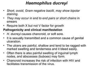
Haemophilus ducreyi
• Short,ovoid, Gram negative bacilli, may show bipolar
staining.
• They may occur in end to end pairs or short chains in
smears
• Require both X but not V factor for growth
Pathogenicity and clinical manifestation
• H. ducreyi causes chancroid, or soft sore.
• It is sexually transmitted and a common cause of genital
ulceration.
• The ulcers are painful, shallow and tend to be ragged with
marked swelling and tenderness and it bleed easily.
• Often there is also painful swelling of inguinal lymph
nodes, and abscesses (buboes) may form.
• Chancroid increases the risk of infection with HIV and
facilitates transmission of the virus.
17.
Lab diagnosis:
• Specimens:
–Specimens should be collected from the base and
margins of ulcers following cleansing with a saline
swab (exclude necrotic tissue).
– Specimens for culture must be delivered to the
laboratory with the minimum of delay.
– When this is not possible the swab should be
placed in Amies transport medium and delivered the
same day to the laboratory or sent in an insulated
cool box to reach the laboratory within 48 h.
18.
Culture
• H. ducreyiis difficult to isolate.
• It is grown best from scrapings of the ulcer base on
– chocolate agar containing 1% IsoVitaleX and vancomycin, 3
g/mL, and incubated in 5-10% CO2 at 33-36 °C for 2-3
days.
– GC agar base with added haemoglobin, vancomycin, and
fetal calf serum.
• The organism grows slowly, producing small grey-yellow or
brown colonies usually within 2–4 days.
Biochemical tests
• Slowly oxidase positive (colour develops after 15–20 seconds).
• Catalase, urease, and indole negative.
• Ornithine decarboxylase (ODC) negative.
• delta-Aminolevulinic acid (ALA) negative
19.
Treatment
• There isno permanent immunity following
chancroid infection.
• Treatment with intramuscular ceftriaxone,
oral trimethoprim-sulfamethoxazole, or
oral erythromycin often results in healing
in 2 weeks.
20.
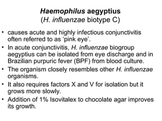
Haemophilus aegyptius
(H. influenzaebiotype C)
• causes acute and highly infectious conjunctivitis
often referred to as ‘pink eye’.
• In acute conjunctivitis, H. influenzae biogroup
aegyptius can be isolated from eye discharge and in
Brazilian purpuric fever (BPF) from blood culture.
• The organism closely resembles other H. influenzae
organisms.
• It also requires factors X and V for isolation but it
grows more slowly.
• Addition of 1% Isovitalex to chocolate agar improves
its growth.
21.
Genus Bordetella
• Minutestrictly aerobic, non motile ,gram
negative coccobacilli that appears singly or in
pairs
• non-spore forming, Catalase +ve
• Fastidious organism (require blood
supplemented medium)
• Bordetella species of clinical significance include
– B. pertussis, B. parapertussis, B. bronchoseptica, …
22.
• Bordetella pertussis,a highly communicable and
important pathogen of humans, causes
whooping cough (pertussis).
• Bordetella parapertussis can cause a similar
disease.
• Bordetella bronchiseptica (Bordetella
bronchicanis) causes diseases in animals such
as kennel cough in dogs and snuffles in rabbits,
and only occasionally causes respiratory
disease and bacteremia in humans, primarily in
immunocompromised hosts.
23.
Bordetella pertussis
• B.pertussis is a small, non-motile,
capsulated Gram negative coccobacillus.
• It may occur singly or in chains
• With toluidine blue stain, bipolar
metachromatic granules can be
demonstrated
24.
Virulence factor
• Adhesins
–Most important adhesin is filamentous haemagglutinin
– Mediate adhesion to ciliated epithelial Cells
• Toxin
– Pertussis toxin by only B. pertussis
– Adenylate cyclase toxin
– Tracheal cytotoxin
– Dermonecrotic toxin
– Endotoxin LPS
• Agglutinogen
• Outer Membrane Protein (OMP)
25.
Pathogenicity and Clinicalmanifestation
• B. pertussis causes whooping cough, an infection of the
mucosa of the upper respiratory tract.
• Toxin from the organisms causes the secretion of mucus
which leads to irritation and the spasms of coughing
associated with the disease.
• There is a marked leucocytosis with an absolute
lymphocytosis.
• Complications of infection include lung damage with
emphysema, secondary infection leading to
bronchopneumonia, bronchiectasis, convulsions and
occasionally brain damage.
• Note: B. parapertussis causes a milder form of whooping
cough.
26.
• After anincubation period of about 2 weeks, the
"catarrhal stage" develops, with mild coughing
and sneezing.
– During this stage, large numbers of organisms are
sprayed in droplets, and the patient is highly infectious
but not very ill.
• During the "paroxysmal" stage, the cough
develops its explosive character and the
characteristic "whoop" upon inhalation.
– This leads to rapid exhaustion and may be associated
with vomiting, cyanosis, and convulsions.
27.
– The "whoop"and major complications occur
predominantly in infants;
– paroxysmal coughing predominates in older children
and adults.
– The white blood count is high (16,000–30,000/ L),
with an absolute lymphocytosis.
• After 3-4 weeks the disease enters
Convalescent stage
– Frequency and severity of the coughing gradually
decrease
– But secondary complications can occur
28.
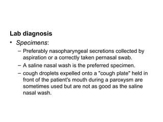
Lab diagnosis
• Specimens:
–Preferably nasopharyngeal secretions collected by
aspiration or a correctly taken pernasal swab.
– A saline nasal wash is the preferred specimen.
– cough droplets expelled onto a "cough plate" held in
front of the patient's mouth during a paroxysm are
sometimes used but are not as good as the saline
nasal wash.
29.
Culture
• The salinenasal wash fluid is cultured on
solid medium agar
• The antibiotics in the media tend to inhibit
other respiratory flora but permit growth of
B pertussis.
• Organisms are identified by
immunofluorescence staining or by slide
agglutination with specific antiserum.
30.
Culture
• For theisolation of B. pertussis must be
cultured as soon as possible after they are
collected.
• A selective and enrichment medium such as
charcoal cephalexin blood agar is
recommended for the primary isolation of B.
pertussis.
• Small, convex, smooth colonies
31.
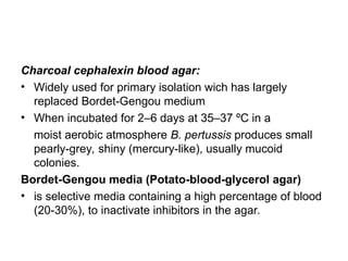
Charcoal cephalexin bloodagar:
• Widely used for primary isolation wich has largely
replaced Bordet-Gengou medium
• When incubated for 2–6 days at 35–37 ºC in a
moist aerobic atmosphere B. pertussis produces small
pearly-grey, shiny (mercury-like), usually mucoid
colonies.
Bordet-Gengou media (Potato-blood-glycerol agar)
• is selective media containing a high percentage of blood
(20-30%), to inactivate inhibitors in the agar.
32.
Treatment
• Sensitive toErythromycin, tetracycline, chloramphenicol
Prevention and control
• adequate active immunization of all infants
33.
Genus Brucella
• Gramnegative, short rods, coccobacilli
• Non-motile, non-spore forming, non-capsulated
• Aerobic, require complex media (amino acid,
thiamine, nicotinamide)
• Growth enhanced (serum/blood)
• Catalase +ve, oxidase +ve
• Many require CO2 for growth
• characteristically located intracellularly
• Cause disease primarily in animals
– Bacteremia and septicaemia in humans
34.
Natural habitats
• Animals– main reservoir
• Transmit from Animal to humans through
– Milk products
– Handling of animals and cultures
– Inhalation of aerosols
35.
• Brucella speciesand their preferred host
– B. abortus-cattle, horse, buffalo
– B. melitensis-goat, sheep, camel
– B.suis-swine, hare, rodents
– B. canis-dogs
– B.ovis-sheep
– B.neotomae-rodents
• B. abortus, B. melitensis, and B.suis causes
human brucellosis
36.
Virulence factor
• Abilityto resist phagocytosis
– Has certain low-molecular substance on bacterial surface
• Ability of intracellular survival
Pathogenesis and clinical manifestation
• Brucellae are intracellular organisms infecting reticuloendothelial cells of
the spleen, liver, kidneys and bone marrow. From these sites, the
bacteria pass into the blood.
• The disease in humans, brucellosis (undulant fever, Malta fever), is
characterized by an acute bacteremic phase followed by a chronic stage
that may extend over many years and may involve many tissues.
• The disease is characterized by fever which may be continuous,
intermittent, undulating or irregular.
• Acute infection may resemble severe influenza with headache, sweating
(especially at night) and generalized pains associated with fatigue and
depression.
37.
• Urogenital symptomsmay occur
• Often the patient is anaemic and
leukopenic with a relative lymphocytosis.
• Untreated infections can become chronic
with musculoskeletal symptoms (back
pain, arthritis, arthralgia).
38.
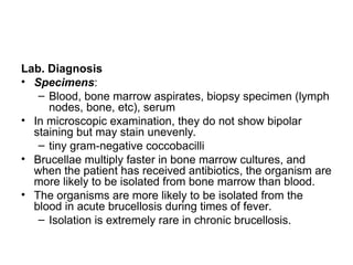
Lab. Diagnosis
• Specimens:
–Blood, bone marrow aspirates, biopsy specimen (lymph
nodes, bone, etc), serum
• In microscopic examination, they do not show bipolar
staining but may stain unevenly.
– tiny gram-negative coccobacilli
• Brucellae multiply faster in bone marrow cultures, and
when the patient has received antibiotics, the organism are
more likely to be isolated from bone marrow than blood.
• The organisms are more likely to be isolated from the
blood in acute brucellosis during times of fever.
– Isolation is extremely rare in chronic brucellosis.
39.
• Brucella speciesbacteria will grow on commonly used media, including
– trypticase soy medium with or without 5% sheep blood,
– brain heart infusion medium, and
– chocolate agar.
– Serum dextrose agar
– Blood culture media (readily grow Brucella species bacteria.)
– Tryptone soya (tryptic soy) diphasic medium (Castaneda)
• Brucellae are aerobic with B. abortus requiring a carbon dioxide enriched
atmosphere.
• Cultures should be kept for 4 weeks with sub-culturing every few days.
• When sub-cultured on solid agar, colonies usually appear 2–3 days after
incubation.
• All cultures should be incubated in 8–10% CO2 at 35–37 °C with sub-
culturing every few days
– It should be observed for 4 weeks before being discarded as negative;
40.
• After afew days of incubation on agar media, the brucellae
form colonies
– They are nonhemolytic.
– variety of colonial forms are produced by Brucella strains
including Small, smooth, convex, mucoid, and rough
colonies.
– They may be colourless or greywhite.
• All further work on such a culture should be done in a
biologic safety cabinet.
• A Christensen urea slant should be inoculated and observed
frequently.
41.
Biochemical test
• Thebacteria are catalase-positive, urease-positive,
and usually oxidase-positive.
• They are indole negative and most strains
hydrolyze urea.
• Except B. ovis, Brucella species reduce nitrate to
nitrite
Serological diagnosis
• Many cases of brucellae infection are diagnosed by
serological test which include the standard
agglutination test and an ELISA procedure for
detection of brucella-specific IgM/IgG.
42.
Treatment
• Brucellae maybe susceptible to tetracyclines or ampicillin.
• For best results, treatment must be prolonged.
• Combined treatment with a tetracycline (such as doxycycline)
and either streptomycin or rifampin is recommended.
Prevention and control
• limitation of spread and possible eradication of animal
infection,
• pasteurization of milk and milk products, and
• reduction of occupational hazards wherever possible.
Editor's Notes
#11 Factors X and V
Factor X is used by H. influenzae to produce essential respiratory enzymes such as cytochromes, catalase, and peroxidase. Factor V is used as an electron carrier in the organism’s oxidation-reduction system.
#13 The lysed erythrocytes in the agar surrounding the S.aureus streak provide X-factor, and staphylococcal cells themselves secrete V-factor during logarithmic growth. Both growth requirements are met for Haemophlus
#28 B pertussis is a common cause of prolonged (4–6 weeks) cough in adults. Rarely, whooping cough is followed by the serious and potentially fatal complication of encephalitis. Several types of adenovirus and Chlamydia pneumoniae can produce a clinical picture resembling that caused by B pertussis.
#31 B. parapertussis grows more rapidly and forms larger colonies than B. pertussis. It produces a pigment in the medium and is able to grow aerobically on blood agar and nutrient agar.
#37 Human brucellosis can occur when brucellae are ingested in raw milk, fresh cheese, cream or other milk products (large numbers of organisms are shed in the milk of infected animals), enter damaged skin or the eye, or are inhaled in aerosols.
#38 Caution: Brucellae are highly infectious (Hazard Risk Group 3) pathogens. Laboratory-acquired infections can occur following accidental inoculation or inhalation of the organisms. Collect blood with great care, minimize the creation of aerosols and whenever possible, carry out procedures which may produce aerosols in a safety cabinet
#40 Subculturing on a slope of glucose tryptone agar with a lead acetate test strip in the neck of the tube is useful in testing for hydrogen sulphide (H2S) production. B. melitensis is H2S negative, most strains of B. abortus and B. suis are H2S positive.