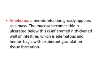
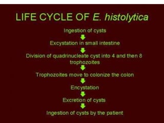
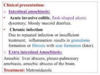
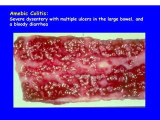
Amoebic colitis
Amoebic colitis
• Infection of the colon with Entamoeba
histolytica
Morphology
• Sites:
• Ascending colon –the most common
• Sigmoid colon
• Rectum
• appendix
• Flask shaped ulcers:
• Early intestinal lesion is microulceration of
mucosa which slowly enlarges.
• Small ulcer with heaped up margin but
extends below the surface n apperars as flask-
shaped.
• Numerous trophozoites can be seen at the
margin of dead n viable tissue.
• Large ulcers show extensive liquefactive
necrosis with shaggy appearance.
• Ameboma: ameobic infection grossly appears
as a mass. The mucosa becomes thin n
ulcerated.Below this is inflammed n thickened
wall of intestine, which is edematous and
hemorrhagic with exuberant granulation
tissue formation.
• Complications
• 1) liver abscess
• 2) effusion;;pleural
• 3) Hepatobronchial fistula
• 4) pericardial effusion
• Clinical picture:
• Pain abdomen with cramp
• Diarrhoea, sometimes with blood.
• Diagnosis: PAS stain: trophozoites of
E.Histolytica are stained by PAS and iron
haematoxylin.
• Thank you..

Amoebic colitis

  • 1.
  • 2.
    Amoebic colitis • Infectionof the colon with Entamoeba histolytica Morphology • Sites: • Ascending colon –the most common • Sigmoid colon • Rectum • appendix
  • 3.
    • Flask shapedulcers: • Early intestinal lesion is microulceration of mucosa which slowly enlarges. • Small ulcer with heaped up margin but extends below the surface n apperars as flask- shaped.
  • 4.
    • Numerous trophozoitescan be seen at the margin of dead n viable tissue. • Large ulcers show extensive liquefactive necrosis with shaggy appearance.
  • 5.
    • Ameboma: ameobicinfection grossly appears as a mass. The mucosa becomes thin n ulcerated.Below this is inflammed n thickened wall of intestine, which is edematous and hemorrhagic with exuberant granulation tissue formation.
  • 6.
    • Complications • 1)liver abscess • 2) effusion;;pleural • 3) Hepatobronchial fistula • 4) pericardial effusion
  • 7.
    • Clinical picture: •Pain abdomen with cramp • Diarrhoea, sometimes with blood. • Diagnosis: PAS stain: trophozoites of E.Histolytica are stained by PAS and iron haematoxylin.
  • 11.