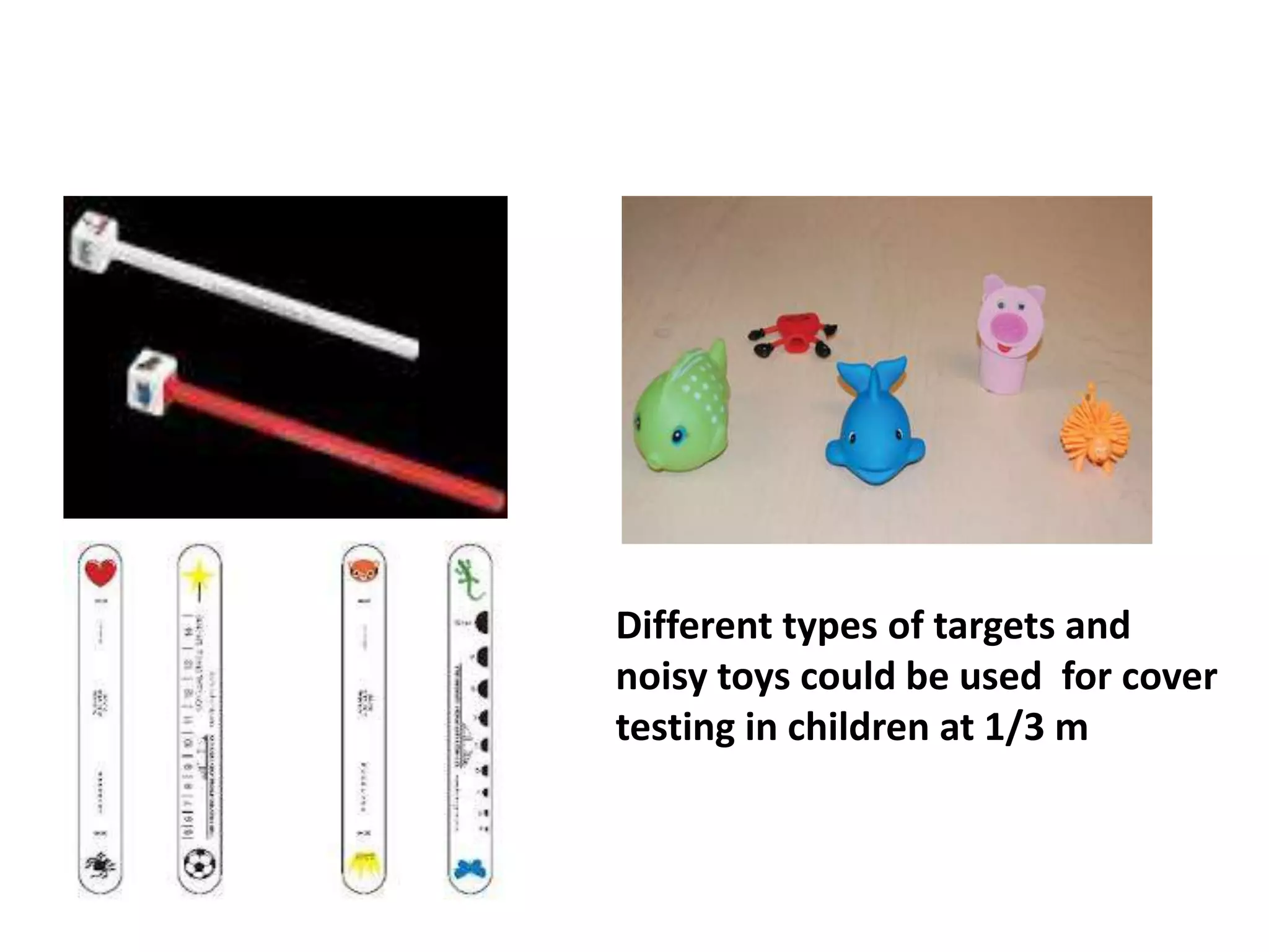
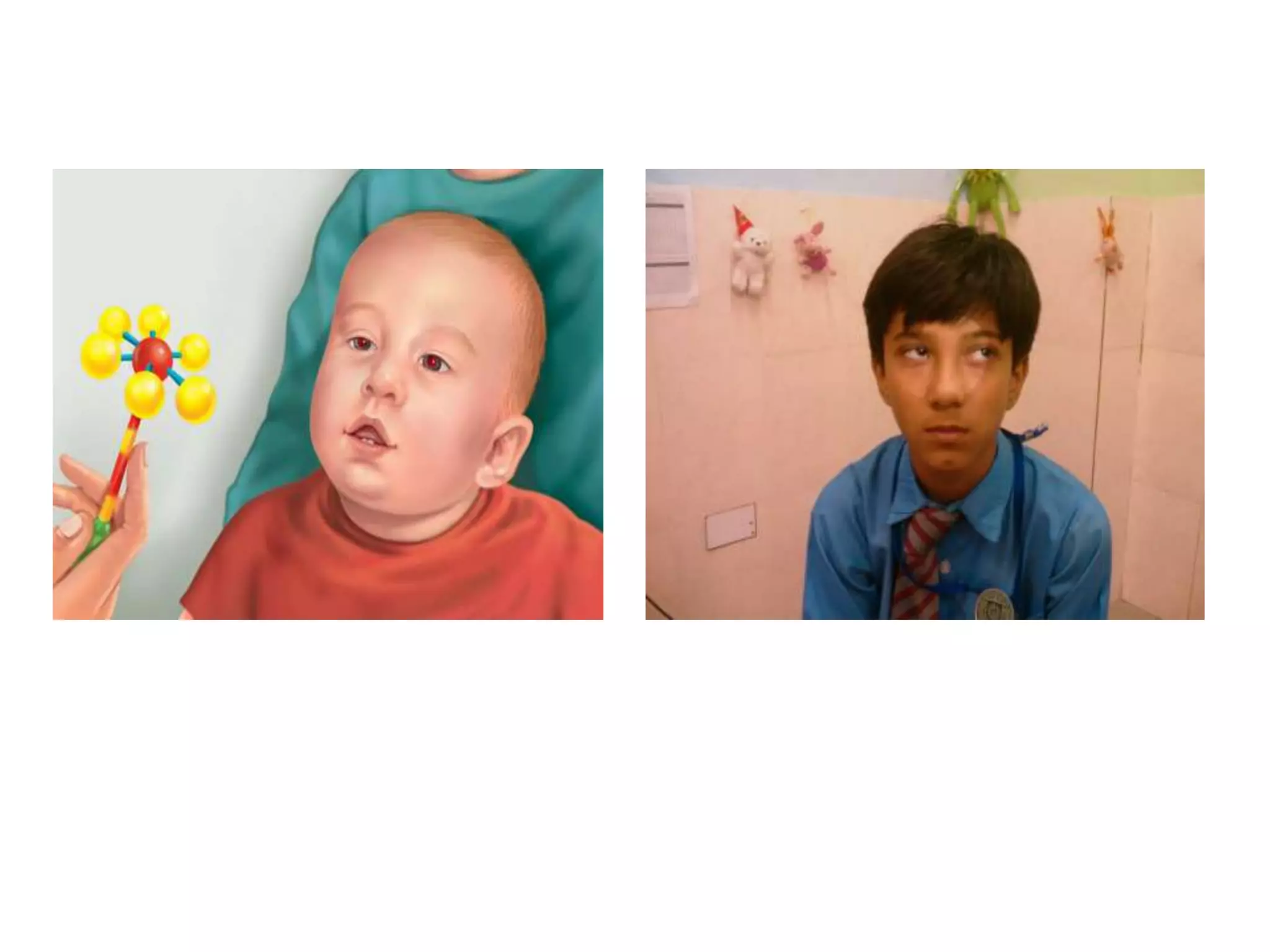
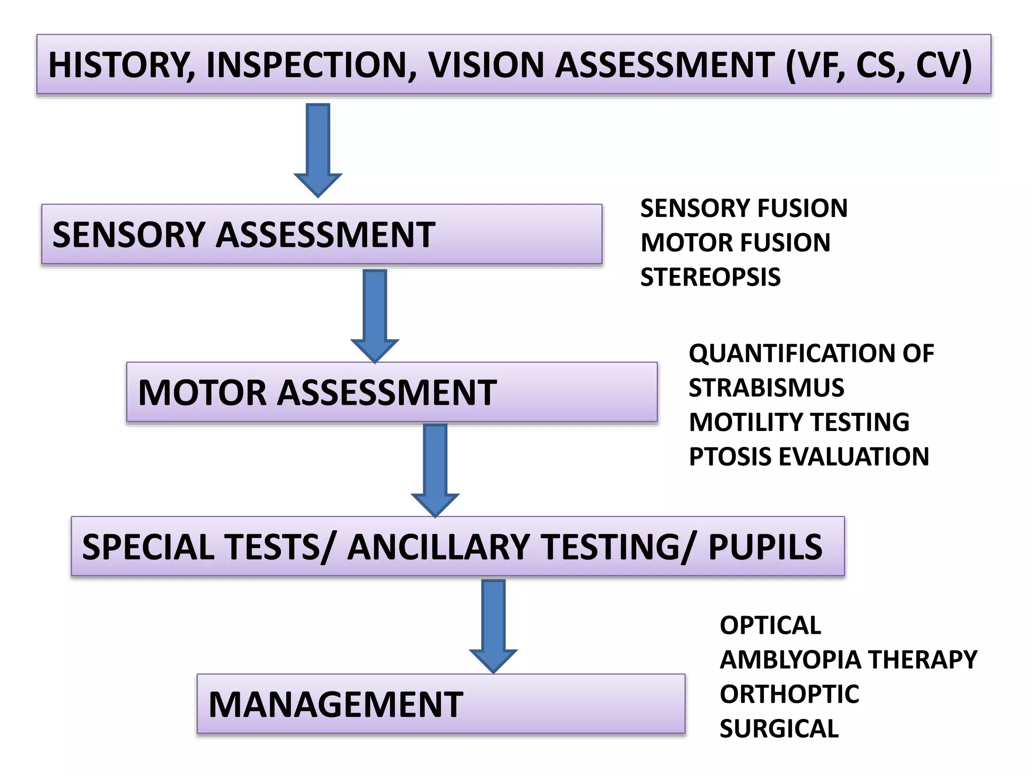
ASSESSMENT OF STRABISMUS IN
CHILDREN
AYESHA SARFRAZ
ORTHOPTIST
BSC(HONS.) ORTHOPTICS, FO (CANADA),
MPHIL SCHOLAR (KEMU)
COAVS,KEMU/MHL
STRABISMUS!
• Strabismus is a visual
problem in which the eyes
are not aligned properly
and point in different
directions. One eye may
look straight ahead, while
the other eye turns inward,
outward, upward, or
downward.
CLASSIFICATION
• Infants: Age <1 year
• Toddlers: 1 year- 4 years
• Children: 5years- 14 years
• Adolescence: 15 years- 17 years
• Adults: 18 years and above
APPROACH TOWARDS
STRABISMUS
 History
 Inspection/ Observations
 Vision assessment
 Refraction
 Sensory Assessment
 Motor Assessment
 Measurements
 Motility testing
 Ptosis/ Proptosis evaluatiion
 Pupil examination
 Special Tests/ Ancillary Testing
 Anterior & Posterior segment evaluation
HISTORY
Case History
There are three main reasons for taking a history before
beginning the examination:
• To acquired detailed information on the patient’s
symptoms, the probable time of onset and the possible
etiology and information concerning any treatment he
may already have had.
• To allow the examiner to establish a good relationship
between himself and the patient and thus gain the
patients confidence and ensure his cooperation.
• To enable the examiner to observe the patient under
normal conditions that may be helpful when making
the diagnosis and prognosis and deciding upon the
treatment.
HISTORY TAKING IN STRABISMUS
• Visual behaviour
– Clumsy? Visually Inattentive? Close viewing distance
• Past History
– History of patching
– History of spectacle wear & Type of strabismus
– History of trauma
– Previous surgery
– Strabismus surgery , Other ocular procedures &
Periocular Surgeries
• Birth History
– Age of gestation
– Birth weight
– Problems during pregnancy
– Type of delivery
– Late Cry Syndrome
• Developmental History
– Developmental Milestones
– Motor and speech development
– Delay of visual maturation
• Family history
– Presence of hereditary forms of strabismus
– Response to strabismus surgery of family
members
• Review of systems
– Neurologic symptoms
– Other systemic abnormalities
INSPECTION
• Epicanthi folds/ Nasal Bridge (Pseudo strabismus)
• Lid Fissure
– Ptosis
– Exophthalmos
• Abnormal Head Posture
• Facial Asymmetry
• Fixation Preference
• Nystagmus
VISION ASSESSMENT
OBSERVATION OF VISUAL RESPONSE
1. Ability to follow target
2. Visually directed reaching
3. Objection to Occlusion
4. Binocular fixation pattern/ Ten Prism Diopter test
5. CSM method.
• CSM – 6/9 – 6/6
• CSNM –6/36 – 6/60
• Unsteady central fixation < 6/60
6. Mirror test
7. Induce Tropia Test
8. Bruckner’s Test/ Red Reflex Test
INDUCED TROPIA TEST
Tests for Resolution Acuity
• Optokinetic nystagmus test(OKN)
• Preferential looking test(PLT) 3-12 months
– Lea Grating paddles
– Teller acuity Cards
• Visually evoked response(VER)
• Catford drum test.
• Cardiff acuity card test.
• Kay Pictures
• Lea Symbols
• ETDRS
• Snellen Chart
Objection to
Occlusion to
rule out
difference of
Vision between
two eyes
Bruckner’s for
Strabismus,
pupil symmetry,
cataract and
retinoblastoma
Estimation of Visual Acuity in Infants
VEP helpful in establishing
the presence of cortical
blindness & give an
estimation of visual acuity
OKN is helpful for sneak
peak into cortical activity
for vision assessment
Visual Acuity in Pre School Children
Lea symbolsKay picture test
Snellen chart for pre school
children
Visual acuity in School Children
Snellen charts for school
age children
ETDRS
Near Vision
1. Reduced Snellen test
2. Reduced Sheridan Gardiner
3. Reduced E test
4. Maclure book
5. Moorfields bar reading book
6. N series test
7. Lea symbols
IF NECESSARY, Go For…
• Visual Field Evaluation
• Contrast Sensitivity Asessment
Hiding Heidi Test Lea numbers
• Color Vision Assessment
Lea Color Vision Panel 16
Refraction
FUNDOSCOPY
CYCLOPLEGIC REFRACTION
PRESCRIBE THE APPROPRIATE
REFRACTIVE CORRECTION
VISION ASSESSMENT
ALSO RULE OUT THE DIFFERENCE OF
VISION BETWEEN TWO EYES FOR
AMBLYOPIA
SENSORY ASSESSMENT
• Fusion
– Sensory Fusion
– Motor Fusion
• Stereopsis
FUSION
Sensory Fusion
• Worth Four Dot Test
– At 6m
– At 1/3m
• Bagolini Glasses
Motor Fusion
Prism Reflex Test
In babies & young children by using 20
prism diopter Base out and look for the
movement of eye.
Prism Fusion range
By using horizontal and vertical prism bars
in cooperative children
Stereopsis
• Lang two pencil Test
• Lang I & II
• Titmus Fly Test
• Randot Stereo Test
• Frisby stereo Test
• TNO stereotest
Stereopsis
MOTOR ASSESSMENT
Assessment of Deviation
• Hirschberg test
• Cover test
• Alternate cover test
• Krimsky test
• Prism Reflection Test
• Alternate Prism cover test
• Prism Under Cover Test
• Synoptophore
Hirscberg Test
Krimsky Test
Cover test ,Uncover test , Alternate
Cover Test & Alternate Prism Cover Test
The cover test for tropias…
Observe the right eye as you cover the left eye…
What would you record this as?
Is it manifest or phoric?
Is there a fixing eye?
And this???
The Cover / Uncover Test…
This eye is exophoric.
Occluder is swiftly moved from one eye to the
other.
Allow the occluder to cover the eye long
enough for the patient to pick up fixation.
A prism could now be introduced.
Cover Test
Cover/ Uncover
APCT…
Information Gained from Cover
Testing
Manifest Deviation
• Type of deviation
• Size of deviation
• Speed to take up fixation
• Effect of accommodation on deviation
• Nystagmus
• Dissociated vertical deviation
• incomitance
Latent Deviation
• Type of Deviation
• Size of deviation
• Rate of recovery to achieve binocular single
vision
• Partial Recovery
Different types of targets and
noisy toys could be used for cover
testing in children at 1/3 m
Synoptophore
• Fusion slides are used
• Alternate cover uncover test is performed
• Patient’s angle of deviation is measured
objectively and subjectively
• To elicit the extent and quality of movement
of each eye
• to determine the presence of comitancy or
incomitancy
• to establish the integrity of the ocular
movement system and their neural pathways.
Ocular Motility Testing
• Use a spot light positioned at primary position
of gaze at 50 cm from the patient
• Always remove the patients glasses
• Give clear instructions to the patient
• An audible and colorful fixation target may
need to be used for young children
While check for the horizontal versions look for the
following:
• Up drift and down drift of the either eye
• Under action or over action of extraocular muscles
• Limitation (restriction of movement)
• Changes in the size of palpebral aperture
• Changes in the pupil size
• Changes in globe position
Ductions are tested monocularly and the
following noted:
 Excrusion
 Behavior of both eyes
 Whether the movement is smooth and jerky
 Nystagmus
 Change in the position of lids and globe
 Effect of fatigue
 Torsional movement
 Any discomfort on movement
 Abnormal head movements
Ductions
When testing vertical movements, look for
• Under action or over action
• Globe changes
• Sign of lid fatigue
• Down movement should be checked both with
and without holding the lid
• Check for A/V pattern in straight up and down
gaze
• Versions and ductions from the primary
position, into each of the diagnostic position
of gaze are assessed.
• Movements are graded on a 9 position scale
-4, -3, -2, -1, 0, +1, +2, +3, +4
• Zero represents a normal movement
Ocular Movements Grading
 One held in the primary position and the other:
to each side of the head for horizontal movement
above and below the head for vertical movements
 The movement, speed and accuracy of both eyes are
compared and any asymmetry between the eyes is noted
Saccadic Movement
Saccade testing in Infants/
Children
Testing of saccadic eye movement s is useful in:
 Comparing movement of the two eyes as this may
show slight limitations
 Suspected myasthenia gravis as fatigue may become
evident
 Suspected internuclear ophtthalmoplegia as
dysmetria may be evident
Note The Pursuit Followed By The
Saccadic Movement OU
• Also known as the Vestibulo Ocular Reflex,
Cephalic Reflex and Doll’s Head Maneuver
Vestibular Eye Movement
• Fast phase is a saccade, the slow phase is a
pursuit
• Move the drum or tape to the right you get
pursuit right for the slow phase followed by a
quick saccade left for the fast phase
Optokinetic Nystagmus
head is rotated briskly by the examiner,
first in the horizontal plane and then in the
vertical plane.
 VOR will result in eye movements equal and
opposite to head movements.
A normal response will indicate that the
muscle function is not impaired.
Doll's Head Test
Clinically, This test can also be very useful in
infants with childhood esotropia where there
is suspected lateral rectus weakness.
Swinging Baby Test
• based on the observation of a conjugate
deviation of the eyes in response to head
movement induced by rotation and is useful with
babies who will not respond to conventional
testing.
• The infant is held upright facing the examiner. The
examiner rotates him/herself and the infant
through 360◦ while observing the infant’s eyes.
• Post-rotational nystagmus in sighted babies will
only persist for a few seconds before being
suppressed.
• One ear is irrigated with cold water, nystagmus is
produced with the fast phase toward the opposite
ear
• When one ear is irrigated with warm water,
nystagmus is produced with the fast phase toward
the same ear
• COWS
• If you inject bilaterally: CUWDS
Induced Vestibular Nystagmus
HISTORY, INSPECTION, VISION ASSESSMENT (VF, CS, CV)
SENSORY ASSESSMENT
MOTOR ASSESSMENT
SENSORY FUSION
MOTOR FUSION
STEREOPSIS
QUANTIFICATION OF
STRABISMUS
MOTILITY TESTING
PTOSIS EVALUATION
SPECIAL TESTS/ ANCILLARY TESTING/ PUPILS
MANAGEMENT
OPTICAL
AMBLYOPIA THERAPY
ORTHOPTIC
SURGICAL
Let’s Start Testing!!!

Strabismus assessment OSP

  • 1.
    ASSESSMENT OF STRABISMUSIN CHILDREN AYESHA SARFRAZ ORTHOPTIST BSC(HONS.) ORTHOPTICS, FO (CANADA), MPHIL SCHOLAR (KEMU) COAVS,KEMU/MHL
  • 2.
    STRABISMUS! • Strabismus isa visual problem in which the eyes are not aligned properly and point in different directions. One eye may look straight ahead, while the other eye turns inward, outward, upward, or downward.
  • 3.
    CLASSIFICATION • Infants: Age<1 year • Toddlers: 1 year- 4 years • Children: 5years- 14 years • Adolescence: 15 years- 17 years • Adults: 18 years and above
  • 4.
  • 5.
     History  Inspection/Observations  Vision assessment  Refraction  Sensory Assessment  Motor Assessment  Measurements  Motility testing  Ptosis/ Proptosis evaluatiion  Pupil examination  Special Tests/ Ancillary Testing  Anterior & Posterior segment evaluation
  • 6.
  • 7.
    Case History There arethree main reasons for taking a history before beginning the examination: • To acquired detailed information on the patient’s symptoms, the probable time of onset and the possible etiology and information concerning any treatment he may already have had. • To allow the examiner to establish a good relationship between himself and the patient and thus gain the patients confidence and ensure his cooperation. • To enable the examiner to observe the patient under normal conditions that may be helpful when making the diagnosis and prognosis and deciding upon the treatment.
  • 8.
    HISTORY TAKING INSTRABISMUS • Visual behaviour – Clumsy? Visually Inattentive? Close viewing distance • Past History – History of patching – History of spectacle wear & Type of strabismus – History of trauma – Previous surgery – Strabismus surgery , Other ocular procedures & Periocular Surgeries
  • 9.
    • Birth History –Age of gestation – Birth weight – Problems during pregnancy – Type of delivery – Late Cry Syndrome • Developmental History – Developmental Milestones – Motor and speech development – Delay of visual maturation
  • 10.
    • Family history –Presence of hereditary forms of strabismus – Response to strabismus surgery of family members • Review of systems – Neurologic symptoms – Other systemic abnormalities
  • 11.
  • 12.
    • Epicanthi folds/Nasal Bridge (Pseudo strabismus) • Lid Fissure – Ptosis – Exophthalmos • Abnormal Head Posture • Facial Asymmetry • Fixation Preference • Nystagmus
  • 13.
  • 14.
    OBSERVATION OF VISUALRESPONSE 1. Ability to follow target 2. Visually directed reaching 3. Objection to Occlusion 4. Binocular fixation pattern/ Ten Prism Diopter test 5. CSM method. • CSM – 6/9 – 6/6 • CSNM –6/36 – 6/60 • Unsteady central fixation < 6/60 6. Mirror test 7. Induce Tropia Test 8. Bruckner’s Test/ Red Reflex Test
  • 15.
  • 16.
    Tests for ResolutionAcuity • Optokinetic nystagmus test(OKN) • Preferential looking test(PLT) 3-12 months – Lea Grating paddles – Teller acuity Cards • Visually evoked response(VER) • Catford drum test. • Cardiff acuity card test. • Kay Pictures • Lea Symbols • ETDRS • Snellen Chart
  • 17.
    Objection to Occlusion to ruleout difference of Vision between two eyes Bruckner’s for Strabismus, pupil symmetry, cataract and retinoblastoma
  • 18.
    Estimation of VisualAcuity in Infants VEP helpful in establishing the presence of cortical blindness & give an estimation of visual acuity OKN is helpful for sneak peak into cortical activity for vision assessment
  • 19.
    Visual Acuity inPre School Children Lea symbolsKay picture test
  • 20.
    Snellen chart forpre school children
  • 21.
    Visual acuity inSchool Children Snellen charts for school age children ETDRS
  • 22.
    Near Vision 1. ReducedSnellen test 2. Reduced Sheridan Gardiner 3. Reduced E test 4. Maclure book 5. Moorfields bar reading book 6. N series test 7. Lea symbols
  • 23.
    IF NECESSARY, GoFor… • Visual Field Evaluation
  • 24.
    • Contrast SensitivityAsessment Hiding Heidi Test Lea numbers
  • 25.
    • Color VisionAssessment Lea Color Vision Panel 16
  • 26.
  • 27.
    FUNDOSCOPY CYCLOPLEGIC REFRACTION PRESCRIBE THEAPPROPRIATE REFRACTIVE CORRECTION VISION ASSESSMENT ALSO RULE OUT THE DIFFERENCE OF VISION BETWEEN TWO EYES FOR AMBLYOPIA
  • 28.
  • 29.
    • Fusion – SensoryFusion – Motor Fusion • Stereopsis
  • 30.
    FUSION Sensory Fusion • WorthFour Dot Test – At 6m – At 1/3m • Bagolini Glasses
  • 31.
    Motor Fusion Prism ReflexTest In babies & young children by using 20 prism diopter Base out and look for the movement of eye. Prism Fusion range By using horizontal and vertical prism bars in cooperative children
  • 32.
    Stereopsis • Lang twopencil Test • Lang I & II • Titmus Fly Test • Randot Stereo Test • Frisby stereo Test • TNO stereotest
  • 33.
  • 34.
  • 35.
    Assessment of Deviation •Hirschberg test • Cover test • Alternate cover test • Krimsky test • Prism Reflection Test • Alternate Prism cover test • Prism Under Cover Test • Synoptophore
  • 36.
  • 37.
    Cover test ,Uncovertest , Alternate Cover Test & Alternate Prism Cover Test
  • 38.
    The cover testfor tropias… Observe the right eye as you cover the left eye…
  • 39.
    What would yourecord this as? Is it manifest or phoric? Is there a fixing eye?
  • 40.
  • 41.
    The Cover /Uncover Test… This eye is exophoric.
  • 42.
    Occluder is swiftlymoved from one eye to the other. Allow the occluder to cover the eye long enough for the patient to pick up fixation. A prism could now be introduced.
  • 43.
  • 44.
    Information Gained fromCover Testing Manifest Deviation • Type of deviation • Size of deviation • Speed to take up fixation • Effect of accommodation on deviation • Nystagmus • Dissociated vertical deviation • incomitance
  • 45.
    Latent Deviation • Typeof Deviation • Size of deviation • Rate of recovery to achieve binocular single vision • Partial Recovery
  • 46.
    Different types oftargets and noisy toys could be used for cover testing in children at 1/3 m
  • 47.
    Synoptophore • Fusion slidesare used • Alternate cover uncover test is performed • Patient’s angle of deviation is measured objectively and subjectively
  • 48.
    • To elicitthe extent and quality of movement of each eye • to determine the presence of comitancy or incomitancy • to establish the integrity of the ocular movement system and their neural pathways. Ocular Motility Testing
  • 49.
    • Use aspot light positioned at primary position of gaze at 50 cm from the patient • Always remove the patients glasses • Give clear instructions to the patient • An audible and colorful fixation target may need to be used for young children
  • 50.
    While check forthe horizontal versions look for the following: • Up drift and down drift of the either eye • Under action or over action of extraocular muscles • Limitation (restriction of movement) • Changes in the size of palpebral aperture • Changes in the pupil size • Changes in globe position
  • 52.
    Ductions are testedmonocularly and the following noted:  Excrusion  Behavior of both eyes  Whether the movement is smooth and jerky  Nystagmus  Change in the position of lids and globe  Effect of fatigue  Torsional movement  Any discomfort on movement  Abnormal head movements Ductions
  • 54.
    When testing verticalmovements, look for • Under action or over action • Globe changes • Sign of lid fatigue • Down movement should be checked both with and without holding the lid • Check for A/V pattern in straight up and down gaze
  • 55.
    • Versions andductions from the primary position, into each of the diagnostic position of gaze are assessed. • Movements are graded on a 9 position scale -4, -3, -2, -1, 0, +1, +2, +3, +4 • Zero represents a normal movement Ocular Movements Grading
  • 56.
     One heldin the primary position and the other: to each side of the head for horizontal movement above and below the head for vertical movements  The movement, speed and accuracy of both eyes are compared and any asymmetry between the eyes is noted Saccadic Movement
  • 57.
    Saccade testing inInfants/ Children
  • 58.
    Testing of saccadiceye movement s is useful in:  Comparing movement of the two eyes as this may show slight limitations  Suspected myasthenia gravis as fatigue may become evident  Suspected internuclear ophtthalmoplegia as dysmetria may be evident
  • 59.
    Note The PursuitFollowed By The Saccadic Movement OU
  • 60.
    • Also knownas the Vestibulo Ocular Reflex, Cephalic Reflex and Doll’s Head Maneuver Vestibular Eye Movement
  • 61.
    • Fast phaseis a saccade, the slow phase is a pursuit • Move the drum or tape to the right you get pursuit right for the slow phase followed by a quick saccade left for the fast phase Optokinetic Nystagmus
  • 62.
    head is rotatedbriskly by the examiner, first in the horizontal plane and then in the vertical plane.  VOR will result in eye movements equal and opposite to head movements. A normal response will indicate that the muscle function is not impaired. Doll's Head Test
  • 63.
    Clinically, This testcan also be very useful in infants with childhood esotropia where there is suspected lateral rectus weakness.
  • 64.
    Swinging Baby Test •based on the observation of a conjugate deviation of the eyes in response to head movement induced by rotation and is useful with babies who will not respond to conventional testing. • The infant is held upright facing the examiner. The examiner rotates him/herself and the infant through 360◦ while observing the infant’s eyes. • Post-rotational nystagmus in sighted babies will only persist for a few seconds before being suppressed.
  • 66.
    • One earis irrigated with cold water, nystagmus is produced with the fast phase toward the opposite ear • When one ear is irrigated with warm water, nystagmus is produced with the fast phase toward the same ear • COWS • If you inject bilaterally: CUWDS Induced Vestibular Nystagmus
  • 67.
    HISTORY, INSPECTION, VISIONASSESSMENT (VF, CS, CV) SENSORY ASSESSMENT MOTOR ASSESSMENT SENSORY FUSION MOTOR FUSION STEREOPSIS QUANTIFICATION OF STRABISMUS MOTILITY TESTING PTOSIS EVALUATION SPECIAL TESTS/ ANCILLARY TESTING/ PUPILS MANAGEMENT OPTICAL AMBLYOPIA THERAPY ORTHOPTIC SURGICAL
  • 68.

Editor's Notes

  • #16 Induced tropia test for assessing visual acuity. (A) A prism is held with its base down in front of the right eye. This shifts the image in this eye superiorly. In this photograph the patient is continuing to view through the left eye. (B) A hand is placed in front of the left eye, and the right eye moves up to fi xate on the image, which has been shifted superiorly by the prism. (C) After the hand is removed, the right eye remains up, indicating that this eye continues to maintain fi xation. If both eyes behave in a similar fashion, the vision is equal or nearly equal between the two eyes.
  • #18 Red reflex testing with the direct ophthalmoscope, which also screens for strabismus and pupil symmetry. (A) The examiner sits about 3 ft away from the patient. The dial on the ophthalmoscope (arrow) is used to focus on the patient’s face. The patient sits comfortably in her parent’s lap. (B) If needed, a small toy can be placed on top of the ophthalmoscope to get the child’s attention. (C) Red refl ex results. This patient has esotropia of the left eye (note the lateral displacement of the left corneal light refl ex compared to the centered refl ex on the right [arrows]). The red refl exes are otherwise normal, without sign of a cataract or retinoblastoma.