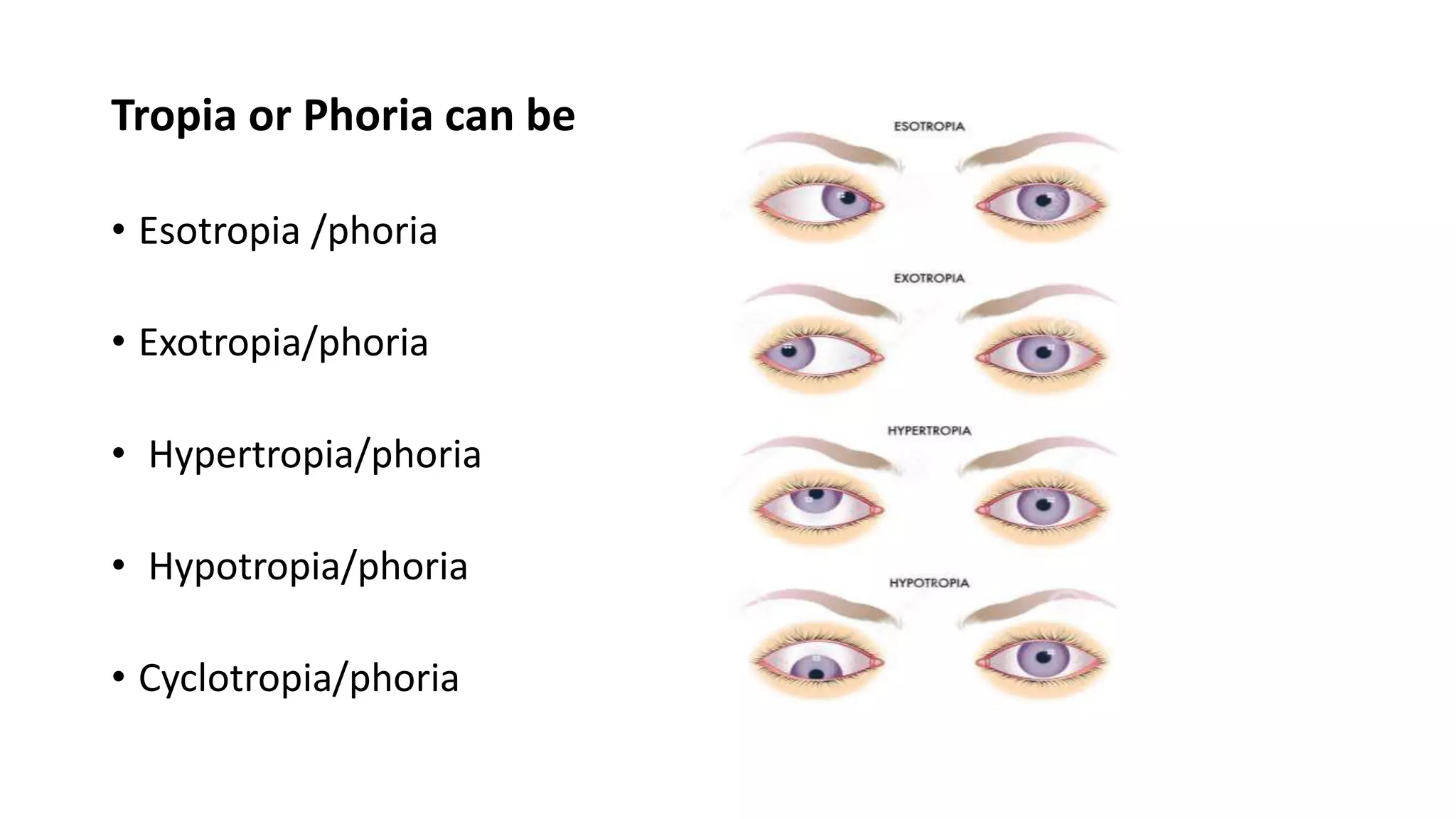
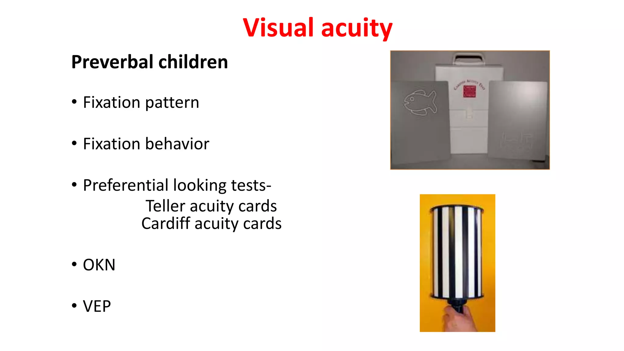
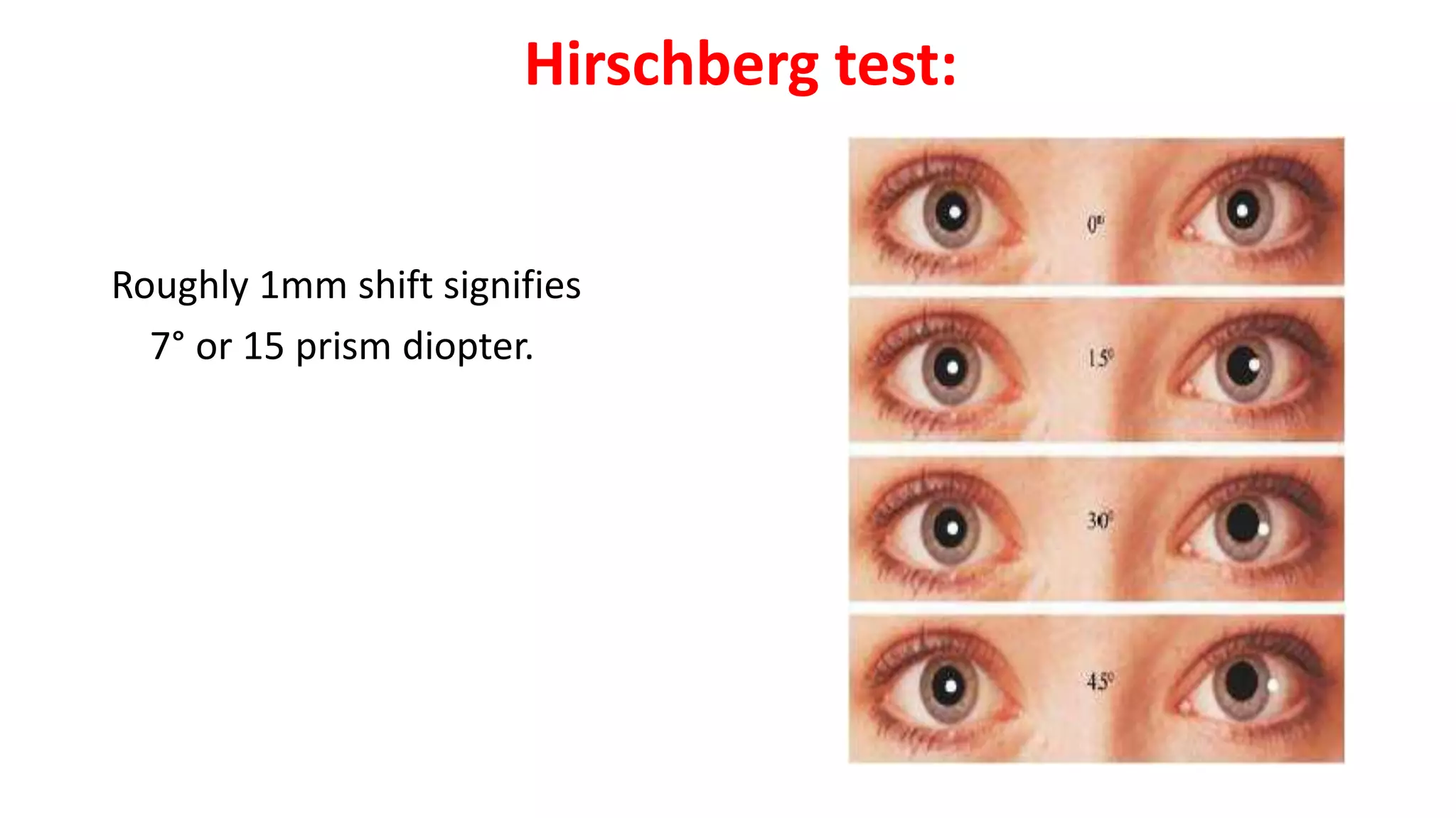
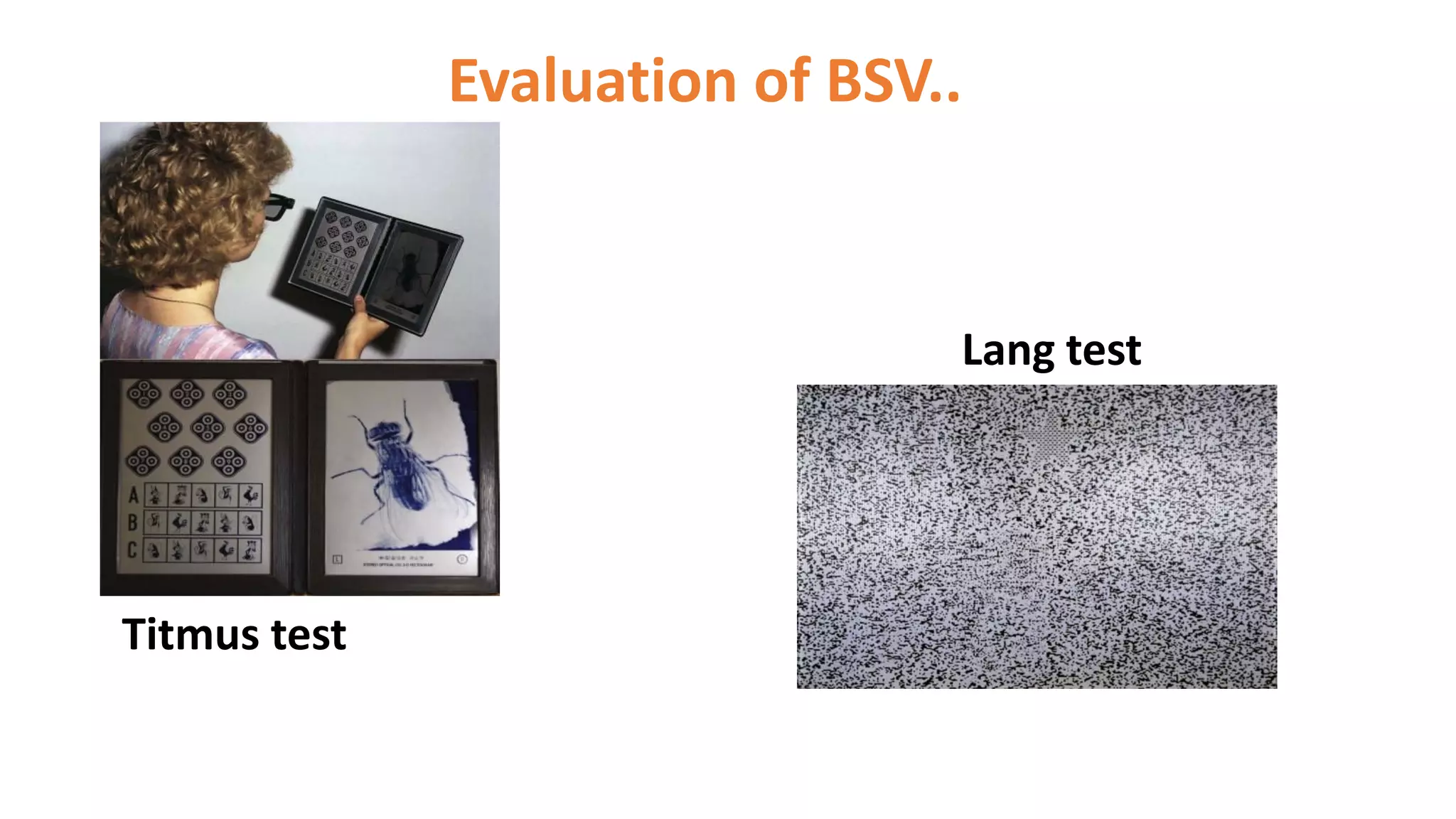
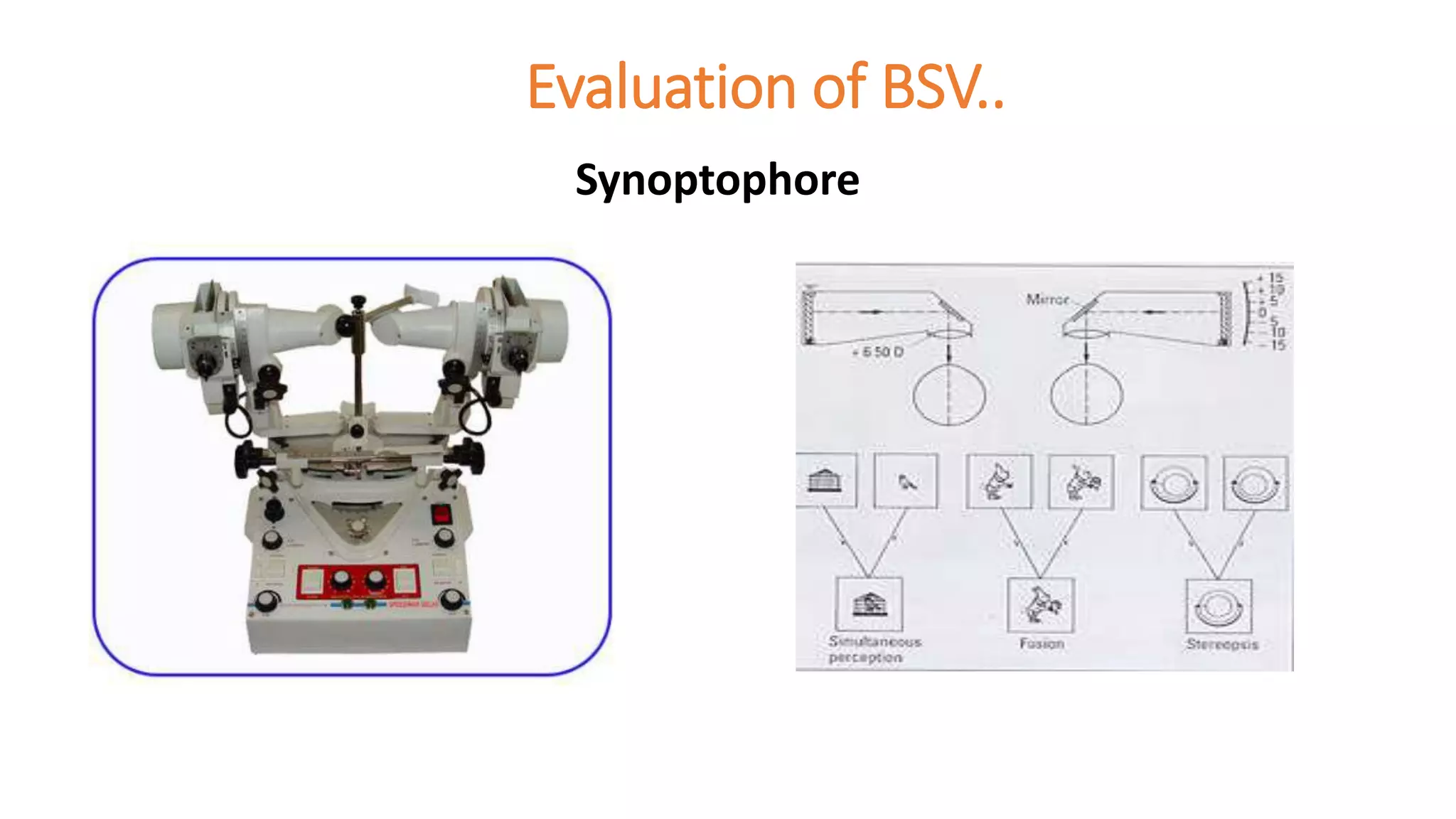
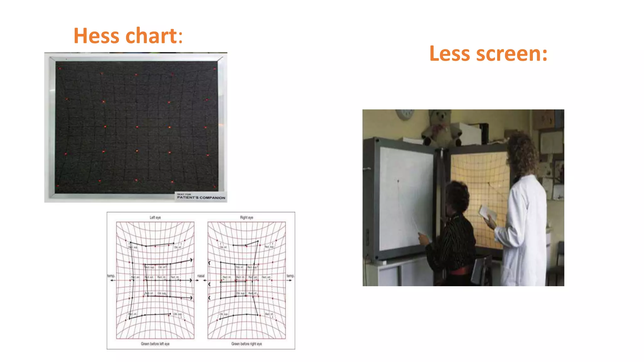
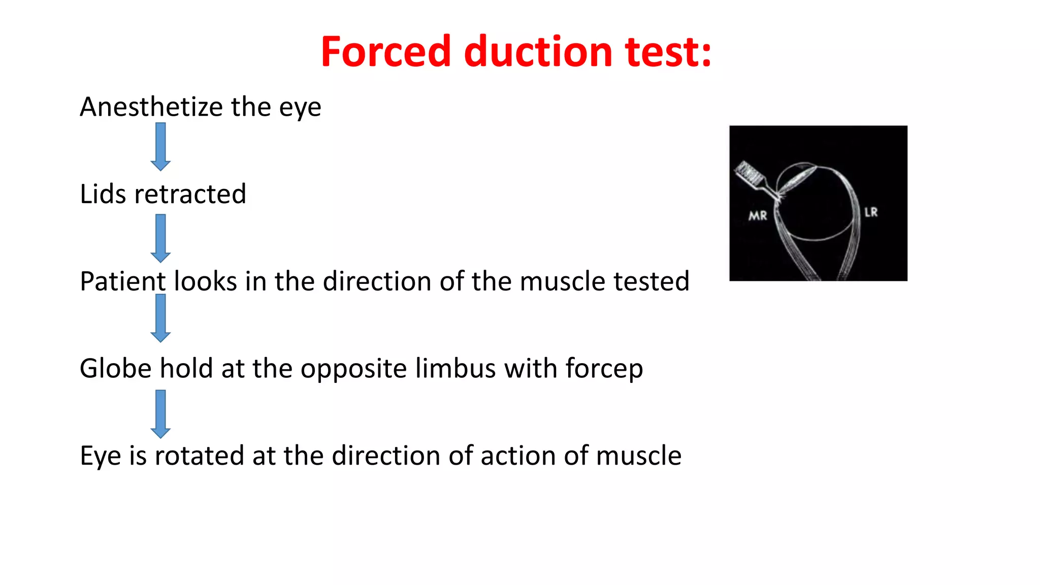
Assessment of Strabismus
Dr. Arup krishna Choudhury
FCPS, DO, MBBS
vitreo-Retina Fellow (IIEI&H)
Strabismus is the condition when
visual axes of two eyes do not meet at
the point of fixation
• Comitant: Although misaligned they retain relation in all direction of
gaze
• Incomitant: Deviation is different in all position of gaze
• Tropia: It is manifest ocular misalignment
• Phoria: It is latent ocular deviation
• Alternate : Fixation is retained by alternate eye
• Unilateral: Only one eye habitually fixes
• Intermittent: when deviation remain only for some time
Tropia or Phoria can be
• Esotropia /phoria
• Exotropia/phoria
• Hypertropia/phoria
• Hypotropia/phoria
• Cyclotropia/phoria
Children at risk
• Consanguinity
• Positive family history
• Pre mature and LBW
• Smoking during pregnancy
• Chromosomal abnormalities - DRS
• Uncorrected refractive error
• Cerebral palsy
History
A careful history is important in the diagnosis
• Mother's Obstretical history
• Birth history
• Drug history
• Family history
• Developmental milestones
• Any trauma, especially to head or face
History..
• History of presenting complaints
• Age of onset
• Sudden or gradual
• Constant or intermittent
• Present for distant or near or both
History..
• Unilateral or alternating
• Is it present only when the patient is inattentive or fatigued
• Association with trauma or physical stress
• Old photographs
• History of previous therapy/Rx received
• General health
Examination
• Vision test
• Refraction
• Evaluation of strabismus
• Assessment of binocular vision
• Detailed ophthalmic evaluation
• Special test
Visual acuity
Preverbal children
• Fixation pattern
• Fixation behavior
• Preferential looking tests-
Teller acuity cards
Cardiff acuity cards
• OKN
• VEP
Visual acuity..
For 2-3 years
• Sheridan Gardner test
• Crowded kay picture test
• Coin test
• Miniature toy test
Visual acuity..
Preschool Children
• HOTV chart
• Lea symbols
• Allen figures
Visual acuity..
For school children and adults-
LogMAR
Snellen test type
E chart
Landolt’s test type
Refraction
• Refractive error may be
responsible for the symptoms of
the patient or for the deviation
itself.
• Preferably cycloplegic refraction
Strabismus evaluation
• Hirschberg test:
• Cover tests
• Corneal reflection test
• Subjective tests of ocular alignment
• Ocular motility
Hirschberg test:
Roughly 1mm shift signifies
7° or 15 prism diopter.
Cover tests
• Cover/uncover test
-one eye is covered and observe the movement of uncovered eye to
take up fixation
-position and movement of the covered eye, as the cover is removed
Cover tests..
• Alternate cover test
-one eye or the other is covered throughout the test;
- the movement of the covered eye is noted as the cover is changed
from one eye to the other
Cover tests..
• Cover-uncover test Alternate cover test
Phoria Total deviation
Tropia Eye preference
Information provided by the cover test
• Direction of the deviation
• Difference in the angle from near to distance fixation
• Effect of accommodation
• Patients refractive error
• Comitance or incomitance
Information provided by the cover test
• Characteristics of manifest strabismus; constant or intermittent;
unilateral or alternating
• Speed of recovery
• Presence of latent nystagmus
Recording results of the Cover test..
• Type of deviation; whether manifest or latent
• Direction of deviation ; Horizontal, vertical or combination of the two
• Which eye deviates
• Estimation of size
• Any special features ; incomitance, nystagmus, DVD
Prism cover test
• It combines the alternate cover test with prism
• Indication: measure the angle of total deviation
• Apex of the prism always placed towards the deviation
Prism cover test..
Technique:
• First alternate cover test performed
• Prism of increasing strength are placed in front of one eye
• Alternate cover test continuously performed
• As stronger prism are brought
Prism cover test..
• Amplitude of re-fixation movement gradually decrease
• At a point no movement is seeing
• Increased prism power until movement is in the opposite direction
• Then reduce prism power to find the neutral value.
Prism cover test..
Corneal reflex tests
Krimsky test:
• A prism bar is placed in
front of the fixating eye
• The power increased
• Until the reflections get
symmetrical.
Corneal reflex tests…
• Prism reflection test:
Involves placement of prisms in front of deviating eye
The power increased
Until the reflections are symmetrical
Subjective tests of ocular alignment
Maddox rod test:
Orthophoria: streak passes
through white light.
If streak passes on the left to
the light i.e. esophoria &, if
to the right then exophoria
Double maddox rod test:
MADDOX WING TEST:
Ocular motility
• Ductions: Monocular movement
• Versions: Binocular , simultaneous conjugate movement
• Vergences: Binocular, simultaneous, disjugate movement
Ocular motility..
• 9 position of gaze:
Ocular motility..
Information recorded must include:
• Whether movement is full, limited , or excessive
• Grade of abnormality,usually-4 to+4
• Direction of abnormality
• Any associated signs , like lid changes or presence of nytagmus
Ocular motility..
Grading of limitation and overaction:
• Limitation is usually graded on a scale of -1 to -4
• -4 indicate there is no movement beyond the midline
• -3 indicates that 25% movement remains,
• Over action is graded from +1 to +4
Ocular motility..
• Near point of convergence:
Nearest point on which eye can maintain binocular fixation.
Measured by RAF rule
It should be Less than 10 cm.
• Near point of accommodation:
Nearest point on which eyes can maintain clear focus.
Evaluation of BSV
3 grades
1. Simultaneous perception
2. Fusion
3. Stereopsis
Evaluation of BSV..
Aims of evaluation of binocular function
To determine if the aim of treatment should be restore
binocular single vision or to improve the patients appearance
and ensure that s/he has the best possible visual acuity
Evaluation of BSV..
Test for fusion
Base-out prism test
Evaluation of BSV..
Test for stereopsis
TNO test
Frisby test
Evaluation of BSV..
Lang test
Titmus test
Evaluation of BSV..
Test for sensory anomalies
Worth four-dot test Bagolini striated glasses test
Evaluation of BSV..
Synoptophore
Ophthalmic evaluation
• Anterior segment
• Pupils
• Intra-ocular pressure
• Fundus examination
Special test
• Diplopia charting
• Hess screen test
• Lees screen test
• PARK’S 3 step test
• Forced duction test
Diplopia chart
It is indicated in patients complaining of double vision
Hess chart:
Less screen:
PARK’S 3 step test:
• Use to identify cyclovertical muscle paralysis.
• Performed by measuring the vertical alignment in
1)primary position,
2) In right and left gaze and
3) In head tilt to the right and to the left.
Forced duction test:
Anesthetize the eye
Lids retracted
Patient looks in the direction of the muscle tested
Globe hold at the opposite limbus with forcep
Eye is rotated at the direction of action of muscle
Forced duction test:
Result:
• If free movement is present then the test is negative
• If restricted then the test is positive
Take Home message
• Time consuming
• Needs Patience specially for intermittent exotropia
For children
• Poor cooperation
• Lack of subjective response
Challenges
Assessment of strabismus

Assessment of strabismus

  • 1.
    Assessment of Strabismus Dr.Arup krishna Choudhury FCPS, DO, MBBS vitreo-Retina Fellow (IIEI&H)
  • 2.
    Strabismus is thecondition when visual axes of two eyes do not meet at the point of fixation
  • 3.
    • Comitant: Althoughmisaligned they retain relation in all direction of gaze • Incomitant: Deviation is different in all position of gaze • Tropia: It is manifest ocular misalignment • Phoria: It is latent ocular deviation • Alternate : Fixation is retained by alternate eye • Unilateral: Only one eye habitually fixes • Intermittent: when deviation remain only for some time
  • 4.
    Tropia or Phoriacan be • Esotropia /phoria • Exotropia/phoria • Hypertropia/phoria • Hypotropia/phoria • Cyclotropia/phoria
  • 5.
    Children at risk •Consanguinity • Positive family history • Pre mature and LBW • Smoking during pregnancy • Chromosomal abnormalities - DRS • Uncorrected refractive error • Cerebral palsy
  • 6.
    History A careful historyis important in the diagnosis • Mother's Obstretical history • Birth history • Drug history • Family history • Developmental milestones • Any trauma, especially to head or face
  • 7.
    History.. • History ofpresenting complaints • Age of onset • Sudden or gradual • Constant or intermittent • Present for distant or near or both
  • 8.
    History.. • Unilateral oralternating • Is it present only when the patient is inattentive or fatigued • Association with trauma or physical stress • Old photographs • History of previous therapy/Rx received • General health
  • 9.
    Examination • Vision test •Refraction • Evaluation of strabismus • Assessment of binocular vision • Detailed ophthalmic evaluation • Special test
  • 10.
    Visual acuity Preverbal children •Fixation pattern • Fixation behavior • Preferential looking tests- Teller acuity cards Cardiff acuity cards • OKN • VEP
  • 11.
    Visual acuity.. For 2-3years • Sheridan Gardner test • Crowded kay picture test • Coin test • Miniature toy test
  • 12.
    Visual acuity.. Preschool Children •HOTV chart • Lea symbols • Allen figures
  • 13.
    Visual acuity.. For schoolchildren and adults- LogMAR Snellen test type E chart Landolt’s test type
  • 14.
    Refraction • Refractive errormay be responsible for the symptoms of the patient or for the deviation itself. • Preferably cycloplegic refraction
  • 15.
    Strabismus evaluation • Hirschbergtest: • Cover tests • Corneal reflection test • Subjective tests of ocular alignment • Ocular motility
  • 16.
    Hirschberg test: Roughly 1mmshift signifies 7° or 15 prism diopter.
  • 17.
    Cover tests • Cover/uncovertest -one eye is covered and observe the movement of uncovered eye to take up fixation -position and movement of the covered eye, as the cover is removed
  • 18.
    Cover tests.. • Alternatecover test -one eye or the other is covered throughout the test; - the movement of the covered eye is noted as the cover is changed from one eye to the other
  • 19.
    Cover tests.. • Cover-uncovertest Alternate cover test Phoria Total deviation Tropia Eye preference
  • 20.
    Information provided bythe cover test • Direction of the deviation • Difference in the angle from near to distance fixation • Effect of accommodation • Patients refractive error • Comitance or incomitance
  • 21.
    Information provided bythe cover test • Characteristics of manifest strabismus; constant or intermittent; unilateral or alternating • Speed of recovery • Presence of latent nystagmus
  • 22.
    Recording results ofthe Cover test.. • Type of deviation; whether manifest or latent • Direction of deviation ; Horizontal, vertical or combination of the two • Which eye deviates • Estimation of size • Any special features ; incomitance, nystagmus, DVD
  • 23.
    Prism cover test •It combines the alternate cover test with prism • Indication: measure the angle of total deviation • Apex of the prism always placed towards the deviation
  • 24.
    Prism cover test.. Technique: •First alternate cover test performed • Prism of increasing strength are placed in front of one eye • Alternate cover test continuously performed • As stronger prism are brought
  • 25.
    Prism cover test.. •Amplitude of re-fixation movement gradually decrease • At a point no movement is seeing • Increased prism power until movement is in the opposite direction • Then reduce prism power to find the neutral value.
  • 26.
  • 27.
    Corneal reflex tests Krimskytest: • A prism bar is placed in front of the fixating eye • The power increased • Until the reflections get symmetrical.
  • 28.
    Corneal reflex tests… •Prism reflection test: Involves placement of prisms in front of deviating eye The power increased Until the reflections are symmetrical
  • 29.
    Subjective tests ofocular alignment
  • 30.
    Maddox rod test: Orthophoria:streak passes through white light. If streak passes on the left to the light i.e. esophoria &, if to the right then exophoria
  • 31.
  • 32.
  • 33.
    Ocular motility • Ductions:Monocular movement • Versions: Binocular , simultaneous conjugate movement • Vergences: Binocular, simultaneous, disjugate movement
  • 34.
    Ocular motility.. • 9position of gaze:
  • 35.
    Ocular motility.. Information recordedmust include: • Whether movement is full, limited , or excessive • Grade of abnormality,usually-4 to+4 • Direction of abnormality • Any associated signs , like lid changes or presence of nytagmus
  • 36.
    Ocular motility.. Grading oflimitation and overaction: • Limitation is usually graded on a scale of -1 to -4 • -4 indicate there is no movement beyond the midline • -3 indicates that 25% movement remains, • Over action is graded from +1 to +4
  • 37.
    Ocular motility.. • Nearpoint of convergence: Nearest point on which eye can maintain binocular fixation. Measured by RAF rule It should be Less than 10 cm. • Near point of accommodation: Nearest point on which eyes can maintain clear focus.
  • 38.
    Evaluation of BSV 3grades 1. Simultaneous perception 2. Fusion 3. Stereopsis
  • 39.
    Evaluation of BSV.. Aimsof evaluation of binocular function To determine if the aim of treatment should be restore binocular single vision or to improve the patients appearance and ensure that s/he has the best possible visual acuity
  • 40.
    Evaluation of BSV.. Testfor fusion Base-out prism test
  • 41.
    Evaluation of BSV.. Testfor stereopsis TNO test Frisby test
  • 42.
    Evaluation of BSV.. Langtest Titmus test
  • 43.
    Evaluation of BSV.. Testfor sensory anomalies Worth four-dot test Bagolini striated glasses test
  • 44.
  • 45.
    Ophthalmic evaluation • Anteriorsegment • Pupils • Intra-ocular pressure • Fundus examination
  • 46.
    Special test • Diplopiacharting • Hess screen test • Lees screen test • PARK’S 3 step test • Forced duction test
  • 47.
    Diplopia chart It isindicated in patients complaining of double vision
  • 48.
  • 49.
    PARK’S 3 steptest: • Use to identify cyclovertical muscle paralysis. • Performed by measuring the vertical alignment in 1)primary position, 2) In right and left gaze and 3) In head tilt to the right and to the left.
  • 50.
    Forced duction test: Anesthetizethe eye Lids retracted Patient looks in the direction of the muscle tested Globe hold at the opposite limbus with forcep Eye is rotated at the direction of action of muscle
  • 51.
    Forced duction test: Result: •If free movement is present then the test is negative • If restricted then the test is positive
  • 52.
    Take Home message •Time consuming • Needs Patience specially for intermittent exotropia For children • Poor cooperation • Lack of subjective response Challenges