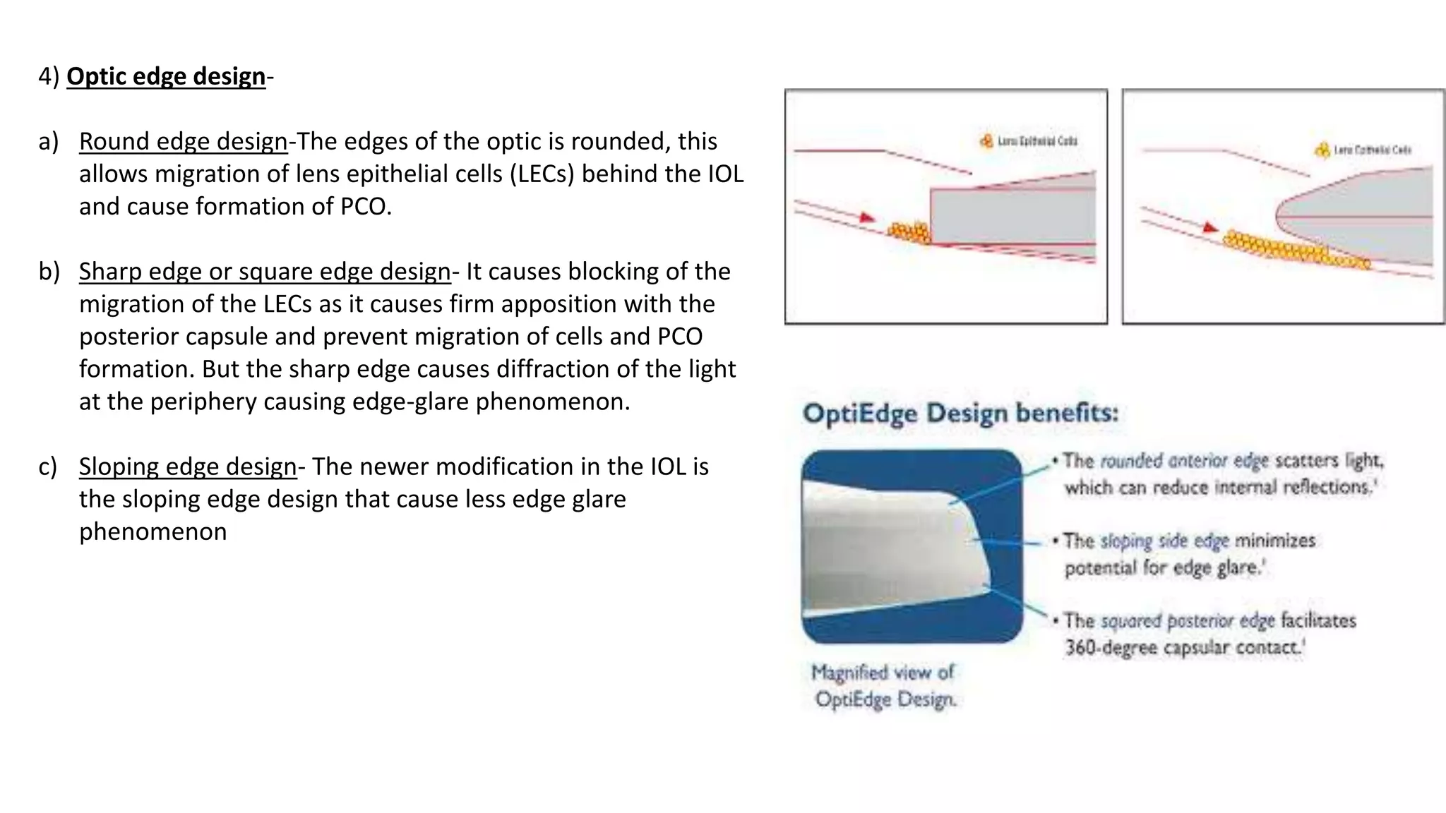
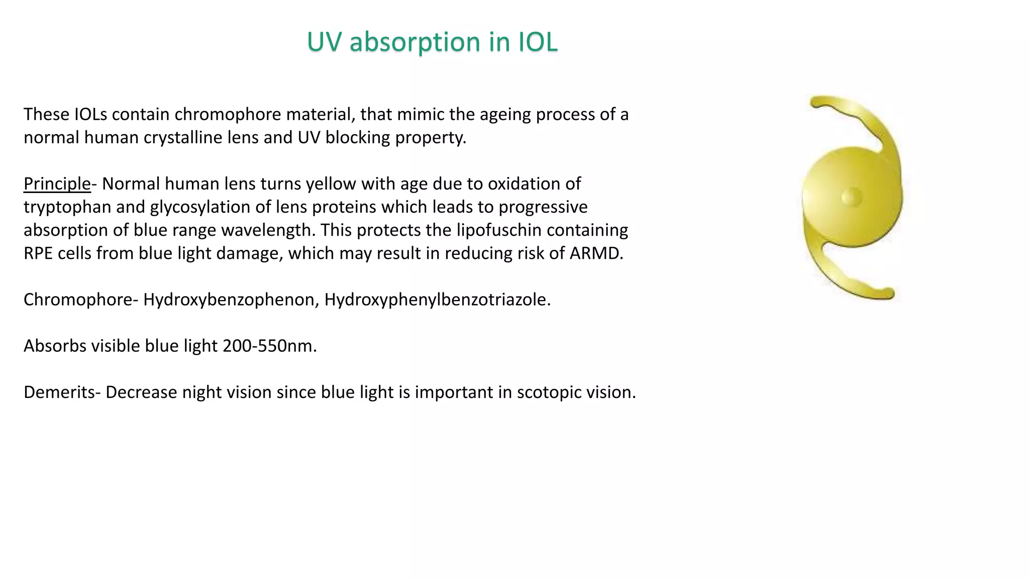
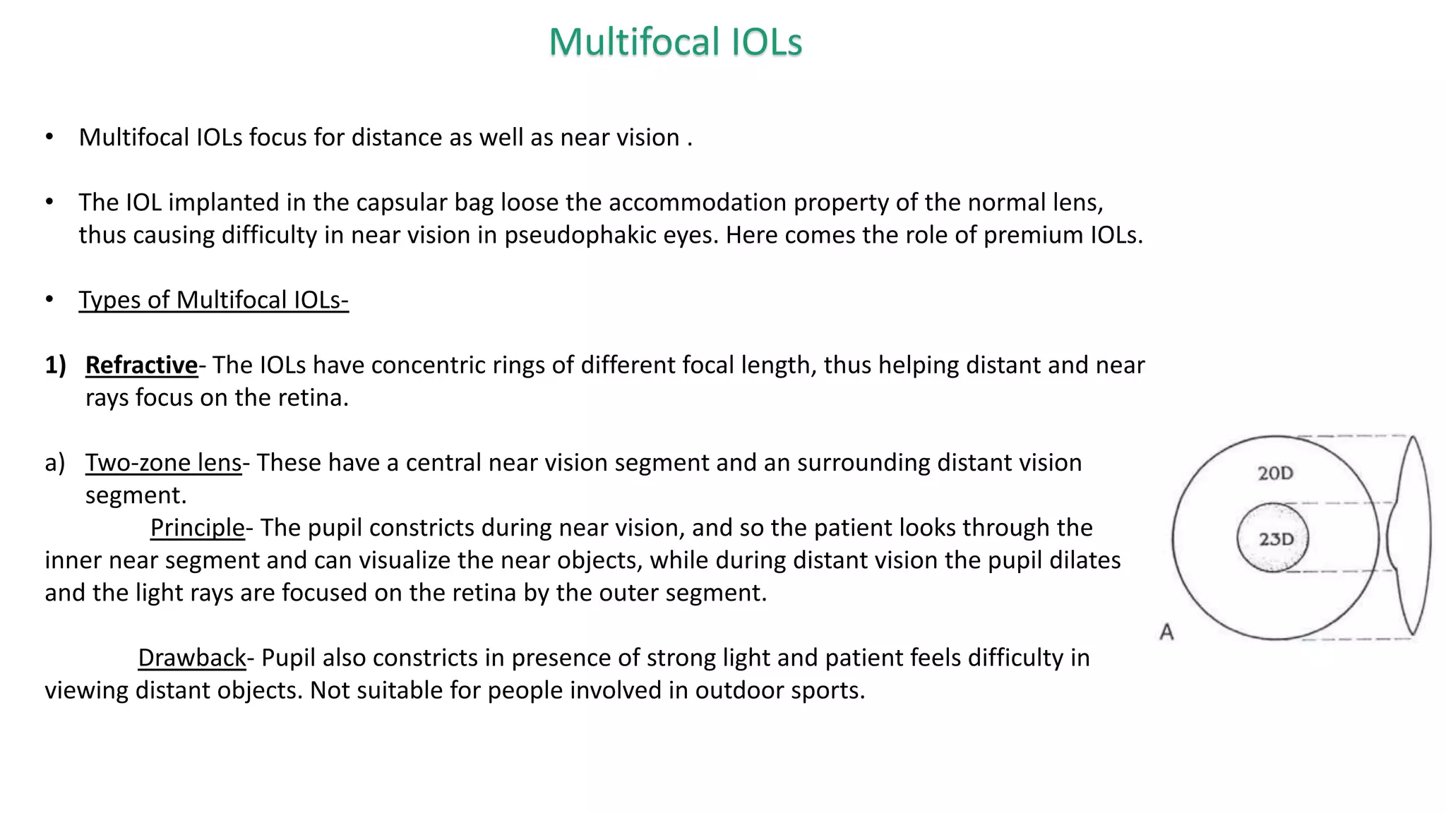
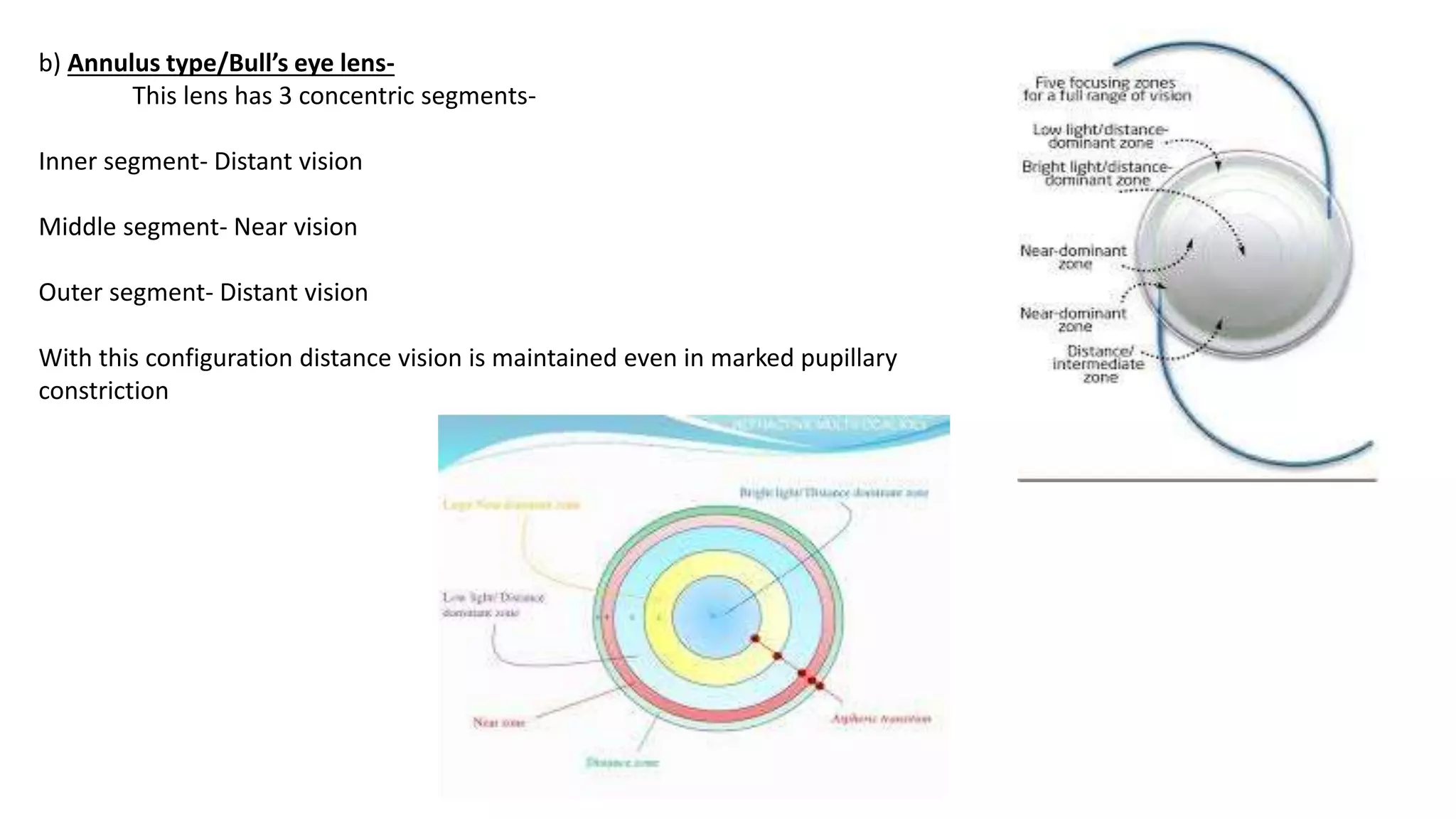
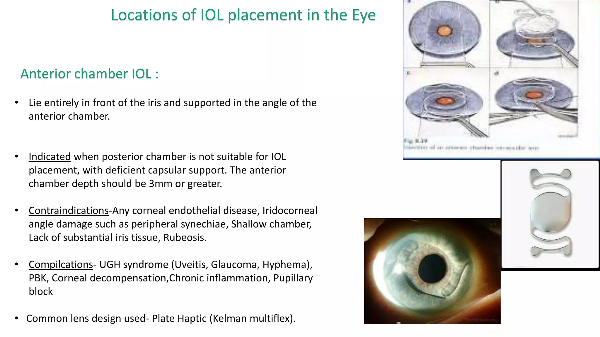
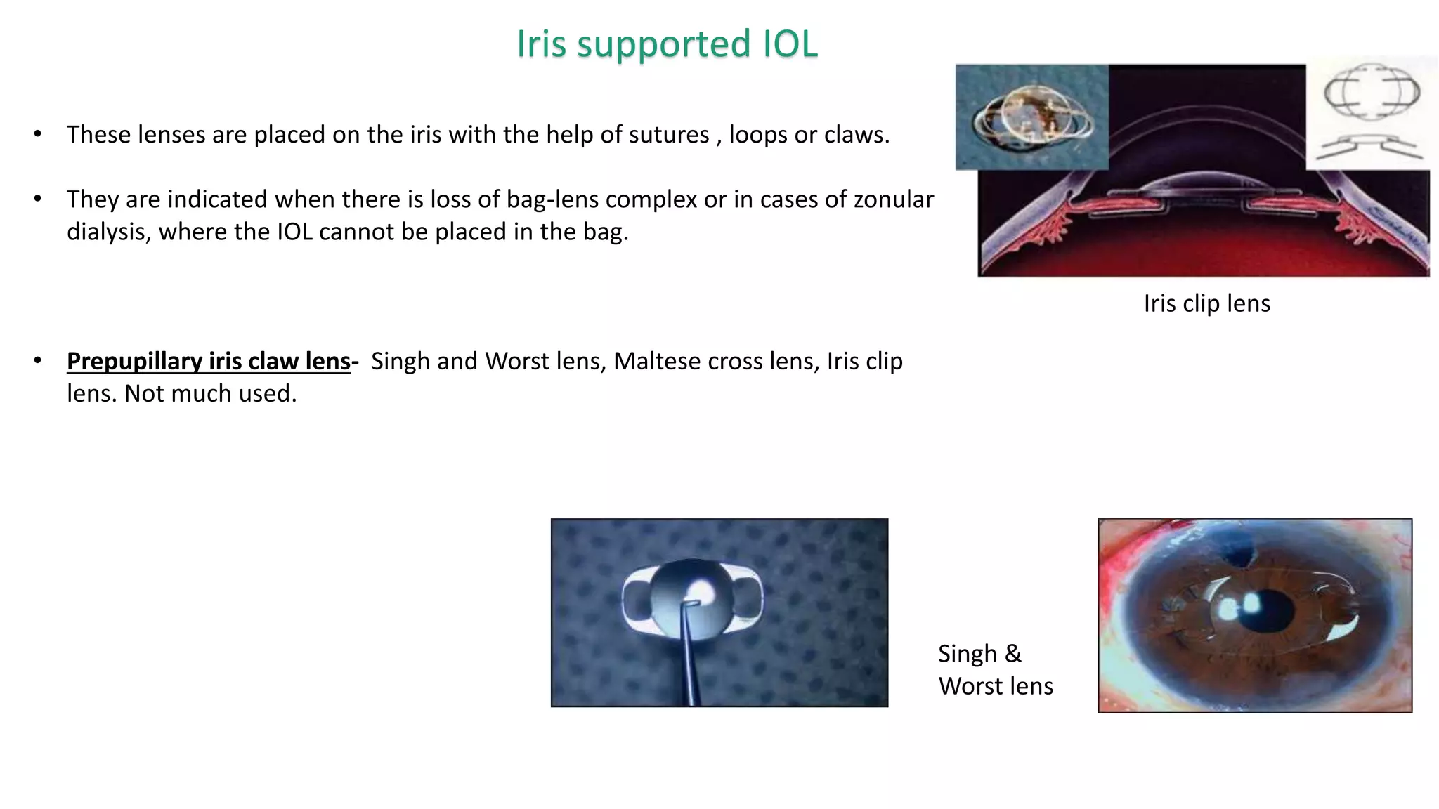
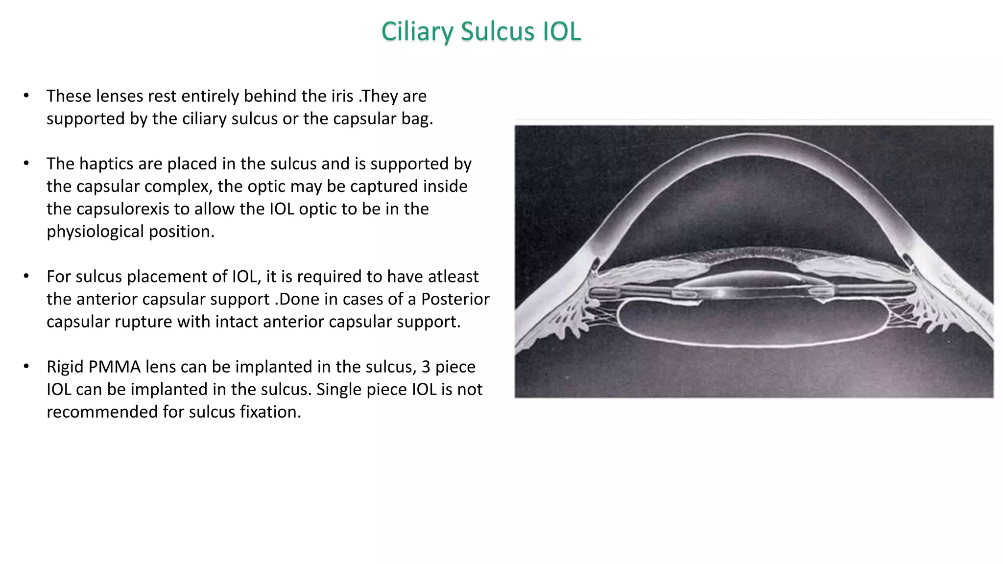
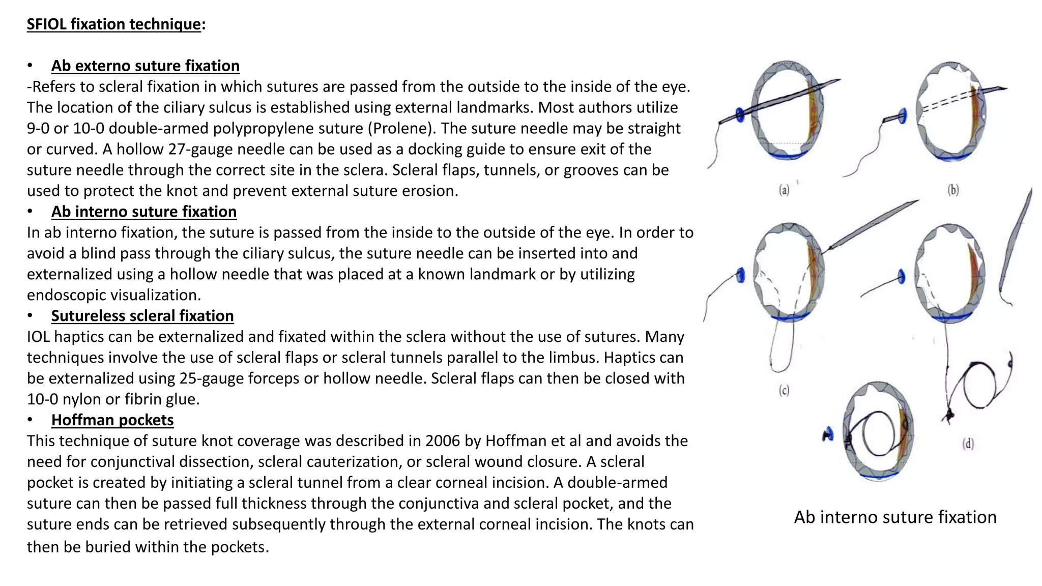
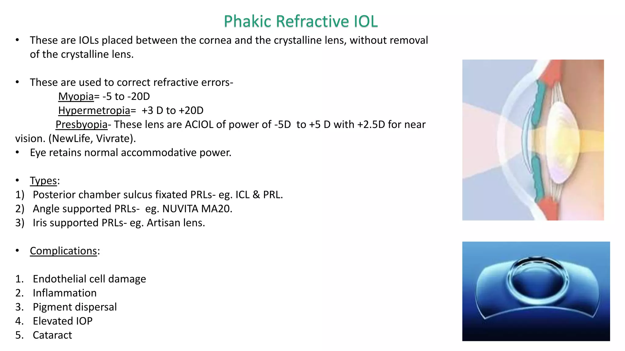
Artificial lenses implanted in the anterior or posterior chamber of the eye in the presence of the natural crystalline lens to correct refractive errors. Phakic IOLs an evolving technique in the field of refractive surgery for the correction of moderate to high refractive errors. Patients with high myopia (above -10 diopters) constitute only about 2% of the myopic population but 13-15% of patients presenting for refractive surgery belong to this group. The increased knowledge on anterior segment anatomy and availability of better imaging technologies along with improved IOL designs and surgical techniques have led to higher success rates with these lenses.
Compared to corneal refractive surgery , phakic IOLs compete favorably for the correction of high ametropias, with excellent predictability, efficacy, safety and quality of vision.
The Right Contact - Up to date information regarding contact lenses, Hyrid lenses, Soft lenses, button materials, gas permable lens and contact lens care products.
ARTIFICIAL INTELLIGENCE IN HEALTHCARE.pdfAnujkumaranit
Artificial intelligence (AI) refers to the simulation of human intelligence processes by machines, especially computer systems. It encompasses tasks such as learning, reasoning, problem-solving, perception, and language understanding. AI technologies are revolutionizing various fields, from healthcare to finance, by enabling machines to perform tasks that typically require human intelligence.
The prostate is an exocrine gland of the male mammalian reproductive system
It is a walnut-sized gland that forms part of the male reproductive system and is located in front of the rectum and just below the urinary bladder
Function is to store and secrete a clear, slightly alkaline fluid that constitutes 10-30% of the volume of the seminal fluid that along with the spermatozoa, constitutes semen
A healthy human prostate measures (4cm-vertical, by 3cm-horizontal, 2cm ant-post ).
It surrounds the urethra just below the urinary bladder. It has anterior, median, posterior and two lateral lobes
It’s work is regulated by androgens which are responsible for male sex characteristics
Generalised disease of the prostate due to hormonal derangement which leads to non malignant enlargement of the gland (increase in the number of epithelial cells and stromal tissue)to cause compression of the urethra leading to symptoms (LUTS
Tom Selleck Health: A Comprehensive Look at the Iconic Actor’s Wellness Journeygreendigital
Tom Selleck, an enduring figure in Hollywood. has captivated audiences for decades with his rugged charm, iconic moustache. and memorable roles in television and film. From his breakout role as Thomas Magnum in Magnum P.I. to his current portrayal of Frank Reagan in Blue Bloods. Selleck's career has spanned over 50 years. But beyond his professional achievements. fans have often been curious about Tom Selleck Health. especially as he has aged in the public eye.
Follow us on: Pinterest
Introduction
Many have been interested in Tom Selleck health. not only because of his enduring presence on screen but also because of the challenges. and lifestyle choices he has faced and made over the years. This article delves into the various aspects of Tom Selleck health. exploring his fitness regimen, diet, mental health. and the challenges he has encountered as he ages. We'll look at how he maintains his well-being. the health issues he has faced, and his approach to ageing .
Early Life and Career
Childhood and Athletic Beginnings
Tom Selleck was born on January 29, 1945, in Detroit, Michigan, and grew up in Sherman Oaks, California. From an early age, he was involved in sports, particularly basketball. which played a significant role in his physical development. His athletic pursuits continued into college. where he attended the University of Southern California (USC) on a basketball scholarship. This early involvement in sports laid a strong foundation for his physical health and disciplined lifestyle.
Transition to Acting
Selleck's transition from an athlete to an actor came with its physical demands. His first significant role in "Magnum P.I." required him to perform various stunts and maintain a fit appearance. This role, which he played from 1980 to 1988. necessitated a rigorous fitness routine to meet the show's demands. setting the stage for his long-term commitment to health and wellness.
Fitness Regimen
Workout Routine
Tom Selleck health and fitness regimen has evolved. adapting to his changing roles and age. During his "Magnum, P.I." days. Selleck's workouts were intense and focused on building and maintaining muscle mass. His routine included weightlifting, cardiovascular exercises. and specific training for the stunts he performed on the show.
Selleck adjusted his fitness routine as he aged to suit his body's needs. Today, his workouts focus on maintaining flexibility, strength, and cardiovascular health. He incorporates low-impact exercises such as swimming, walking, and light weightlifting. This balanced approach helps him stay fit without putting undue strain on his joints and muscles.
Importance of Flexibility and Mobility
In recent years, Selleck has emphasized the importance of flexibility and mobility in his fitness regimen. Understanding the natural decline in muscle mass and joint flexibility with age. he includes stretching and yoga in his routine. These practices help prevent injuries, improve posture, and maintain mobilit
Flu Vaccine Alert in Bangalore Karnatakaaddon Scans
As flu season approaches, health officials in Bangalore, Karnataka, are urging residents to get their flu vaccinations. The seasonal flu, while common, can lead to severe health complications, particularly for vulnerable populations such as young children, the elderly, and those with underlying health conditions.
Dr. Vidisha Kumari, a leading epidemiologist in Bangalore, emphasizes the importance of getting vaccinated. "The flu vaccine is our best defense against the influenza virus. It not only protects individuals but also helps prevent the spread of the virus in our communities," he says.
This year, the flu season is expected to coincide with a potential increase in other respiratory illnesses. The Karnataka Health Department has launched an awareness campaign highlighting the significance of flu vaccinations. They have set up multiple vaccination centers across Bangalore, making it convenient for residents to receive their shots.
To encourage widespread vaccination, the government is also collaborating with local schools, workplaces, and community centers to facilitate vaccination drives. Special attention is being given to ensuring that the vaccine is accessible to all, including marginalized communities who may have limited access to healthcare.
Residents are reminded that the flu vaccine is safe and effective. Common side effects are mild and may include soreness at the injection site, mild fever, or muscle aches. These side effects are generally short-lived and far less severe than the flu itself.
Healthcare providers are also stressing the importance of continuing COVID-19 precautions. Wearing masks, practicing good hand hygiene, and maintaining social distancing are still crucial, especially in crowded places.
Protect yourself and your loved ones by getting vaccinated. Together, we can help keep Bangalore healthy and safe this flu season. For more information on vaccination centers and schedules, residents can visit the Karnataka Health Department’s official website or follow their social media pages.
Stay informed, stay safe, and get your flu shot today!
Explore natural remedies for syphilis treatment in Singapore. Discover alternative therapies, herbal remedies, and lifestyle changes that may complement conventional treatments. Learn about holistic approaches to managing syphilis symptoms and supporting overall health.
Lung Cancer: Artificial Intelligence, Synergetics, Complex System Analysis, S...Oleg Kshivets
RESULTS: Overall life span (LS) was 2252.1±1742.5 days and cumulative 5-year survival (5YS) reached 73.2%, 10 years – 64.8%, 20 years – 42.5%. 513 LCP lived more than 5 years (LS=3124.6±1525.6 days), 148 LCP – more than 10 years (LS=5054.4±1504.1 days).199 LCP died because of LC (LS=562.7±374.5 days). 5YS of LCP after bi/lobectomies was significantly superior in comparison with LCP after pneumonectomies (78.1% vs.63.7%, P=0.00001 by log-rank test). AT significantly improved 5YS (66.3% vs. 34.8%) (P=0.00000 by log-rank test) only for LCP with N1-2. Cox modeling displayed that 5YS of LCP significantly depended on: phase transition (PT) early-invasive LC in terms of synergetics, PT N0—N12, cell ratio factors (ratio between cancer cells- CC and blood cells subpopulations), G1-3, histology, glucose, AT, blood cell circuit, prothrombin index, heparin tolerance, recalcification time (P=0.000-0.038). Neural networks, genetic algorithm selection and bootstrap simulation revealed relationships between 5YS and PT early-invasive LC (rank=1), PT N0—N12 (rank=2), thrombocytes/CC (3), erythrocytes/CC (4), eosinophils/CC (5), healthy cells/CC (6), lymphocytes/CC (7), segmented neutrophils/CC (8), stick neutrophils/CC (9), monocytes/CC (10); leucocytes/CC (11). Correct prediction of 5YS was 100% by neural networks computing (area under ROC curve=1.0; error=0.0).
CONCLUSIONS: 5YS of LCP after radical procedures significantly depended on: 1) PT early-invasive cancer; 2) PT N0--N12; 3) cell ratio factors; 4) blood cell circuit; 5) biochemical factors; 6) hemostasis system; 7) AT; 8) LC characteristics; 9) LC cell dynamics; 10) surgery type: lobectomy/pneumonectomy; 11) anthropometric data. Optimal diagnosis and treatment strategies for LC are: 1) screening and early detection of LC; 2) availability of experienced thoracic surgeons because of complexity of radical procedures; 3) aggressive en block surgery and adequate lymph node dissection for completeness; 4) precise prediction; 5) adjuvant chemoimmunoradiotherapy for LCP with unfavorable prognosis.
Pulmonary Thromboembolism - etilogy, types, medical- Surgical and nursing man...VarunMahajani
Disruption of blood supply to lung alveoli due to blockage of one or more pulmonary blood vessels is called as Pulmonary thromboembolism. In this presentation we will discuss its causes, types and its management in depth.
Title: Sense of Taste
Presenter: Dr. Faiza, Assistant Professor of Physiology
Qualifications:
MBBS (Best Graduate, AIMC Lahore)
FCPS Physiology
ICMT, CHPE, DHPE (STMU)
MPH (GC University, Faisalabad)
MBA (Virtual University of Pakistan)
Learning Objectives:
Describe the structure and function of taste buds.
Describe the relationship between the taste threshold and taste index of common substances.
Explain the chemical basis and signal transduction of taste perception for each type of primary taste sensation.
Recognize different abnormalities of taste perception and their causes.
Key Topics:
Significance of Taste Sensation:
Differentiation between pleasant and harmful food
Influence on behavior
Selection of food based on metabolic needs
Receptors of Taste:
Taste buds on the tongue
Influence of sense of smell, texture of food, and pain stimulation (e.g., by pepper)
Primary and Secondary Taste Sensations:
Primary taste sensations: Sweet, Sour, Salty, Bitter, Umami
Chemical basis and signal transduction mechanisms for each taste
Taste Threshold and Index:
Taste threshold values for Sweet (sucrose), Salty (NaCl), Sour (HCl), and Bitter (Quinine)
Taste index relationship: Inversely proportional to taste threshold
Taste Blindness:
Inability to taste certain substances, particularly thiourea compounds
Example: Phenylthiocarbamide
Structure and Function of Taste Buds:
Composition: Epithelial cells, Sustentacular/Supporting cells, Taste cells, Basal cells
Features: Taste pores, Taste hairs/microvilli, and Taste nerve fibers
Location of Taste Buds:
Found in papillae of the tongue (Fungiform, Circumvallate, Foliate)
Also present on the palate, tonsillar pillars, epiglottis, and proximal esophagus
Mechanism of Taste Stimulation:
Interaction of taste substances with receptors on microvilli
Signal transduction pathways for Umami, Sweet, Bitter, Sour, and Salty tastes
Taste Sensitivity and Adaptation:
Decrease in sensitivity with age
Rapid adaptation of taste sensation
Role of Saliva in Taste:
Dissolution of tastants to reach receptors
Washing away the stimulus
Taste Preferences and Aversions:
Mechanisms behind taste preference and aversion
Influence of receptors and neural pathways
Impact of Sensory Nerve Damage:
Degeneration of taste buds if the sensory nerve fiber is cut
Abnormalities of Taste Detection:
Conditions: Ageusia, Hypogeusia, Dysgeusia (parageusia)
Causes: Nerve damage, neurological disorders, infections, poor oral hygiene, adverse drug effects, deficiencies, aging, tobacco use, altered neurotransmitter levels
Neurotransmitters and Taste Threshold:
Effects of serotonin (5-HT) and norepinephrine (NE) on taste sensitivity
Supertasters:
25% of the population with heightened sensitivity to taste, especially bitterness
Increased number of fungiform papillae
TEST BANK for Operations Management, 14th Edition by William J. Stevenson, Ve...kevinkariuki227
TEST BANK for Operations Management, 14th Edition by William J. Stevenson, Verified Chapters 1 - 19, Complete Newest Version.pdf
Couples presenting to the infertility clinic- Do they really have infertility...Sujoy Dasgupta
Dr Sujoy Dasgupta presented the study on "Couples presenting to the infertility clinic- Do they really have infertility? – The unexplored stories of non-consummation" in the 13th Congress of the Asia Pacific Initiative on Reproduction (ASPIRE 2024) at Manila on 24 May, 2024.
2. References
• Ophthalmology – Yanoff, Duker. Vol 1. Page- 283-374
• Theory and Practice of Optics & Refraction. AK Khurana. Fourth edition. Page 306-346.
• Advanced IOL fixation techniques, David F Chang. Vol 1 and Vol 2
• Lens and Cataract –AAO series Section 11
• “Intraocular Lens Implantation” – Albert and Jackobics, Chapter 116.
• “Intraocular Lens Materials and Design”- Oliver Findl
• “Scleral fixated intraocular lens”- Sarah Brown. AAO July 2019
• “Multifocal IOL Surgery” - Jan Willam van der Linden
3. Contents• IOL Definition and History.
• Generations of IOL.
• Parts of an IOL.
• IOL Designs.
• IOL Materials.
• Aspheric IOLs.
• Premium IOLs- Multifocal IOL, Accomodative IOL, Toric IOL.
• IOL placement sites in the Eye.
• Special IOL placement types.
• IOL Power Calculation.
• IOL complications.- PCO
• Recent advances.
4. Definition & History
Intraocular lens (IOLs) are lens implanted in the eye as a part of treatment of cataract
or refractive errors.
History of IOL advent:
• First documented cataract surgery was in 800BC by Sushrut (India), in which the
cataractous lens was pushed back in the vitreous cavity by a sharp instrument,
yielded poor results with 70% eyes going into blindness.
• In the year 1795, Casamata (Italy) used glass lens as IOL.
• On 29th November 1949, Sir Harold Ridley successfully placed in the bag Poly methyl
Methacrylate (PMMA) lens in the eye after performing an ECCE in a 49year old
woman in England.
• Since then Ridley lens became popular and gained acceptance, but many prominent
Ophthalmologists including Sir Duke-Edlers criticized it for causing glaucoma, uveitis
and frequent dislocation. Since then the IOL advanced in technology providing
better visual outcome, better patient compliance and less complications.
Sir Harold Ridley
Ridley IOL
5. Generations of IOL
1) First generation IOL- Ridley lenses.
Posterior chamber lens. Cause Uveitis, Glaucoma and IOL dislocation
2) Second generation IOL- Early ACIOL.
Rigid/Semi-rigid IOL. Cause corneal decompensation and UGH syndrome.
3) Third generation IOL- Iris supported IOL.
Cause less corneal decompensation, but causes iris shaffing, pupillary
distortion, inflammation.
4) Fourth generation IOL- Modern Anterior Chamber IOL.
Flexible loop design, more stable, better results, less complications.
5) Fifth generation IOL- Posterior chamber rigid PMMA lenses.
6) Sixth generation IOL- Posterior chamber Foldable IOL
7) Seventh generation IOL- Multifocal IOL.
8) Eight generation IOL- Accomodative IOL, Toric IOL, Phakic refractive IOL.
6. Parts of an IOL
1) Optic- Clear part of the lens that focuses light
on the retina.
2) Haptic- Filament attached to the optic that
hold the lens in position in the eye.
Size-
The average size of capsular bag is of 10.4mm. The
IOL haptic to haptic distance is 13mm, it is slightly
oversized to provide centripetal force for lens
stabilization and this lens can also be placed in the
sulcus if required. The optic is 6mm in size.
7. Properties of an IOL
An ideal IOL should be –
• Transparent.
• High optical resolution.
• Non-reactive (inert).
• Non-toxic.
• Non-biodegradable.
• Optically compatible.
8. Concept of Biocompatibility of IOL
• The performance of an IOL is determined by the material, design and its reaction with
the ocular tissue.
• Uveal biocompatibility- Defined as the reaction of the uvea to the IOL. Monocytes and
macrophages migrate through the uvea’s vessel wall into the aqueous and over the IOL
surface.
Hydrophilic IOLs, Hydrogel IOLs have better uveal compatibility.
• Capsular biocompatibility- Defined as the reaction of the LECs (lens epithelial cells) and
the capsule to the IOL design and material. The A cells (anterior capsular cells)
proliferate onto the IOL optic from the anterior capsular rim and lay down collagen
causing whitening and contraction of the capsule. The LECs secretes cytokines (IL-1,IL-6,
TGF beta) which leads to differentiation into myofibroblast leading to capsular
contraction and phimosis. This encompasses pathogenesis of PCO also.
Hydrophobic IOLs have better capsular compatibility.
Silicon IOLs with sharp edge optics have both excellent uveal and capsular biocompatibility.
• In case of Uveitis Hydrophilic lens is preferred.
9. IOL designs
1) Plate Haptic- It was used in the first foldable silicon IOL design, it is still used
by many some manufacturers. The haptic has a plate style design.
The major drawback of plate-style design is the incomplete fusion of the anterior
and posterior capsule that leaves space along the plate haptic axis and cause
migration of LECs onto the posterior capsule causing PCO.
Plate haptics provide better stability in postoperative period and prevent IOL
rotation in the bag in case of silicon IOL.
Some manufacturers use plate-loop haptic design, which allows better
adaptability to capsule bag size variations and also provides stability to the IOL.
10. 2) Open Loop-
a) Single-piece IOL- These IOLs are produced from a single material, i.e, optic
and haptic is of the same material.
Single piece IOL are resistant to damage when used in injectors. They are
commonly preferred for in the bag placement IOL.
Next generation one piece IOL such as Technis 1 piece IOL, incorporate a 360
degree square edge design.
b) Multi-piece IOL- These IOL are produced from 2 materials, the optic and the
haptics are produced separately and then joined. Most common haptic material
is PMMA, Polyvinylidene(PVDF), Polyimide (Elastimide) and Polypropelene
(Prolene).
i) J loop design- Results in pin point contact with the capsular bag. The J loop
in the bag causes stress folds in the posterior capsule, which eventually
disappear by 1 month post-operatively. Preferred for sulcus placement.
ii) C loop design- More preferred for in the bag lens placement.
TechnisSingle-piece IOL
11. 3) Haptic Angulation-
The optic-haptic angulation are present in some lenses. This cause
better apposition of the lens with the posterior capsule causing less
space between them and hence reducing chances of PCO.
a) Planar- They have a straight optic-haptic plane with no angulation.
Eg- Single piece IOLS.
b) Angulated- The angulation between the optic and haptics is around
5-10 degree. Eg- Multipiece IOLs.
c) Offset Haptics- The haptics are placed behind the optics . Eg. Technis
12. 4) Optic edge design-
a) Round edge design-The edges of the optic is rounded, this
allows migration of lens epithelial cells (LECs) behind the IOL
and cause formation of PCO.
b) Sharp edge or square edge design- It causes blocking of the
migration of the LECs as it causes firm apposition with the
posterior capsule and prevent migration of cells and PCO
formation. But the sharp edge causes diffraction of the light
at the periphery causing edge-glare phenomenon.
c) Sloping edge design- The newer modification in the IOL is
the sloping edge design that cause less edge glare
phenomenon
13. IOL Materials
1) Rigid PMMA IOLs- The most common used material for IOL. It is rigid,
chemically stable compound.. The specific gravity is about 1.2 and is
much closer to neutral buoyancy.
Properties:
1) High optical quality.
2) PMMA has relatively high index of refraction (n=1.47-1.49)
3) Hard-rigid lens.
4) Proven biocompatibility.
5) Hydrophobic
6) Good laser resistance.
7) ACIOL and Iris fixated IOLs are PMMA lens and is very inert to uveal
tissue
8) High chance of PCO formation and requirement of a large wound for
lens insertions are the drawback.
9) Used in the bag IOL in low cost setting and also in the sulcus due to its
good centration and resistance to tilt.
14. 2) Silicon- Silicon polymers have lower index of refraction (n=1.43) than PMMA (n=1.49)
and must be thicker for the same refractive correction. Made from a number of
formulations of polyorgano-siloxane.
Properties:
1) The optic insertion requires larger insertion because of large optic size.
2) Hydrophobic material. Angle of contact with water 99 degree
3) Good memory
4) Increased tendency of bacterial adhesion.
5) Lowest threshold for lasers.
6) Glistening occur.
15. 3) Hydrophobic Acrylic-
Properties:
• Copolymers of acrylate and methacrylate
• Absorbs minimal water (4% water content)
• Most successful foldable IOLs today
• Angle of contact with water is 73°
• Used in 3-piece or single piece designs
• Refractive index=1.44 to 1.55
• Easy handling but is prone to mechanical damage
• Requires atleast a 2.2-mm incision
• Low PCO rates
• Good resistance to YAG laser
• Photopsias, Glistenings occur commonly
16. 4) Hydrophilic Acrylic IOL-
Properties:
• Mixture of hydroxyethylmethacrylate (poly- HEMA) and hydrophilic
acrylic monomer
• Refractive index=1.43
• 18 -26% water content
• Contact angle with water is lower than 50°
• Suitable for single piece design
• Easiest to handle; less mechanical and less YAG laser damage
• Sub-2-mm incisions required
• Higher PCO rate
• Low resistance to capsular contraction
• Calcium deposits
17. 5) Hydrogel-
Properties:
• Hydrate to form soft swollen rubbery mass (18% to 38% water content);
Copolymers of methacrylate esters (2-Hydroxy ethyl methacrylate-HEMA)
• Refraction index=1.43-1.48
• Good laser resistance.
• Good bio-comptability.
• Good optical quality.
• Easy handling.
• Require sub 2mm incision for insertion.
• Used to make Rollable IOLs.
• Implanted by Phaconit technique.
• eg. Acrismart lens, Ultrachoice 1.0, Medennium smart lens, Slimflex lens.
18. These IOLs contain chromophore material, that mimic the ageing process of a
normal human crystalline lens and UV blocking property.
Principle- Normal human lens turns yellow with age due to oxidation of
tryptophan and glycosylation of lens proteins which leads to progressive
absorption of blue range wavelength. This protects the lipofuschin containing
RPE cells from blue light damage, which may result in reducing risk of ARMD.
Chromophore- Hydroxybenzophenon, Hydroxyphenylbenzotriazole.
Absorbs visible blue light 200-550nm.
Demerits- Decrease night vision since blue light is important in scotopic vision.
UV absorption in IOL
19. Aspheric IOLs
• They are designed to reduce the spherical aberration(SA)
• Spherical aberration occurs when parallel rays of light do not
focus on one point.
• Positive SA- Peripheral rays of light focus in front of the central
rays.
• Negative SA- Peripheral rays of light focus behind the central
rays.
• The cornea has positive SA which in young patients gets
compensated by negative SA of the lens, but with age the lens
thickens and develops a positive SA. So in old age the eye
assumes a total positive SA leading to reduced contrast
sensitivity.
• Conventional spherical IOLs have positive SA, resulting in
reduced contrast sensitivity under mesopic and scotopic
condition, Aspheric IOLs have shown improvement in contrast
sensitivity under low luminance and high spatial frequency.
20. Types of Aspheric IOLS-
• Anterior prolate surface
– Tecnis
• Posterior prolate surface
– Acrysof IQ
• Both Anterior and Posterior prolate surfaces
– Akreos AO, SofPort AO
21. Disadvantages of Aspheric IOLs-
• Need perfect centration, decentration greater than 0.5mm will
decrease functional vision
• Decreaseddepth perception
• More expensive
• Need corneal topography for optimal results
• Not much difference in photopic conditions and in older age
group
• Not for previous hyperopic refractive surgery
23. • Multifocal IOLs focus for distance as well as near vision .
• The IOL implanted in the capsular bag loose the accommodation property of the normal lens,
thus causing difficulty in near vision in pseudophakic eyes. Here comes the role of premium IOLs.
• Types of Multifocal IOLs-
1) Refractive- The IOLs have concentric rings of different focal length, thus helping distant and near
rays focus on the retina.
a) Two-zone lens- These have a central near vision segment and an surrounding distant vision
segment.
Principle- The pupil constricts during near vision, and so the patient looks through the
inner near segment and can visualize the near objects, while during distant vision the pupil dilates
and the light rays are focused on the retina by the outer segment.
Drawback- Pupil also constricts in presence of strong light and patient feels difficulty in
viewing distant objects. Not suitable for people involved in outdoor sports.
Multifocal IOLs
24. b) Annulus type/Bull’s eye lens-
This lens has 3 concentric segments-
Inner segment- Distant vision
Middle segment- Near vision
Outer segment- Distant vision
With this configuration distance vision is maintained even in marked pupillary
constriction
25. 2) Diffractive IOLs-
• It utilizes the the principle of wavefront optics of light.
• The diffraction optics lens combines a standard convex curvature placed on the front
surface with approximately 25 annular zones cut on the posterior surface with microscopic
steps between coterminous annuli. The step height is in the range of the wavelength of
light.
• Such lens design produces 2 diffractive orders in which the incoming waves of light will be
in phase, resulting in discrete optical foci of equal intensity.
• 82% of the light is found in 2 major foci (41% of light is in phase and 41% of light is for near
vision). Rest 18% is lost.
• If the lens gets decentered or pupil is eccentric still the lens would be able to focus distant
and near rays.
• Drawbacks of Multifocal IOL-
1) The intermediate vision is compromised.
2) Glare and halos caused by randomly diffracted rays can occur, night driving is difficult.
3) Contrast sensitivity and scotopic vision are compromised.
4) Perception of rings around point source of light.
26. Further modifications in Diffractive IOLs to overcome the drawbacks-
1) Height and spacing of diffractive rings are reduced to reduce peripheral scatter and glare.
2) Rings are made smooth rounded edges to reduce glare. Eg. Acri.LISA (Zeiss)
3) Partial optic diffractive lens, only cover the central 3mm of the lens. Eg. Restor (Alcon)
4) Full optic diffractive lens. Eg. Technis (AMO)
5) Apodisation-
In this the diffractive rings are progressively sloped greater from centre to periphery ,
this changes the angle of diffraction of light affording good intermediate vision.
Eg. – iDIFF Plus, AcriDIFF., ReSTOR.
Apodisation
27. Accomodative IOLs
• These IOLs afford both near and distance vision with the help of haptics which
can flex and the lens expands and contracts within the capsular bag, along with
the contraction of the ciliary muscles.
• Ciliary muscle contraction IOL moves forward 0.7mm anterior movement
cause 1D increase in refractive power.
• The patient achieves about J3 vision with these lenses which is sufficient for
driving and reading newspaper.
• In comparision to multifocal lenses there is no loss of contrast sensitivity, colour
distortion and no halos at night.
• With blended vision concept; one eye is made slightly myopic and the other
emmetropic with accommodative IOLs, the patient achieves good near,
intermediate and distant vision.
28. Types-
1)Single optic Accommodative IOLs-
Eg. Cystalens, Kelmantetraflex.
Designed to translate anteriorly with accommodative effort. The forward
movement increases effective lens power.
2) Dual optics Accommodative IOLs-
Eg. Visiogen
The optic consists of 2 lenses – a high plus anterior lens and a minus posterior
lens which are separated by a spring haptic.
In non-accommodative phase- the tension in the capsular bag and zonules
keeps the two optics in close proximity, whereas spring haptic is collapsed and
exhibit potential energy.
With accommodative effort the zonules relax, the capsular bag expands and the
spring exhibits kinetic energy, the optics separate the anterior plus lens moves
forward producing higher optical power and aids in near vision.
Visiogen
29. Toric IOLs
• These IOLs provide the opportunity to correct corneal astigmatism, offering
patients with pre-existing astigmatism optimal distance vision without the use of
spectacles or contact lenses with a cylinder correction.
• Indications-
1) Patient with regular astigmatism.
2) Good visual potential with no other ocular pathology.
• STAAR surgical IOL was the first FDA approved toric IOL (plate-haptic)
• Technique of Toric lens placement-
1) Pre-operative marking of the horizontal axis with the patient in upright position
to correct for cyclotorsion, the axis can also be marked by using a bubble marker.
2) Rhexis should be well-centered 5.5-6mm in size with anterior flap overlap.
3) Intraoperatively the pre-operative horizontal marks are used to position an
angular gradation instrument, gross alignment is done followed by OVD removal
and final alignment is done. The markings on the IOL indicates flat meridian or
plus cylinder axis of the IOL.
30. • Newer techniques to ensure accurate IOL alignment-
1) Iris fingerprinting- Detailed images of iris is taken preoperatively, the desired
alignment is drawn in this image.
2) Intraoperative wavefront abberometry.
3) Verion image guided system uses real time eye tracking based on iris and blood
vessels charecteristics.
• Rotational stability- Is a crucial factor. For every 1degree rotation 3.3% astigmatic
correction is lost, at 30 degree rotation complete lens power is lost.
• IOL design & material used- Hydrophobic acrylic lens is preferred due to highest
bioadhesion property. 13mm IOL diameter with plate or loop haptic design is
preferred.
31. Locations of IOL placement in the Eye
• Lie entirely in front of the iris and supported in the angle of the
anterior chamber.
• Indicated when posterior chamber is not suitable for IOL
placement, with deficient capsular support. The anterior
chamber depth should be 3mm or greater.
• Contraindications-Any corneal endothelial disease, Iridocorneal
angle damage such as peripheral synechiae, Shallow chamber,
Lack of substantial iris tissue, Rubeosis.
• Compilcations- UGH syndrome (Uveitis, Glaucoma, Hyphema),
PBK, Corneal decompensation,Chronic inflammation, Pupillary
block
• Common lens design used- Plate Haptic (Kelman multiflex).
Anterior chamber IOL :
32. Iris supported IOL
• These lenses are placed on the iris with the help of sutures , loops or claws.
• They are indicated when there is loss of bag-lens complex or in cases of zonular
dialysis, where the IOL cannot be placed in the bag.
• Prepupillary iris claw lens- Singh and Worst lens, Maltese cross lens, Iris clip
lens. Not much used.
Iris clip lens
Singh &
Worst lens
33. Retropupillary iris claw lens- Fixed/clawed behind the iris. Cosmetically these are
more acceptable give good visual outcome. Retro-pupillary fixation offers the
advantage with physiological posterior chamber implantation, resulting in a deeper
anterior chamber and a lower intraoperative and postoperative risk of corneal de-
compensation than anterior fixation. The lobster claw IOL allow direct fixation of IOL
in the iris stroma.
Advantages:
• Away from angle structures
• Rate of dislocation was less
• Less contact with corneal endothelium
Disadvantages:
• Iris chaffing and pigment dispersion
• Pupillary distortion
• Transillumination defects
• Chronic inflammation
• Distortion on pupillary dilatation
• Endothelial decompensation
Retropupillary attachment of IOL
34. Ciliary Sulcus IOL
• These lenses rest entirely behind the iris .They are
supported by the ciliary sulcus or the capsular bag.
• The haptics are placed in the sulcus and is supported by
the capsular complex, the optic may be captured inside
the capsulorexis to allow the IOL optic to be in the
physiological position.
• For sulcus placement of IOL, it is required to have atleast
the anterior capsular support .Done in cases of a Posterior
capsular rupture with intact anterior capsular support.
• Rigid PMMA lens can be implanted in the sulcus, 3 piece
IOL can be implanted in the sulcus. Single piece IOL is not
recommended for sulcus fixation.
36. Scleral fixated IOL (SFIOL)
• The IOL is placed in the sulcus with scleral support sutures to support the
IOL, this is the preferred position of IOL fixation in case of loss of capsular
bag complex during surgery or in case of a large zonular dialysis.
• The IOL generally comes with eyelets that allow the passage of the sutures
through it for attachment.
The Alcon CZ70BD PMMA lens contains eyelets along the haptics that
facilitate suture fixation.
The Bausch & Lomb Akreos AO60 hydrophilic acrylic lens contains 4 eyelets
through which suture can be passed, providing 4 point fixation .
The Bausch & Lomb enVista MX60 IOL is a hydrophobic acrylic IOL that
contains eyelets at the 2 haptic-optic junctions
• Suture material used:
1) Poly-propelene sutures –most commonly used
2) Gore-tex- non-absorbable, polytetrafluoroethylene monofilament suture.
Gore-Tex has greater tensile strength and has been reported to have
lower suture breakage rates when used in the eye
37. SFIOL fixation technique:
• Ab externo suture fixation
-Refers to scleral fixation in which sutures are passed from the outside to the inside of the eye.
The location of the ciliary sulcus is established using external landmarks. Most authors utilize
9-0 or 10-0 double-armed polypropylene suture (Prolene). The suture needle may be straight
or curved. A hollow 27-gauge needle can be used as a docking guide to ensure exit of the
suture needle through the correct site in the sclera. Scleral flaps, tunnels, or grooves can be
used to protect the knot and prevent external suture erosion.
• Ab interno suture fixation
In ab interno fixation, the suture is passed from the inside to the outside of the eye. In order to
avoid a blind pass through the ciliary sulcus, the suture needle can be inserted into and
externalized using a hollow needle that was placed at a known landmark or by utilizing
endoscopic visualization.
• Sutureless scleral fixation
IOL haptics can be externalized and fixated within the sclera without the use of sutures. Many
techniques involve the use of scleral flaps or scleral tunnels parallel to the limbus. Haptics can
be externalized using 25-gauge forceps or hollow needle. Scleral flaps can then be closed with
10-0 nylon or fibrin glue.
• Hoffman pockets
This technique of suture knot coverage was described in 2006 by Hoffman et al and avoids the
need for conjunctival dissection, scleral cauterization, or scleral wound closure. A scleral
pocket is created by initiating a scleral tunnel from a clear corneal incision. A double-armed
suture can then be passed full thickness through the conjunctiva and scleral pocket, and the
suture ends can be retrieved subsequently through the external corneal incision. The knots can
then be buried within the pockets.
Ab interno suture fixation
38. Contraindications of SFIOL placement :
1)High myopia (thin sclera).
2) Scleritis or Scleromalacia
Complications of scleral-fixated IOLs:
1) corneal edema
2) Increased chance of Cystoid macular oedema.
3) intraocular hemorrhage (due to the passage of suture through
uveal tissue),
4) suture erosion and infection (due to externalized or exposed
sutures),
5) IOL dislocation or tilt.
39. In the bag IOL
• Most physiologic and preferred site of IOL implantation in the eye.
• The IOL is placed in the capsular bag, formed after anterior capsulorhexis
and removal of the lens nucleus and the cortex.
• It is supported in the bag by the posterior capsule and is held in position.
• Advantages:
• Proper anatomical site
• Symmetrical loop placement
• Minimal magnification (<2%;ACIOL 2-5%)
• Low incidence of lens decentration and dislocation
• Maximal distance from the posterior iris pigment epithelium, iris
root, and ciliary processes
• Loop material alteration is less likely
• Safer for children and young individuals
41. Aniridia IOLs
• These lenses are devised to cosmetically cover the defects of aniridia or
partial iris loss in case of trauma or intra-op iris injury.
• For partial iris damage- A ring with one black segment added to it can be
implanted on the damaged region.
• For extensive iris damage- Multisegmented ring is available. These lenses are
best used if these are secured within capsular bag or when suturing of the
lens is done.
42. Piggyback IOLs
• An eye with a shorter axial length requires stronger IOL.
• As IOL power becomes too high the lens becomes spheric and
image quality decreases, so 2 lens are used to correct the
refractive error and maintain good quality vision.
• One IOL is placed in the bag and the second IOL is preferably
placed in the ciliary sulcus.
• In the bag IOL- 3 piece Acrylic Hydrophobic IOL is preferred.
• In the sulcus IOL- Large diameter silicon IOL is preferred.
• Complications-
– Inter-lenticular opacification
• Interpseudophakic Elshnig’spearls
• Red Rock syndrome
– Unpredictable final IOL position
43. Implantable Collamer Lens
• Pre-crystalline lens made of silicon and collamer.
• Copolymer of HEMA (99%) + Porcine (1%).
• Overall size-11-13mm, Optic size- 4.5-5.5mm.
• Optic of the PCIOL is vaulted in such a fashion to
avoid contact with the crystalline lens and to allow
aqueous to flow over the crystalline lens.
• Used in myopes -3D to -20 D, age group of 21-45
years of age.
44. Phakic Refractive IOL
• These are IOLs placed between the cornea and the crystalline lens, without removal
of the crystalline lens.
• These are used to correct refractive errors-
Myopia= -5 to -20D
Hypermetropia= +3 D to +20D
Presbyopia- These lens are ACIOL of power of -5D to +5 D with +2.5D for near
vision. (NewLife, Vivrate).
• Eye retains normal accommodative power.
• Types:
1) Posterior chamber sulcus fixated PRLs- eg. ICL & PRL.
2) Angle supported PRLs- eg. NUVITA MA20.
3) Iris supported PRLs- eg. Artisan lens.
• Complications:
1. Endothelial cell damage
2. Inflammation
3. Pigment dispersal
4. Elevated IOP
5. Cataract
45. Implantable Miniature Telescope
• Miniature implantable Galilean telescopes.
• Implanted in the posterior chamber.
• Contains microlenses in the optic that give 3x magnification at the macular
central visual field.
• Newer generation miniature telescopes contain 2 mirrors instead of lenses to
provide magnification with minimum loss of peripheral vision.
• Used to treat macular degenerative disorders.
46. IOL Power calculation
• Precise IOL power calculation is essential for good optical outcomes.
• Generations of IOL calculation formulae :
First Generation - SRK- 1 and Binkhorst formulae
Second Generation – SRK-2
Third Generation – SRK T, Holladay. Hoffer-Q
Fourth Generation – Holladay 2, HAIGIS
Fifth Generation- Hoffer H-5
• Theoretical formula- Mathematical principles revolving around the schematic
eye.
• Regression formula- Derieved from looking at post-operative outcomes
retrospectively.
48. Biometry in special situations
1) Aphakia:
Immersion technique is the method of choice.
In Biometers, options are available for aphakic mode where in the calculation compensate for the
change in speed of the sound waves in cataract lens or aqueous or vitreous.
In ACIOL or Scleral fixated IOL, the appropriate A constant is used.
2) Keratoconus:
Formulas which consider only axial length and not Keratometry to calculate lens power gives better
results.
K reading has less of this effect in the Hoffer Q formula.
Also, overestimation is not a factor with the Haigis formula as it does not use the K reading in estimating
the lens power.
49. 3) Eyes filled with Silicon oil/ Vitrectomized eyes:
The refractive index of the oil is much less than that of the vitreous. Usage of a
standard sound velocity can give an error of upto 8mm.
Difficulty of measuring the AL can be overcome by increasing the ‘systemgain’.
Error in AL measurement occurs as ultrasound travels slower in silicon oil compared
to vitreous and thus taking a longer time to reach the probe which is interpreted as
longer wavelength.
TAL = 1133/1550 × AAL.
As silicone oil alters the optics of the eye due to its refractive index, further adjustments
in IOL power are required. However it is less affected in optical biometer
Usually, IOL required is 2 –3 D stronger than indicated by standard
power calculation.
50. 4) Paediatric Eyes:
As myopia increases rapidly in pediatric age group, goal should be under-correction.
Undercorrection of 60-75% is recommended depending upon the child’s age.
5)PrimaryPiggyBacklens:
Haigis or Hoffer Q formulae is used.
Divide the power between the IOL and reduce 1 D for sulcus placed IOL.
6) Secondary Piggy Back Lens:
Patients with refractive error following the primary IOL implantation.
Calculated based on refractive error
Holladay’srefractive formula.
52. Optical Biometers
1) IOL Master 700-
• Based on swept source OCT technology. It provides an image-based
measurement, allowing to view the complete longitudinal section of eyeball.
• No of points tested –6 points in hexagonal
pattern
• Zone of cornea tested –Diameter of 2.3mm
2) Lenstar-
• BasedonLowcoherence optical reflectometry (LCOR). Superluminescent diode laser
of 820nm is used.
• No of points tested –32 points in two circles (16 each)
• Zone of cornea tested –Inner circle diameter –
1.65mm
Outer circle diameter –2.3mm
53. Complications of an IOL
• Lens malposition:
– Pupillary capture of the IOL- May occur following postoperative iritis or proliferation of remains on lens
fibre
– Decentration- Sunset syndrome (inferior subluxation of lens), Sunrise syndrome(superior subluxation
of lens)
– Windshield wiper syndrome- It is when a small IOL is placed in the sulcus, the superior haptic moves left
and right with movement of the head.
– Lost lens syndrome- Refers to complete dislocation of the IOL in the vitreous cavity.
• Posterior capsular opacity (PCO)
• IOL surprise- It is the residual refractive error left behind after IOL implantation, due to wrong biometry
readings.
• Dysphotopsias- Unwanted image or flashes seen by the patient after uneventful cataract surgery
– Positive : night time glare and halos
– Negative : black ring in the field
• Glare and glistening- Glare is because of diffraction of light from the edge of the lens and glistening is
because of aqueous imbibition on the IOL material
• Cystoid macular oedema (CME)- CME is more common after IOL implantation. Incidence is more in
case of ACIOL.
54. Posterior Capsular Opacity
• PCO remains a common problem after cataract surgery with implantation of an
IOL.
Clinically 2 types-
1) Regeneratory type- More common. Caused by the residual LECs from the the lens
equator region, E cells, migrating and proliferating into the space between
posterior capsule and the IOL, forming layers of lens material and Elschnig pearls.
2) Fibrotic PCO- Caused by LECs from the anterior capsule A-cells that undergo
transformation into myofibroblasts and gain access to the posterior capsule,causing
whitening and wrinkling of the cells.
PCO relation with IOL-
• Hydrophobic Acrylic has least PCO causing effect, followed by Silicon lens followed
by Hydrophilic acrylic and PMMA.
• Square edge design prevents migration of LECs behind the IOL, inhibiting PCO.
• 3 piece lens has a angulation and cause better IOL-capsule contact, inhibiting PCO.
Treatment- YAG laser capsulotomy
55. Recent advances
1) Light adjusted IOL
• Developed by Calhoun Labs with Zeiss Meditech.
• These lenses are designed to correct residual refractive error post implantation in the eye.
• 3 piece, silicone polymer lens with PMMA haptics, square edge design.
• Near UV light is irradiated on the IOL with the help of a slit lamp. When a portion of the lens is
irradiated with near-UV light, it polymerizes the macromers in that portion. That creates an excess
concentration of macromers in the nonirradiated portion and sets up a diffusion gradient over which
the free macromers move from the concentrated area to the less concentrated area.
• Adjustment of 2D Hypermetropia, Myopia or Astigmatism can be corrected.
56. 2) Injectable Gel IOLs
• Also known as Phaco-Ersatz.
• After femtosecond cataract surgery, the lens is broken down in many
pieces and is aspirated out.
• The lens is introduced into the eye through this small opening.
• The lens is hydrophobic, thermoplastic, acrylic gel that fills the capsular
bag.
• Main advantage of restoring accommodation.
• Cannot be used in case of posterior capsular rupture.
• Flexoptic (AMO), Fluidvision (Belmont CA), NuLens(NuLens)