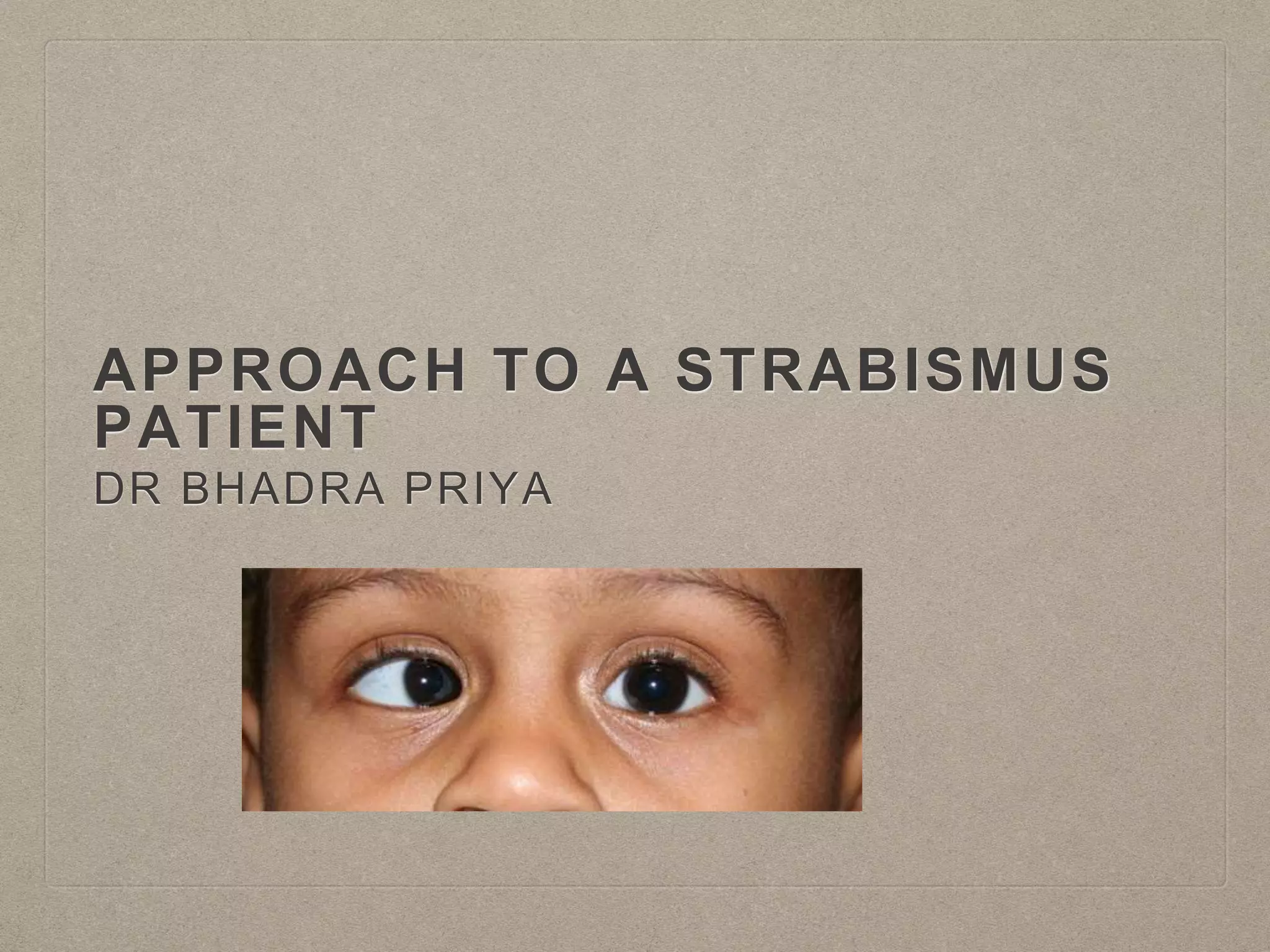
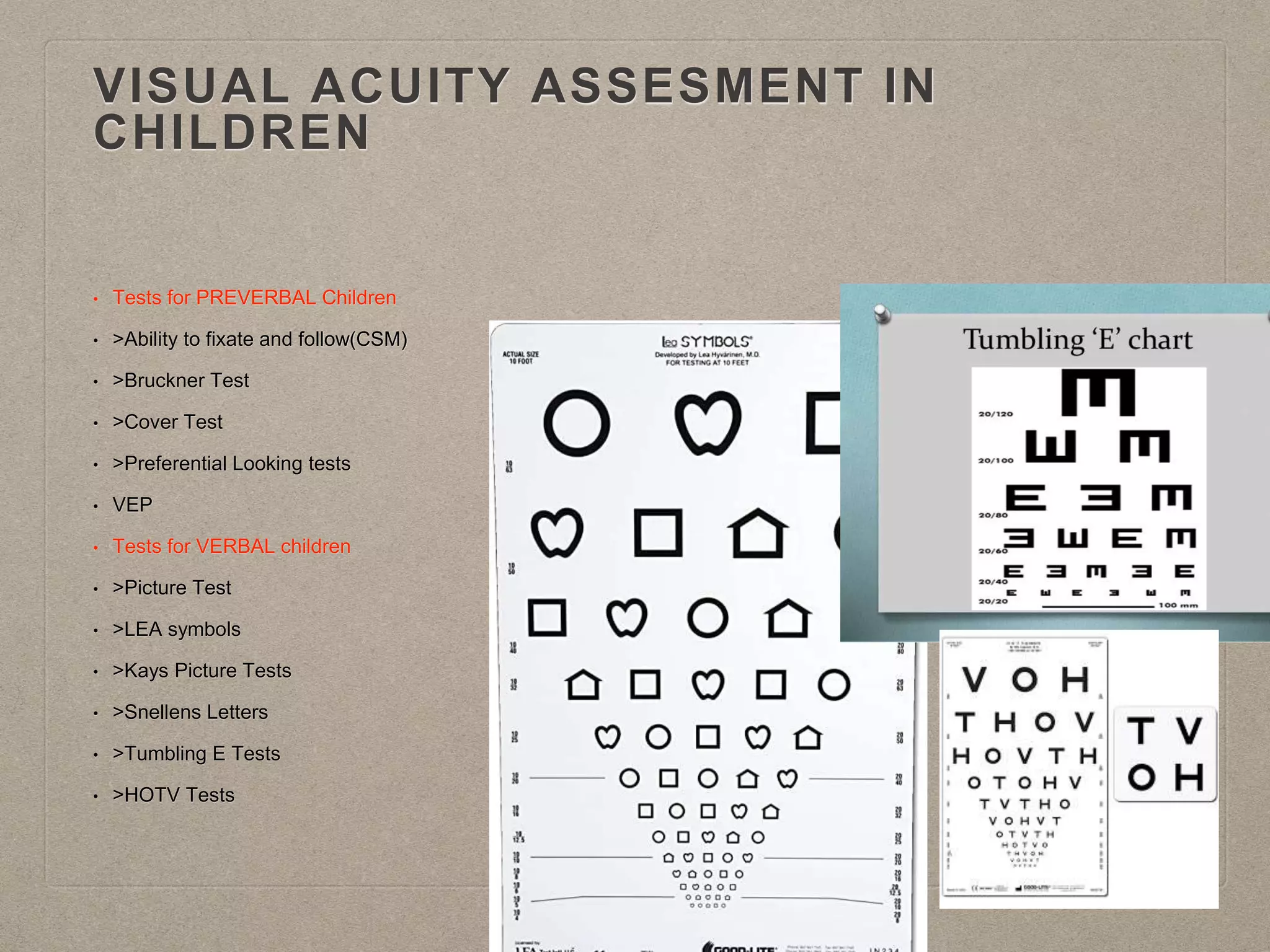
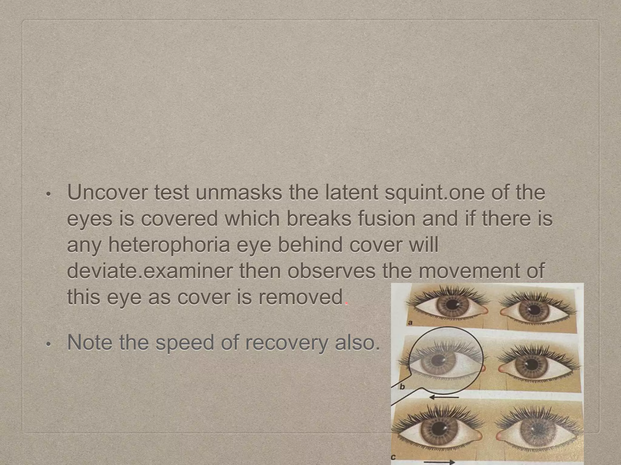
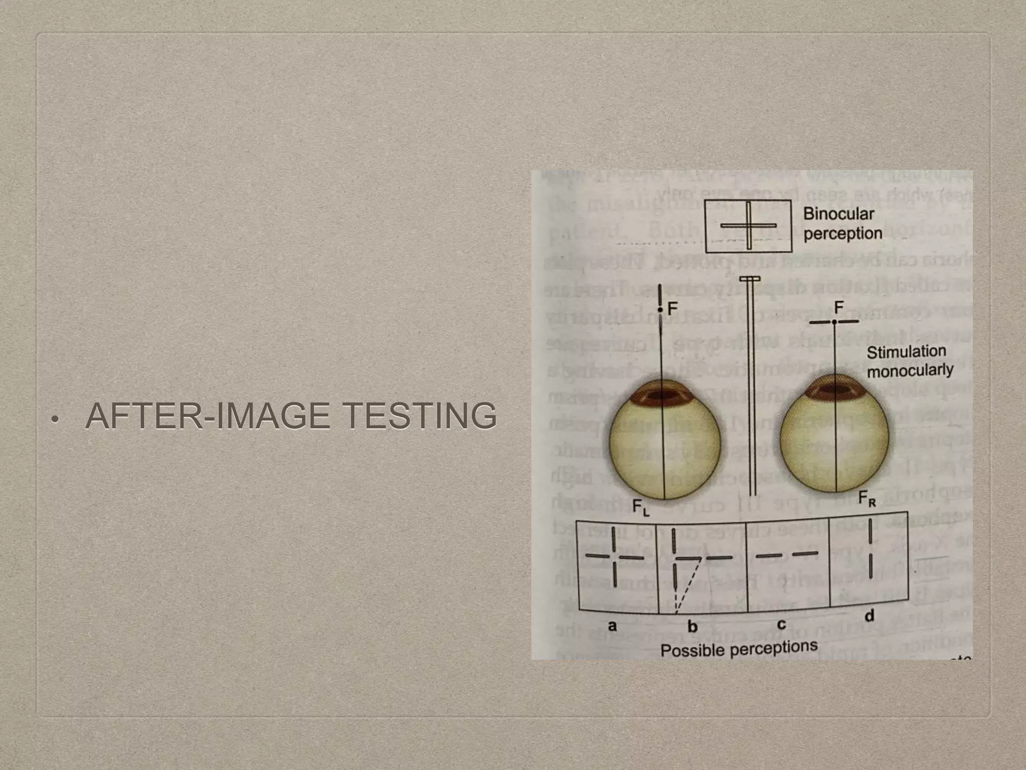
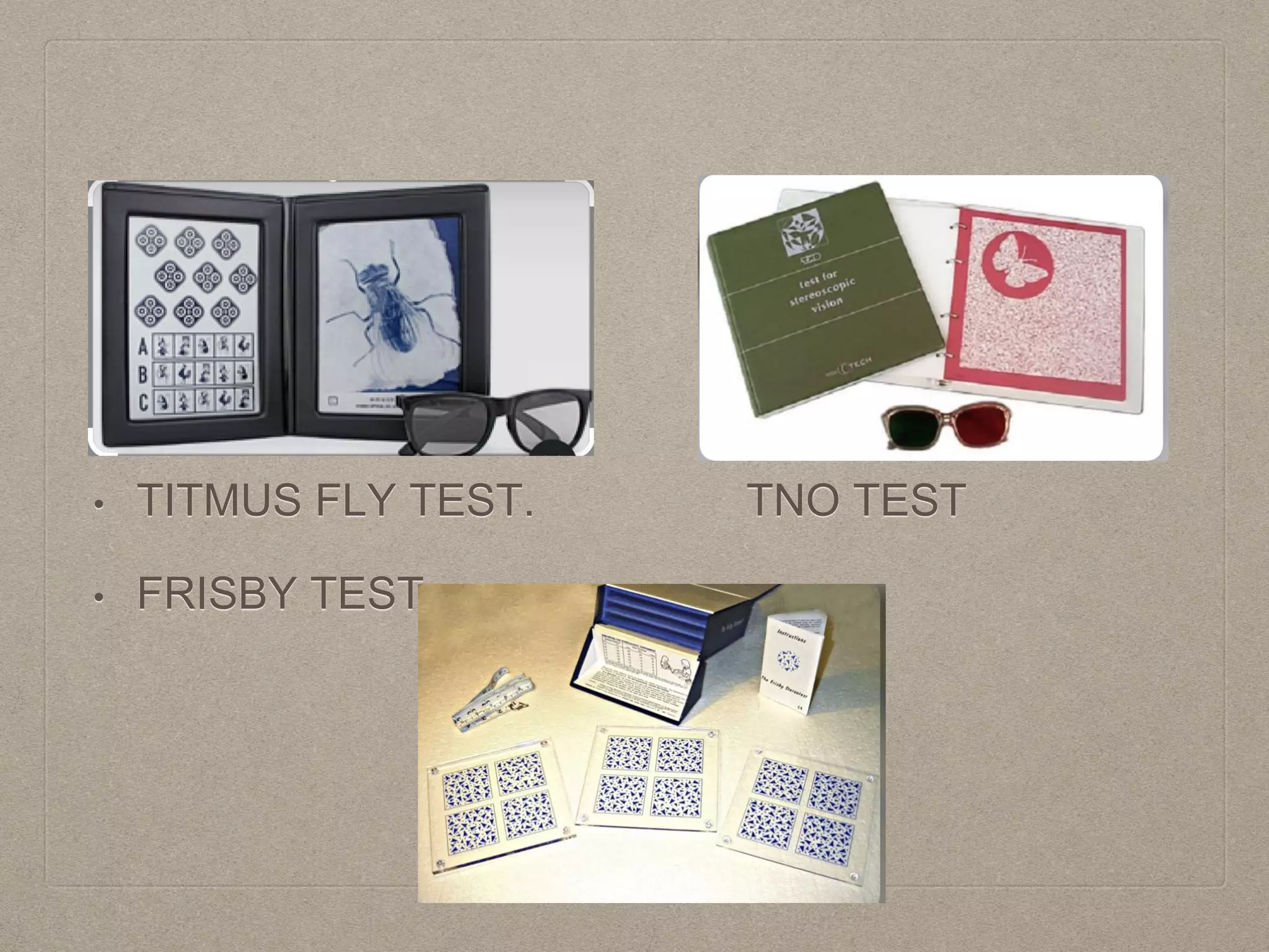
APPROACH TO A STRABISMUS
PATIENT
DR BHADRA PRIYA
• GOALS
• 1.History Taking
• 2.Measuring and characterising the deviation
• 3.Assessing the binocular status
• 4.Establishing the diagnosis
• 5.Management
• HISTORY TAKING
• >Time of onset
• >Onset of strabismus-acute or gradual
• >Frequency of deviation-constant or intermittent
• >Eye dominance-unilateral or alternating
• >Progression/diurnal variation of strabismus or relation of strabismus to any gaze
• >Presence of diplopia- onset-sudden/gradual/painful/progression or resolution, nature- horizontal/ vertical /
torsional
• >Abnormal head posture
• >Family history and birth history
• >Treatment history with history of patching/ glasses/ surgery
• >Use of glasses and its effect on deviation
• >History of trauma
PEDIATRIC EYE EXAMINATION
• Paediatric examination
requires patience.
• While taking history from
parents examine for any
AHP and eye alignment.
• Do non touch tests first
like reflex tests, fixation,
DDO
POINTS IN STRABISMUS EXAMINATION
• 1.Visual acuity assessment, Refraction and
detection of amblyopia.
• 2.Inspection
• 3.Motor Evaluation
• 4.Sensory Evaluation
• 5.Supplementary tests
VISUAL ACUITY ASSESMENT IN
CHILDREN
• Tests for PREVERBAL Children
• >Ability to fixate and follow(CSM)
• >Bruckner Test
• >Cover Test
• >Preferential Looking tests
• VEP
• Tests for VERBAL children
• >Picture Test
• >LEA symbols
• >Kays Picture Tests
• >Snellens Letters
• >Tumbling E Tests
• >HOTV Tests
• Child with significant
asymmetry between vision
of two eyes, resists
occlusion of better eye.
• Do remote occlusion or
use high plus lens for
fogging while measuring
visual acuity in patients
with nystagmus.
CYCLOPLEGIC REFRACTION
AGE CATEGORY DRUG OF CHOICE
<2 YEARS
All children
With esotropia
Atropine 1%e/o
2-5 years
Without esotrpia
With esotropia
C- T-C
Atropine
5-8years
Myopia
Hyperopia&Ast.
Tropicamide1%
C-T-C
8-12 years
With eso
Without eso
CTC
Tropicamide
INSPECTION
• Observe patients visual behaviour, eye alignment, eye
movements, fixation and head posture.
• ptosis/ pseudoptosis
• Rule out pseudo strabismus- epicanthal fold/too narrow
or wide IPD.
• HEAD POSTURE- Components 1.Head tilt
2. Face Turn
3. Chin elevation/depression
ANOMALOUS HEAD POSTURE
• The characteristics of AHP are noted at distance, near, face
turn, chin elevation or depression, head turn or a
combination of these.
• Cause- Ocular, Musculoskeletal or Neurological.
• Most patients develop AHP to gain some level of BSV or
fusion. Thus, diplopia may be noted in primary position but
not in preferred head position.
• Patching one eye should improve AHP due to strabismus
but not other causes.
MOTOR EXAMINATION
• Angle of deviation can be tested by
• 1.Light reflex tests
• 2. Cover tests
• 3.Prism bar tests
BRUCKNER REFLEX
• Screening test used in
children.
• Performed by using DDO.
• Red reflex is equal in both
eyes normally.
• HIRSCHBERG TEST
• Assess the alignment of eye by central reflex on cornea.
• Temporal - esotropia
• Nasal - exotropia
• Inferior- hypertropia
• Superior- hypotropia
POSITION OF
CORNEAL
REFLECTION
ANGLE OF
DEVIATION
On margin of pupil 15degree/ 30PD
Halfway between
margins of pupil
and limbus
30degree/50PD
On the limbus 45degree/90PD
COVER UNCOVER TEST
Cover uncover test is used to pick up tropias.
In cover tests target is shown to both eyes and examiner covers
the apparently fixating eye.the strabismic eye is observed for
tropia shift.
In case of no shift, cover the opposite eye and observe the
uncovered eye for tropia.
No movement of either eyes implies orthotropia with no manifest
squint however it may not rule out phorias
• Uncover test unmasks the latent squint.one of the
eyes is covered which breaks fusion and if there is
any heterophoria eye behind cover will
deviate.examiner then observes the movement of
this eye as cover is removed.
• Note the speed of recovery also.
• While recording the results of tests include:
• >type of deviation
• >estimation of its degree
• >whether manifest, constant or intermittent
• >speed of recovery(rapid, moderate, slow or delayed)
• >any special conditions like DVD.
PRISM BAR TESTS
• Prism Diopter: It is a unit of measurement of
deflection of light rays caused by a prism.
• 1PD: It is a power of prism that deflects the rays of
light at one meter by one cm.
• 1PD=1/2 degree approx
• PREREQUISITES:
• >Patient should be able to fixate at the
accomodative target.
• >co operative patient
• >Refractive correction should be worn by the
patient.
• PRISM BAR COVER TEST
• >objective measurement
• >33cm, 6m, far distance
• >Cover test is done to estimate the angle of
deviation.
• >Prism of approx power is then placed over the
squinting eye(manifest d) or either eye(latent
d).(Apex towards deviation/Base out)
• >alt cover test is performed over prism.
• >Point of neutralisation is no movement on alt
cover test.
• Accuracy of measurement can be checked by
increasing the prism strength till there is reverse
movement and then decreasing till there is no
movement.
• Deviation measured with distance and near fixation
• Deviation with and without glasses
• Deviation in 9 cardinal positions
• Deviation in 25degree up gaze and 35degree downgaze
• Deviation with right and left eye fixating alternately
• Deviation after prolonged cover
KRIMSKY TEST
• OCULAR MOVEMENTS
• Conjugate Movements- Versions
• Dysjugate Movements- Vergence
• Duction
• Saccades
• Pursuit
MUSCLE
PRIMARY
ACTION
SECONDARY
ACTION
TERTIARY
ACTION
MR ADDUCTION
LR ABDUCTION
SR ELEVATION ADDUCTION INTORSION
IR DEPRSSION ADD EXTORSION
SO INTORSION DEPRESSION ABDUCTION
IO EXTORSION ELEVATION ABDUCTION
Grading:
+4 to -4 with 0 indicating Normal
+4 max overaction
-4 severe underaction
• SUPPLEMENTARY TESTS
• >Measuring AC/A ratio
• >Torsion Measurement
• >Parks 3 step test and head tilt test
• MEASURING AC/A RATIO
• >AC/A ratio is the amount of accommodative
convergence exerted in response to one unit of
accomodation.
• >When to measure?
• >High AC/A ratio means over convergence
• >Low AC/A ratio means undercovergence
>HETEROPHORIA METHOD
AC/A=IPD+N-D/DA
Example:Distance=ET41. Near=ET50
IPD=52mm
Nearest distance target=1/3m=3Dacc
AC/A=5.2+(50-41)/3=8.2 (High)
• LENS GRADIENT METHOD
• AC/A=Deviation without lens-Deviation with
lens/. Lens in Dioptres
• Example: Deviation without lens=XT6
• Deviation with lens=ET15
• AC/A=-6-15/3=7
• MEASURMENT OF
TORSION
• OBJECTIVE TORSION
MEASUREMENT BY
FUNDUS
PHOTOGRAPHY
• SUBJECTIVE MEASUREMENT:
• Double Maddox Rod Test
EXAMINATION OF SENSORY STATUS
• Is binocularity present?
• Is there diplopia present?type?
• Type of correspondence?
• Is Suppression present, extent and grade?
• Amblyopia?
• stereopsis?
• DIPLOPIA
• >Binocular/Monocular
• >use dissociating tests- Red green goggles,
Bagolinis glasses, Maddox rod tests
• Types of correspondence
• Bifoveal correspondance is called NRC.
• A correspondance between fovea of one eye and
extrafoveal point of other eye is called ARC
• ARC is sensory adaptation of immature visual
system to an abnormal motor position of eye.
• 2 types of ARC- Harmonious SA=0
• Unharmonious SA>0 but < OA
• OA is measured by prism bar cover test.
• SA can be measured by After image tests
• Bagolini’s striated
glasses
• Suppression
• unilateral/alternating
• Facultative/ Obligatory
• Extent(area of suppression)
• Depth(severity)
4D PRISM TEST
• Differentiates bifocal fixation from a central suppression scotoma.
• BAGOLINI’S STRIATED GLASSES
• WORTH FOUR DOT TEST
• SYNAPTOPHORE
• >Special slides for testing SP, Fusion and
stereopsis
• AFTER-IMAGE TESTING
• STEREOPSIS
• Stereopsis occurs when two retinal images, slightly
disparate because of normally different views
provided by the horizontal separation of two eyes
are cortically integrated.
• TITMUS FLY TEST. TNO TEST
• FRISBY TEST
• DIPLOPIA CHARTING
• Is the diplopia horizontal or vertical?
• In which direction of gaze does it increase?
• To which eye does the outer image belong?
• Is there a predominant tilt?
• In which position of gaze does the tilt increase maximally?
• Interpretation:
• Diplopia increases in the direction of action of paralysed muscle.
• Peripheral image belongs to the deviated eye.
• Separation of image is maximum in the direction of action of paralysed
muscle.
• Esodeviation- uncrossed images
• Exodeviation- crossed images.
• vertical separation with uncrossed image- oblique muscle
• Vertical separation with crossed image-vertical recti involved.
• FORCE DUCTION TEST
• Mechanical restriction(+FDT) vs paralysis(- FDT)
FDT FOR SUPERIOR OBLIQUES
• FORCE GENERATION TEST
• FGT assesses if the muscle has some strength to enable eye movements.
• Patient is asked to look in the field of limitation and examiner applies
traction in the opposite direction and evaluates resistance.
• Interpretation:
• If a muscle contracts normally observer is unable to rotate the globe in the
opposite direction.
• If a muscle is paretic examiner can rotate the globe but notes resistance
• In a palsied muscle no resistance is felt in the opposite direction.
• Fundus Examination
• Look for any sensory cause of strabismus.
• Eg. RD, PHPV, RB
• Any torsion should be ruled out.
• ORDER OF EXAMINATION
• 1.History
• 2.Observe:AHP, Facial asymmetry, Lid changes, systemic abnormality
• 3.Visual acuity and refraction and r/o amblyopia.
• 4.Ocular alignment and movements
• 5.Binocular status testing.
• 6.Diplopia/FDT/FGT
• 7.Supplementary tests: AC/A ratio
THANK YOU…

approach to a strabismus patient

  • 1.
    APPROACH TO ASTRABISMUS PATIENT DR BHADRA PRIYA
  • 2.
    • GOALS • 1.HistoryTaking • 2.Measuring and characterising the deviation • 3.Assessing the binocular status • 4.Establishing the diagnosis • 5.Management
  • 3.
    • HISTORY TAKING •>Time of onset • >Onset of strabismus-acute or gradual • >Frequency of deviation-constant or intermittent • >Eye dominance-unilateral or alternating • >Progression/diurnal variation of strabismus or relation of strabismus to any gaze • >Presence of diplopia- onset-sudden/gradual/painful/progression or resolution, nature- horizontal/ vertical / torsional • >Abnormal head posture • >Family history and birth history • >Treatment history with history of patching/ glasses/ surgery • >Use of glasses and its effect on deviation • >History of trauma
  • 4.
    PEDIATRIC EYE EXAMINATION •Paediatric examination requires patience. • While taking history from parents examine for any AHP and eye alignment. • Do non touch tests first like reflex tests, fixation, DDO
  • 5.
    POINTS IN STRABISMUSEXAMINATION • 1.Visual acuity assessment, Refraction and detection of amblyopia. • 2.Inspection • 3.Motor Evaluation • 4.Sensory Evaluation • 5.Supplementary tests
  • 6.
    VISUAL ACUITY ASSESMENTIN CHILDREN • Tests for PREVERBAL Children • >Ability to fixate and follow(CSM) • >Bruckner Test • >Cover Test • >Preferential Looking tests • VEP • Tests for VERBAL children • >Picture Test • >LEA symbols • >Kays Picture Tests • >Snellens Letters • >Tumbling E Tests • >HOTV Tests
  • 7.
    • Child withsignificant asymmetry between vision of two eyes, resists occlusion of better eye. • Do remote occlusion or use high plus lens for fogging while measuring visual acuity in patients with nystagmus.
  • 8.
    CYCLOPLEGIC REFRACTION AGE CATEGORYDRUG OF CHOICE <2 YEARS All children With esotropia Atropine 1%e/o 2-5 years Without esotrpia With esotropia C- T-C Atropine 5-8years Myopia Hyperopia&Ast. Tropicamide1% C-T-C 8-12 years With eso Without eso CTC Tropicamide
  • 9.
    INSPECTION • Observe patientsvisual behaviour, eye alignment, eye movements, fixation and head posture. • ptosis/ pseudoptosis • Rule out pseudo strabismus- epicanthal fold/too narrow or wide IPD. • HEAD POSTURE- Components 1.Head tilt 2. Face Turn 3. Chin elevation/depression
  • 10.
    ANOMALOUS HEAD POSTURE •The characteristics of AHP are noted at distance, near, face turn, chin elevation or depression, head turn or a combination of these. • Cause- Ocular, Musculoskeletal or Neurological. • Most patients develop AHP to gain some level of BSV or fusion. Thus, diplopia may be noted in primary position but not in preferred head position. • Patching one eye should improve AHP due to strabismus but not other causes.
  • 11.
    MOTOR EXAMINATION • Angleof deviation can be tested by • 1.Light reflex tests • 2. Cover tests • 3.Prism bar tests
  • 12.
    BRUCKNER REFLEX • Screeningtest used in children. • Performed by using DDO. • Red reflex is equal in both eyes normally.
  • 13.
    • HIRSCHBERG TEST •Assess the alignment of eye by central reflex on cornea. • Temporal - esotropia • Nasal - exotropia • Inferior- hypertropia • Superior- hypotropia
  • 14.
    POSITION OF CORNEAL REFLECTION ANGLE OF DEVIATION Onmargin of pupil 15degree/ 30PD Halfway between margins of pupil and limbus 30degree/50PD On the limbus 45degree/90PD
  • 15.
    COVER UNCOVER TEST Coveruncover test is used to pick up tropias. In cover tests target is shown to both eyes and examiner covers the apparently fixating eye.the strabismic eye is observed for tropia shift. In case of no shift, cover the opposite eye and observe the uncovered eye for tropia. No movement of either eyes implies orthotropia with no manifest squint however it may not rule out phorias
  • 16.
    • Uncover testunmasks the latent squint.one of the eyes is covered which breaks fusion and if there is any heterophoria eye behind cover will deviate.examiner then observes the movement of this eye as cover is removed. • Note the speed of recovery also.
  • 17.
    • While recordingthe results of tests include: • >type of deviation • >estimation of its degree • >whether manifest, constant or intermittent • >speed of recovery(rapid, moderate, slow or delayed) • >any special conditions like DVD.
  • 18.
    PRISM BAR TESTS •Prism Diopter: It is a unit of measurement of deflection of light rays caused by a prism. • 1PD: It is a power of prism that deflects the rays of light at one meter by one cm. • 1PD=1/2 degree approx
  • 19.
    • PREREQUISITES: • >Patientshould be able to fixate at the accomodative target. • >co operative patient • >Refractive correction should be worn by the patient.
  • 20.
    • PRISM BARCOVER TEST • >objective measurement • >33cm, 6m, far distance • >Cover test is done to estimate the angle of deviation. • >Prism of approx power is then placed over the squinting eye(manifest d) or either eye(latent d).(Apex towards deviation/Base out) • >alt cover test is performed over prism. • >Point of neutralisation is no movement on alt cover test. • Accuracy of measurement can be checked by increasing the prism strength till there is reverse movement and then decreasing till there is no movement.
  • 21.
    • Deviation measuredwith distance and near fixation • Deviation with and without glasses • Deviation in 9 cardinal positions • Deviation in 25degree up gaze and 35degree downgaze • Deviation with right and left eye fixating alternately • Deviation after prolonged cover
  • 22.
  • 23.
    • OCULAR MOVEMENTS •Conjugate Movements- Versions • Dysjugate Movements- Vergence • Duction • Saccades • Pursuit
  • 24.
    MUSCLE PRIMARY ACTION SECONDARY ACTION TERTIARY ACTION MR ADDUCTION LR ABDUCTION SRELEVATION ADDUCTION INTORSION IR DEPRSSION ADD EXTORSION SO INTORSION DEPRESSION ABDUCTION IO EXTORSION ELEVATION ABDUCTION
  • 25.
    Grading: +4 to -4with 0 indicating Normal +4 max overaction -4 severe underaction
  • 27.
    • SUPPLEMENTARY TESTS •>Measuring AC/A ratio • >Torsion Measurement • >Parks 3 step test and head tilt test
  • 28.
    • MEASURING AC/ARATIO • >AC/A ratio is the amount of accommodative convergence exerted in response to one unit of accomodation. • >When to measure? • >High AC/A ratio means over convergence • >Low AC/A ratio means undercovergence
  • 29.
  • 30.
    • LENS GRADIENTMETHOD • AC/A=Deviation without lens-Deviation with lens/. Lens in Dioptres • Example: Deviation without lens=XT6 • Deviation with lens=ET15 • AC/A=-6-15/3=7
  • 31.
    • MEASURMENT OF TORSION •OBJECTIVE TORSION MEASUREMENT BY FUNDUS PHOTOGRAPHY
  • 32.
    • SUBJECTIVE MEASUREMENT: •Double Maddox Rod Test
  • 33.
    EXAMINATION OF SENSORYSTATUS • Is binocularity present? • Is there diplopia present?type? • Type of correspondence? • Is Suppression present, extent and grade? • Amblyopia? • stereopsis?
  • 34.
    • DIPLOPIA • >Binocular/Monocular •>use dissociating tests- Red green goggles, Bagolinis glasses, Maddox rod tests
  • 35.
    • Types ofcorrespondence • Bifoveal correspondance is called NRC. • A correspondance between fovea of one eye and extrafoveal point of other eye is called ARC • ARC is sensory adaptation of immature visual system to an abnormal motor position of eye.
  • 36.
    • 2 typesof ARC- Harmonious SA=0 • Unharmonious SA>0 but < OA • OA is measured by prism bar cover test. • SA can be measured by After image tests • Bagolini’s striated glasses
  • 37.
    • Suppression • unilateral/alternating •Facultative/ Obligatory • Extent(area of suppression) • Depth(severity)
  • 38.
    4D PRISM TEST •Differentiates bifocal fixation from a central suppression scotoma.
  • 39.
  • 40.
  • 41.
    • SYNAPTOPHORE • >Specialslides for testing SP, Fusion and stereopsis
  • 42.
  • 43.
    • STEREOPSIS • Stereopsisoccurs when two retinal images, slightly disparate because of normally different views provided by the horizontal separation of two eyes are cortically integrated.
  • 44.
    • TITMUS FLYTEST. TNO TEST • FRISBY TEST
  • 45.
    • DIPLOPIA CHARTING •Is the diplopia horizontal or vertical? • In which direction of gaze does it increase? • To which eye does the outer image belong? • Is there a predominant tilt? • In which position of gaze does the tilt increase maximally?
  • 46.
    • Interpretation: • Diplopiaincreases in the direction of action of paralysed muscle. • Peripheral image belongs to the deviated eye. • Separation of image is maximum in the direction of action of paralysed muscle. • Esodeviation- uncrossed images • Exodeviation- crossed images. • vertical separation with uncrossed image- oblique muscle • Vertical separation with crossed image-vertical recti involved.
  • 47.
    • FORCE DUCTIONTEST • Mechanical restriction(+FDT) vs paralysis(- FDT)
  • 48.
  • 49.
    • FORCE GENERATIONTEST • FGT assesses if the muscle has some strength to enable eye movements. • Patient is asked to look in the field of limitation and examiner applies traction in the opposite direction and evaluates resistance. • Interpretation: • If a muscle contracts normally observer is unable to rotate the globe in the opposite direction. • If a muscle is paretic examiner can rotate the globe but notes resistance • In a palsied muscle no resistance is felt in the opposite direction.
  • 50.
    • Fundus Examination •Look for any sensory cause of strabismus. • Eg. RD, PHPV, RB • Any torsion should be ruled out.
  • 51.
    • ORDER OFEXAMINATION • 1.History • 2.Observe:AHP, Facial asymmetry, Lid changes, systemic abnormality • 3.Visual acuity and refraction and r/o amblyopia. • 4.Ocular alignment and movements • 5.Binocular status testing. • 6.Diplopia/FDT/FGT • 7.Supplementary tests: AC/A ratio
  • 52.