Downloaded 309 times
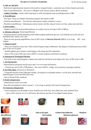


Café-au-lait spots, neurofibromas, Lisch nodules, and axillary freckling are characteristic of neurofibromatosis type 1. Plexiform neurofibromas appear as subcutaneous elastic tumors over the face, scalp, neck and chest. Adenoma sebaceum presents as numerous discrete smooth papules over the butterfly area of the face and nasolabial folds. Shagreen patches are irregular cobblestone-like plaques in the lumbosacral area, a characteristic of tuberous sclerosis. Ocular and cutaneous telangiectasias occur in Ataxia telangiectasia, appearing as dilated blood vessels over


