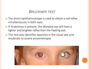
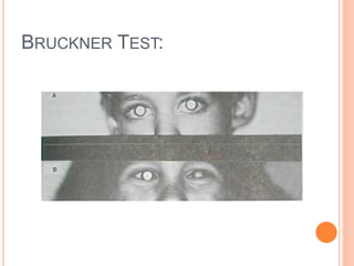
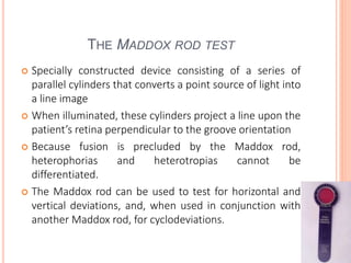
strabismus , gaze , ocular movements , classification etc
presented by senior optometrist & orthoptician at Sagarmatha Choudhary Eye Hospital, SCEH, LAHAN (NEPAL )
He explain details about the binocular gaze , EOMs, etc & work up of a patient of squint etc.
Call Girl Number in Vashi Mumbai📲 9833363713 💞 Full Night Enjoy
Assessment of ocular alignment
1. ASSESSMENT OF OCULAR ALIGNMENT
AND EVALUATION OF EYE MOVEMENTS
RAY ARBIND
Senior Optometrist
SCEH, Eye hospital, Lahan, Nepal
Moderator-Dr.Vinit Kumar
2. POSITIONS OF GAZE
Primary position of gaze: The position of the eyes when
fixating straight ahead on an object at infinity.
For practical purposes, infinity is considered to be 20 ft (6
m), and for this position the head should be straight.
Cardinal positions: Those 6 positions of gaze in which the
prime mover is 1 muscle of each eye, together called
yoke muscles.
3. Midline positions: Straight up and straight down from
primary position.
These latter 2 gaze positions help determine the
elevating and depressing capabilities of the eye, but they
do not isolate any 1 muscle because 2 elevator and 2
depressor muscles affect midline gaze positions.
4. Diagnostic positions of gaze has been applied to the
composite of these 9 gaze positions: the 6 cardinal
positions, straight up and down, and primary position.
For patients with vertical strabismus the diagnostic
positions of gaze include forced head tilt to the right and
left
5. DEFINITIONS
Phoria: a latent visual axis deviation held in check by
fusion.
Tropia: a manifest visual axis deviation.
Intermittent tropia: an intermittent visual axis deviation
that may exist only in certain gaze positions or target
distances.
Ductions: monocular movements into various gaze
positions; each eye views in the same direction.
Vergences: binocular eye movements into opposite gaze
directions.
6. INTRODUCTION
Subjective methods: useful for cooperative,
communicative older patients.
Objective methods: must be used in younger patients or
those less cooperative.
7. LABORATORY METHODS
Most laboratory tests are objective.
Electro-oculogram (EOG)
is generated by alterations in direct current between the
front and back of the eye as the eye rotate
Insulated wire placed in a silicone rubber limbal annulus
(eye coil) generates a current in response to a magnetic
field;
This very precise technique may be used to detect
horizontal, vertical, and torsional changes in eye position.
8. CLINICAL METHODS
The position of the patient’s head when it is mobilized in
free space is carefully noted
Atypical head positions may indicate:
Restrictive or paralytic strabismus
The examiner must differentiate between head turns,
tilts, and vertical head positions
9. Patient is seated in the examination chair
The patient’s lid position is noted;
lid asymmetry is usually found in patients who have
vertical strabismus
If hypotropia is present and the non-fixing eye is lower
than the fixing eye, the lid position is lower in the
nonfixing eye; this is termed pseudoptosis if the lid
regains normal position when the previously hypotropic
eye fixes in primary position.
Epicanthal skin folds that extend over the nasal sclera in
a small child may simulate esotropia.
Vertical displacement of an orbit may simulate vertical
strabismus, and hypertelorism may simulate exotropia.
10. OBJECTIVE CLINICAL METHODS
Ocular alignment tests can be grouped into 4 basic types:
Cover tests,
Corneal light reflex tests,
Dissimilar image tests, and
Dissimilar target tests
11. COVER TESTS
These objective tests detect and measure horizontal and
vertical strabismus,
but they cannot measure torsional deviations and detect
only some and not all torsional deviations
The cover test detects tropias – constant visual axis
deviations.
The uncovered eye should be observed for movement as
its fellow is covered with a paddle, thumb, or remote
occluder
12. COVER TESTS
Necessities for cover testing-
Eye movement capability, image formation and
perception, foveal fixation in each eye, attention, and
cooperation
There are 3 types of cover tests:
The cover-uncover test,
The alternate cover test, and
The simultaneous prism and cover test
All can be performed with fixation at distance or near
13.
14. THE MONOCULAR COVER-UNCOVER
TEST
Most important test for
detecting the presence of
manifest strabismus
Differentiating a heterophoria
from a heterotropia
As 1 eye is covered, the
examiner watches carefully for
any movement in the opposite,
noncovered eye;
such movement indicates the
presence of a heterotropia.
15. With movement of the noncovered eye assumed to be
absent, movement of the covered eye in one direction just
after the cover is applied and a movement in the opposite
direction (a fusional movement) as the cover is removed
indicate a heterophoria
If the patient has a heterophoria, the eyes will be straight
before and after the cover-uncover test;
The deviation that appears
during the test is a result
of interruption of binocular
vision
16. A patient with a heterotropia-starts out with a deviated
eye and ends up (after the test) with either the same or
the opposite eye deviated (if the opposite eye is the
deviated one, the condition is termed alternating
heterotropia)
Some patients with heterophoria the eyes are straight
before testing but they dissociate into a manifest
deviation(heterotropia) after the occlusion interrupts
binocular vision
17. Small toys are suitable for young children, but bright white
lights are avoided as the patient cannot accommodate on the
contours of a light.
Tropias established by the cover test may be measured using
the simultaneous prism and cover test; a prism of appropriate
strength held in the appropriate direction is introduced before
one eye as its fellow is covered
Prism strength is increased until eye movement ceases; this
prism strength corresponds to the size of the strabismus.
The test is then repeated with the prism before the other eye.
18. The uncover test requires observation of the covered eye
as the cover is removed.
If that eye is deviated under cover, it may regain fixation
or may remain deviated.
The former implies the presence of a phoria, a latent
deviation held in check by sensory fusion, or an
intermittent tropia; the latter implies a tropia with
fixation preference for the fellow eye.
Phorias may be detected more directly using the
alternate cover test, in which each eye is occluded
alternately to dissociate the visual axes maximally.
19. THE ALTERNATE COVER TEST (PRISM AND
COVER TEST)
Measures the total deviation, regardless of whether it is
latent or manifest
20. Testing should be performed at both distance and near
fixation
The deviation is quantified by using prism to eliminate
eye movement as occluder is switched from eye to
eye(prism alternate cover test)
21. It may be necessary to use both horizontally and
vertically placed prisms
The amount of prism power required is the measure of
deviation
Two horizontal or 2 vertical prisms should not be
superimposed on each other because this can induce
significant measurement errors
A more accurate method for measuring large deviations
is to place prisms in front of each eye,
However,It is acceptable to superimpose a horizontal
prism on a vertical prism before the same eye if
necessary
22. Care must be taken to permit time for each eye to reside
behind the cover
(the cover must not be ‘fanned’ before the eyes).
Appropriately held prisms enable quantitation of the
Phoria
For most clinical purposes, measurements within 2 D are
sufficiently accurate.
Cover test measurements are influenced by the presence
of eccentric fixation;
Its presence must be investigated in patients who have
severe amblyopia.
23. The eye behind the prism is the ‘fixing’ eye.
If the cause of strabismus is paralytic or restrictive,
patients may have greater cover test measurements
when the paretic or restricted eye fixes in a given gaze
position (secondary deviation) than when the sound eye
fixes (primary deviation).
24. Strabismus should be detected and measured in primary
position at distance and near fixation and in gaze up,
down, right, and left 30° from primary position.
The nine ‘diagnostic gaze positions’ include the above
plus,
Up and right, up and left, down and right, and down and
left;
these are useful to measure cyclovertical muscle palsies.
For oblique muscle dysfunction, measurements are
taken with the head tilted 30° right and left at distance
fixation
25. The alternate cover test measures the total deviation
It doesn’t distinguish between latent (heterophoria) and
manifest(heterotropia)
26. For more accurate results,
Plastic prisms should be held with back surface(the
surface closest to patient) in the patients frontal
plane,regardless of size of deviation
If the patients head is tilted,the prism must be tilted
accordingly
With incomittant (paretic and restrictive) strabismus,the
clinician can measure the primary and secondary
deviations by holding the prism over the paretic or
restricted eye and the sound eye ,respectively
27. The simultaneous prism and cover test is helpful in
determining the manifest deviation without
occlusion(only the heterotropia)
The prism is placed infront of deviating eye and covering
the fixating eye at the same time
Test is repeated using increasing prism power until
deviated eye no longer shifts
28. LIGHT REFLEX TESTS
The main tests of this type are the
Hirschberg,
Modified Krimsky,
Bruckner, and
Major amblyoscope methods
29. THE HIRSCHBERG METHOD
Is based on the premise that 1 mm of decentration of the
corneal light reflection corresponds to approximately 7degree
or 15 ,of ocular deviation of the visual axis
Therefore, a light reflex at the pupillary margin is about 2 mm
from the pupillary center (with a 4-mm pupil), which
corresponds to 15degree, or approximately 30 , of deviation.
30. A reflex in the mid-iris region is about 4 mm from the
pupillary center, which is roughly 30degree, or 60Δ of
deviation; similarly, a reflex at the limbus is about
45degree, or 90Δ of deviation
31. THE KRIMSKY METHOD
Uses reflections produced on both corneas by a penlight
and is ideally used at near fixation.
The original method involved placing prisms in front of
the deviating eye
More common modifications today involve holding
prisms before the fixating eye or split between the 2 eyes
32. By adjusting the prisms to center the corneal reflection in
the deviated eye, it is possible to approximate and
quantitate the near deviation
33. Prism deflect light toward their base, but the patient views
the light as deflected toward the prism apex
The prism diopter is defined as the strength of prism
necessary to deflect a light beam 0.4 inches (1 cm) at 3.3 ft
(1 m) distance
As a tangent function, the prism diopter is not linear and
increases in size as the deviation increases, but for small
deviations 1° approximates 1.7Δ.
34. The angle kappa can affect light reflex measurements.
Angle kappa is the angle between the visual axis and the
anatomical pupillary axis of the eye
If the fovea is temporal to the pupillary axis (as is usually the
case), the corneal light reflection will be slightly nasal to the
center of the cornea
This is termed positive angle kappa and simulates exodeviation.
35. If the position of the fovea is nasal to the pupillary axis,
the corneal light reflection will be slightly temporal to the
center of the cornea
This is termed negative angle kappa and simulates
esodeviation
36. BRUCKNER TEST
The direct ophthalmoscope is used to obtain a red reflex
simultaneously in both eyes.
If strabismus is present, the deviated eye will have a
lighter and brighter reflex than the fixating eye.
This test also identifies opacities in the visual axis and
moderate to severe anisometropia.
39. DISSIMILAR IMAGE TESTS
1-target tests in which the image of the target is made
artificially dissimilar in both eyes.
Because one image falls on the fovea of one eye and on a
non foveal region of the other eye in strabismic patients;
esotropic patients perceive the images homonymously
and exotropic patients perceive them as crossed.
All vertical deviations result in vertically crossed diplopia.
The 3 most commonly used of these tests are the
Maddox rod test, the double Maddox rod test, and the
red glass test.
40. THE MADDOX ROD TEST
Specially constructed device consisting of a series of
parallel cylinders that converts a point source of light into
a line image
When illuminated, these cylinders project a line upon the
patient’s retina perpendicular to the groove orientation
Because fusion is precluded by the Maddox rod,
heterophorias and heterotropias cannot be
differentiated.
The Maddox rod can be used to test for horizontal and
vertical deviations, and, when used in conjunction with
another Maddox rod, for cyclodeviations.
41. To test for horizontal deviations, the Maddox rod is
placed in front of one eye (for eg,right eye) with the
cylindres positioned horizontally
The patient fixates a point source of light and then sees a
vertical line with the right eye and a point sorce of white
light with the left eye
If the light superimposes the line, orthophoria is present
If the light is to the left of the line,an esodeviation is
present
If the light is to the right ,an exodeviation is present
42. To measure the amount of
deviation the examiner finds
the prism that superimpose
the point source of line
43. THE DOUBLE MADDOX ROD TEST
Is used to determine cyclodeviations
Maddox rods are placed in a trial frame or phoropter
Positioned in front of each eye, with the rods aligned
vertically so that the patient sees two horizontal lines
The patient or examiner rotates the axes of the rods until
the lines are perceived to be parallel.
44. To facilitate the patient's recognition of the 2 lines, it is
often helpful to dissociate the lines by placing a small
prism base-up or base-down in front of 1 eye
The angle of rotation that causes the line image to
appear parallel determines the magnitude and direction
(intorsion or extorsion)of cyclotropia
45. RED GLASS TEST
A red glass is placed in front of the right eye.
Same purpose as the Maddox rod test but is not
applicable to cyclodeviations
The red glass test requires the patient to alert the
examiner when the red light viewed behind a red filter
before the right eye and a white light viewed with the left
eye are superimposed or displaced one from the other
46. Fusion is disrupted by the red glass and thus horizontal
and vertical phorias are uncovered and measured;
Torsional deviations are not detected by this method.
The gaze position of maximal image separation is a clue
to the identity of paretic or restricted muscles.
47. DISSIMILAR TARGET TESTS
Based on the patient's response to the dissimilar images
created by each eye viewing a different target;
the deviation is measured first with 1 eye fixating and
then with the other.
In contrast to dissimilar image tests, diplopia is created
by having the patient observe 2 different targets
simultaneously, with each target seen by 1 eye only.
Esotropic patients will have crossed diplopia, and
exotropic patients homonymous diplopia.
48. Several dissimilar target tests, but the 3 most frequently
encountered
Lancaster red-green projection test,
the Hess screen test,
and the major amblyoscope test.
49. THE LANCASTER RED-GREEN
TEST
Uses red-green goggles that can be reversed, a red-slit
projector, a green-slit projector, and a screen ruled into
squares
The patient's head is held steady; by convention, the test
is begun with the red filter in front of the right eye.
The Lancaster red–green test uses a screen marked in 2°
increments viewed from a distance of 6.6 ft (2 m).
The examiner projects a red slit onto the screen, and the
patient is asked to place the green slit so that it appears
to coincide with the red slit.
The relative positions of the 2 streaks are then recorded
50. The test is repeated for the diagnostic positions of gaze,
The goggles are then reversed so that the deviation with
the fellow eye fixating can be recorded.
51. THE HESS SCREEN TEST
Contains a tangent pattern displayed on a dark grey background.
Red lights that can be individually illuminated by a control panel indicate
the cardinal positions of gaze within a central field (15° from primary
position) and a peripheral field (30°); each square represents 5° of ocular
rotation.
The eyes are dissociated by the use of reversible goggles incorporating a
red and a green lens, the red lens in front of the fixating eye and the green
lens the non-fixating eye.
Red points of lights are illuminated at selected positions on the screen.
The patient holds a green pointer, and is asked to superimpose a green light
over each red light in turn.
In orthophoria the two lights should be more or less superimposed in all
positions of gaze.
The goggles are then reversed and the procedure repeated.
Software is available that facilitates the plotting of a Hess chart using a
standard desktop computer screen.
54. MAJOR AMBLYOSCOPE METHOD
uses separate target illumination, which can be moved to center
the corneal light reflection.
The amount of deviation is then read directly from the scale of
the amblyoscope
The instrument is adjusted to the patients interpupillary
distance in the usual manner, the correcting spectacles are
worn.
Targets are used which ensure foveal fixation.
The subjective angle is determined and the readings taken from
the prism diopter scale.
Minus lenses usually-3DS are inserted in the lens holder of the
56. OCULAR ROTATIONS
Generally, when eye movement are assessed,
versions are tested first.
The examiner should pay particular attention to the
movements of both eyes in to the 9 diagnostic
positions of gaze.
Limitations of movement into these positions and
asymmetry of excursion of the 2 eyes should be noted.
Spinning the child, or provoking the doll's head
phenomenon,may be helpful in eliciting the vestibular -
stimulated eye movements.
If versions are not full, duction movements should be
tested for each eye separately
57. CONVERGENCE
Alignment at near is usually measured at 13 inch (33
cm) directly in front of the patient in the horizontal
plane.
Comparison of the alignment in the primary position at
both distance and near fixation helps assess the
accommodative convergence (synkinetic near) reflex.
The near point of convergence is determined by
placing a fixation object at 40 cm in the midsagittal
plane of the patient's head.
As the subject fixates on the object, it is moved toward
the subject until 1eye loses fixation and turns out.
58. The point at which this action occurs is the near
point of convergence.
The eye that is able to maintain fixation is
considered to be the dominant eye.
The normal near point of convergence is 8-10 cm or
less.
This determination does not distinguish between
fusional and accommodative convergence,
And any heterophoria that is present should be
taken into account and corrected for algebraically
59. ACCOMMODATIVE
CONVERGENCE/ACCOMMODATION RATIO
The accommodative convergence/accommodation (Ac/A)
ratio is defined as the amount of convergence (in prism
diopters) per unit change in accommodation(diopter)
There are 2 methods of clinical measurement
The gradient method and
The heterophoria method
60. THE GRADIENT METHOD
Arrives at the AC/A ratio by dividing the change in
deviation in prism diopters by the change in lens power
An accommodative target must be used, and the working
distance (typically at 1/3 m or 6 m) is held constant.
Plus or minus lenses (eg, + 1, +2, +3, - 1, -2, -3) are used
to vary the accommodative requirement.
This method measures the stimulus AC/A ratio, which is
not necessarily identical to the response AC/A ratio.
The latter can be determined only with the use of an
optometer that records the change in accommodation
actully produced.
61. THE HETEROPHORIA METHOD
The distance and near deviations are used,along with the
interpupillary distance,to calculate AC/A ratio
If the patient is more exotropic or less esotropic at near,
Too little convergence, or a low AC/A ratio, is present;
If the patient is more esotropic or less exotropic at near,a
high AC/A ratio is present.
In accommodative esotropia,an increase of esotropia of
1O or more from distance to near fixation is considered
to represent a high AC/A ratio.
62. An abnormally high AC/A ratio can be managed optically,
pharmacologically, or surgically.
For example, plus lens spectacles for hyperopia reduce
accommodation and therefore reduce accommodative
convergence.
This principle is the mainstay of the medical management of
esotropia.
Bifocals reduce or eliminate the need to accommodate for
near fixation.
This optical management may be used for excess convergence
at near-that is, an esodeviation greater at near.
Underplussed or overminused spectacles create the need for
greater-than-normal accommodation.
63. This excess accommodation creates more accommodative
convergence and is occasionally used to reduce an
exodeviation.
Long-acting cholinesterase inhibitors (eg, echothiophate
iodide) can be used to decrease accommodative
convergence.
These drugs act directly on the ciliary body, facilitating
transmission at the myoneural junction.
They reduce the central demand for accommodative
innervation and thus reduce the amount of convergence
induced by accommodation
64. FUSIONAL VERGENCE
Vergences move the 2 eyes in opposite directions.
Fusional vergences are motor responses used to
eliminate horizontal, vertical, or torsional image disparity.
They can be grouped by the following functions:
Fusional convergence eliminates bitemporal retinal
disparity and controls an exophoria
Fusional divergence eliminates binasal retinal disparity
and controls an esophoria.
65. Vertical fusional vergence controls a hyperphoria or
hypophoria.
Torsional fusional vergence controls incyclophoria or
excyclophoria.
Fusional vergences can be measured by using a
haploscopic device (major amblyoscope),a rotary prism,
or a bar prism, and gradually increasing the prism power
until diplopia occurs.
Accommodation must be controlled during fusional
vergence testing.
67. Fusional vergences can be altered by the following:
Compensatory mechanisms: As a tendency to deviate
evolves,the patient gradually develops a larger-than-normal
fusional vergence for that deviation.
Very large fusional vergences are common in
compensated,long-standing vertical deviations and in
exodeviations.
68. Change in visual acuity: Improved acuity improves the fusional
vergence mechanism
The treatment of reduced vision may change a symptomatic
intermittent deviation to an asymptomatic heterophoria.
State of awareness: Fatigue, illness, or drug and alcohol
ingestion may decrease the fusional vergence
mechanism,converting a heterophoria to a heterotropia,
Orthoptics: The magnitude of the fusional vergence
mechanism (mainly fusional convergence) may be increased by
exercises.
This treatment works best for near fusional convergence,
particularly for the relief of the symptoms of convergence
insufficiency
69. Optical stimulation of fusional vergence:
In controlled accommodative esotropia, reducing the
strength of the hyperopia or bifocal correction induces
an esophoria that stimulates fusional divergence.
The power of prisms used to control diplopia may be
gradually reduced to stimulate a compensatory fusional
vergence
70. To test fusional convergence, the prism is held base-in
before a patient’s eye; to test fusional divergence, the
prism is held base-out.
Positive vertical vergence requires a base-down prism
placed before the right eye, and negative vertical
vergence requires one placed before the left eye
71. MECHANICAL TESTS OF EYE
MOVEMENT LIMITATION
Patients who have duction limitations may suffer from
paralysis or paresis, or mechanical restriction of full
duction movement, or both.
Forced duction testing and active force generation test
may help to differentiate.
72. FORCED DUCTION TEST
The forced duction test is an attempt by the examiner to
move a patient’s eye farther in a given direction than the
patient can move it.
Topical anesthetic is placed on the appropriate limbal
location (generally 180° away from the duction
limitation) with a small cotton swab and the limbal
conjunctiva is grasped firmly with a toothed forceps.
The patient is asked to rotate the eye fully in the
direction of the limited duction.
73. • An attempt is then made by the examiner to rotate the eye
beyond the position attained by the patient while avoiding
globe retraction
• Care must be taken not to abrade the cornea. Patients who
have pure nerve palsy exhibit no restriction to full movement
by the examiner; patients who have pure restriction
(dysthyroid orbitopathy, entrapment of ocular contents after
blowout fracture) exhibit restricted movements (sometimes
termed a positive forced duction test).
74. ACTIVE FORCE GENERATION TEST
Active force generation testing may be used to evaluate the
ability of a muscle to move the eye against a resisting force.
The forceps is placed at the limbus of the anesthetized globe in
the meridian of the muscle whose duction is limited and the
patient requested to rotate the eye in the direction of the limited
duction;
the examiner judges through the forceps the relative amount of
force generated.
Strain gauges have been devised that enable quantitation of this
force.
In Fig-patient has maximal adduction in the left eye.
The examiner can gauge the
relative abduction
strength as the patient attempts
to move her eye to theleft.
75. SPECIAL TESTS
MOTOR TESTS
Special motor tests include forced ductions, active force
generation, and saccadic velocity.
Saccadic velocity can be recorded using a special
instrument that graphically records the speed and
direction of eye movement
This test is useful to differentiate paralysis from
restriction
A paralyzed muscle generates a reduced saccadic velocity
throughout the movement of the involved eye, whereas
a restricted muscle produces an initially normal velocity
that rapidly decelerates when the eye reaches the limit
of its movement
76. ASSESMENT OF THE FIELD OF SINGLE
BINOCULAR VISION
May be tested on either a Goldmann perimeter or a
tangent screen.
These tests are useful for following the recovery of a
paretic muscle or for measuring the outcome of surgery
to alleviate diplopia.
A small white test object is followed by both eyes in the
various cardinal positions throughout the visual field.
When the patient indicates that the test object is seen
double,the point is plotted.
77. The examiner then repeats the same procedure until he
or she has plotted the entire visual field,noting the area
in which the patient reported single vision and the area
of double vision.
The field of binocular fixation normally measures about
45deg – 50deg from the fixation point except where it is
blocked by the nose
78. 3-STEP TEST
The 3-step test is an algorithm that can be used to help
identify the paretic cyclovertically acting muscle.
There are 8 cyclovertically acting muscles:
4 work as depressors (2 in each eye), and 4 work as
elevators (2 in each eye).
The 2 depressors of each eye are the inferior rectus (IR)
and superior oblique (SO) muscles;
the 2 elevators of each eye are the superior rectus (SR)
and the inferior oblique (IO) muscles.
79. STEP 1
If the case is one of paralysis, determine which eye is
hypertropic by using the cover-uncover test).
Step 1 narrows the number of possible underacting
muscles from 8 to 4.
Draw an oval around these 2 muscle groups
80. STEP2
Determine whether the vertical deviation is greater in
right gaze or in left gaze.
In this example, the deviation is larger in left gaze.
81. This implicates 1 of the 4 vertically acting muscles used in left
gaze.
Draw an oval around the 4 vertically acting muscles that are
used in left gaze
At the end of step 2, the 2 remaining possible muscles (1 in
each eye) are both intortors or extortors and both superior or
inferior muscles (l rectus and 1 oblique).
82. STEP 3
Known as the Bielschowsky head-tilt test,
Final step involves tilting the head to the right and then
to the left during distance fixation
Head tilt to the right stimulates intorsion of the right eye
(RSR, RSO) and extorsion of the left eye (LIR, LIO).
Head tilt to the left stimulates extorsion of the right eye
(RIR, RIO) and intorsion of the left eye (LSR, LSO).
Normally, the 2 intortors and the 2 extortors of each eye
have opposite vertical actions that cancel each other.
If 1 intortor or 1 extortor is weak, it cannot act
vertically,and the vertical action of the other ipsilateral
torting muscle becomes manifest.
83. To continue with example
When head is tilted to right,the right eye moves upward
as it attempts to intort to maintain fixation,increasing
vertical deviation
This suggest vertical action of right superior rectus
muscle is unoppsed,indicating right superior oblique is
weak and paretic
84. PRISM ADAPTATION TEST
The patient is fitted with prisms of sufficient magnitude
to permit alignment of the visual axes.
In many cases, this step provokes a restoration of sensory
binocular cooperation in a form of fusion and even
stereopsis.
This technique simulates orthotropia and possibly offers
some predictive value of whether fusion may be restored
when the patient undergoes surgical alignment.
85. In some patients, however (especially those with
acquired esotropia), placement of such prisms increases
the deviation.
In such cases, anomalous retinal correspondence based
on the objective angle may drive the eyes to maintain
this adaptive alignment even with prismatic correction.
After wearing such prisms, the patient returns with a
greater angle of deviation.
86. The patient is reexamined every 1-2 weeks and given
larger prism correction, if needed, until the deviation no
longer increases.
Surgery is then performed on the new, larger, prism-
adapted angle.
The Prism Adaptation Study demonstrated a smaller
undercorrection rate, about 10%, when surgery was
based on this deviation compared to standard surgery
87. REFERENCES
Ophthalmology, Yanoff. 4th Edition
American academy of Ophthalmology(paediatric
ophthalmology and strabismus), 2016-2017
Clinical ophthalmology, 8th Edition, Kanski
Albert & Jakobiec, volume 4, 3rd Edition
Strabismus simplified,2nd edition,Pradeep Sharma
89. Because the right superior oblique is weak, the
vertical action of the right superior rectus is
unopposed,
Contraction of this muscle in an attempt to
incycloduct the eye results in an upward movement
of the right eye, thus increasing the vertical
deviation.
Because the oblique muscles are minor elevators
and depressors, the difference in vertical alignment
of the eyes will be smaller during head-tilt testing
when there is a paresis of the vertical rectus
muscles as compared to the oblique muscles.