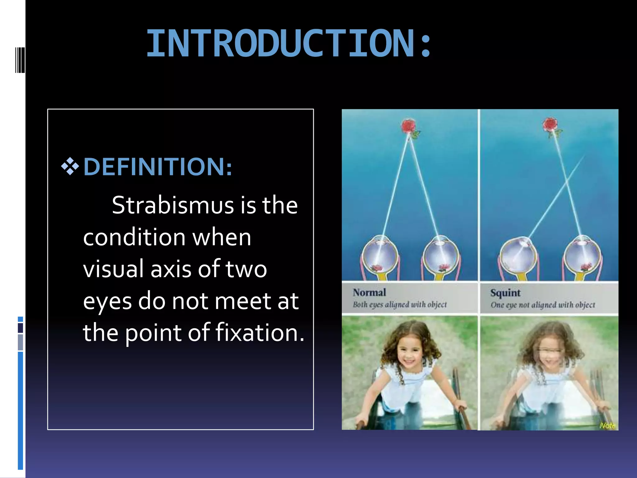
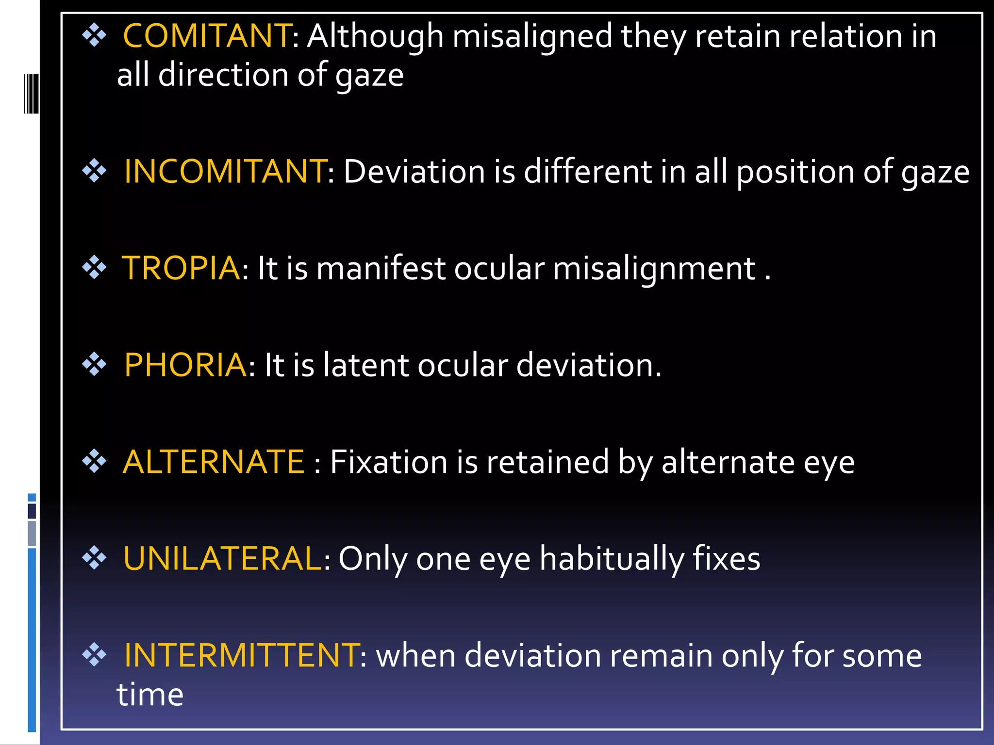
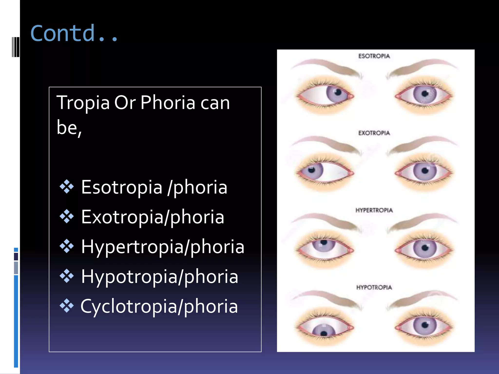
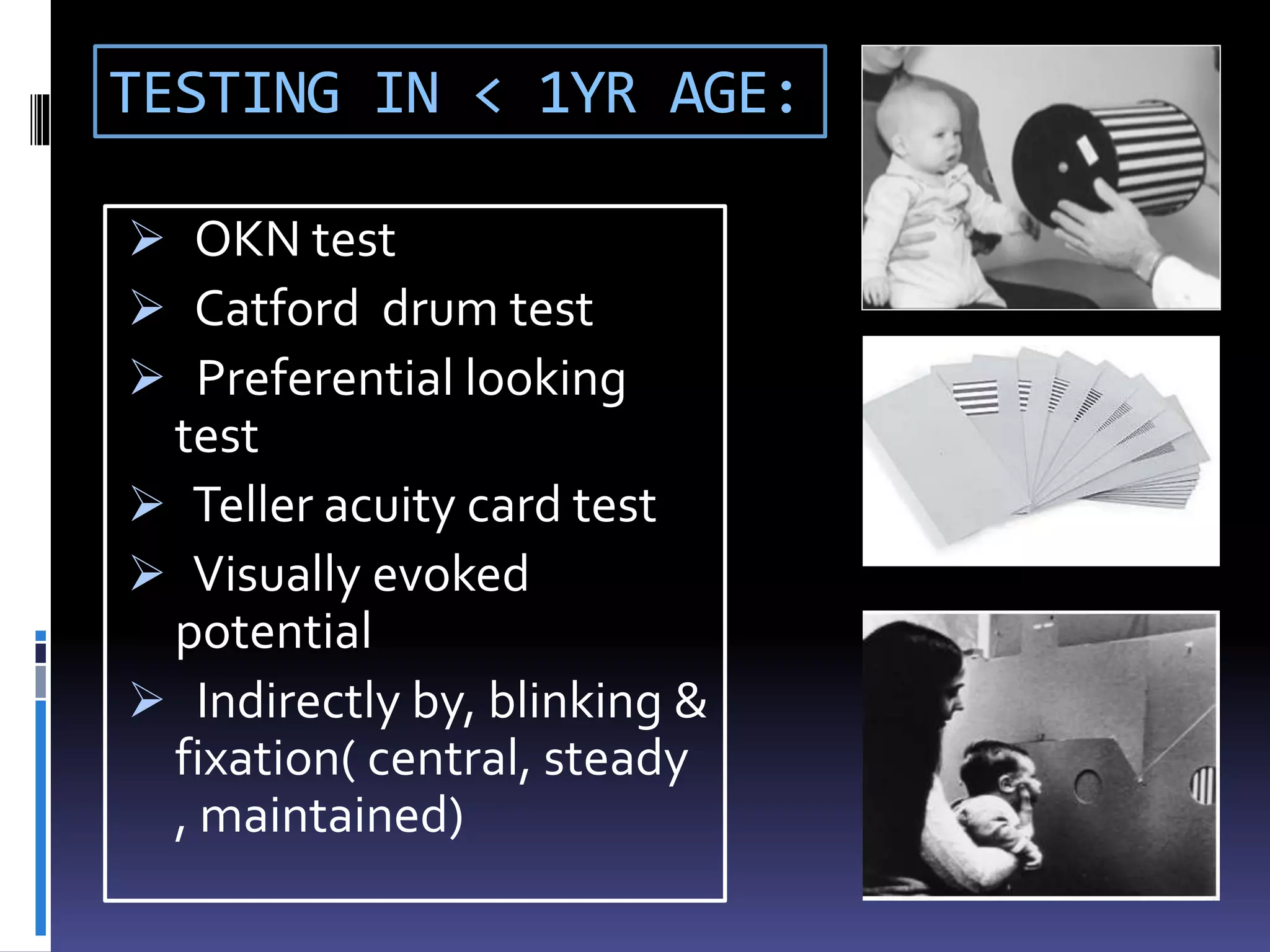
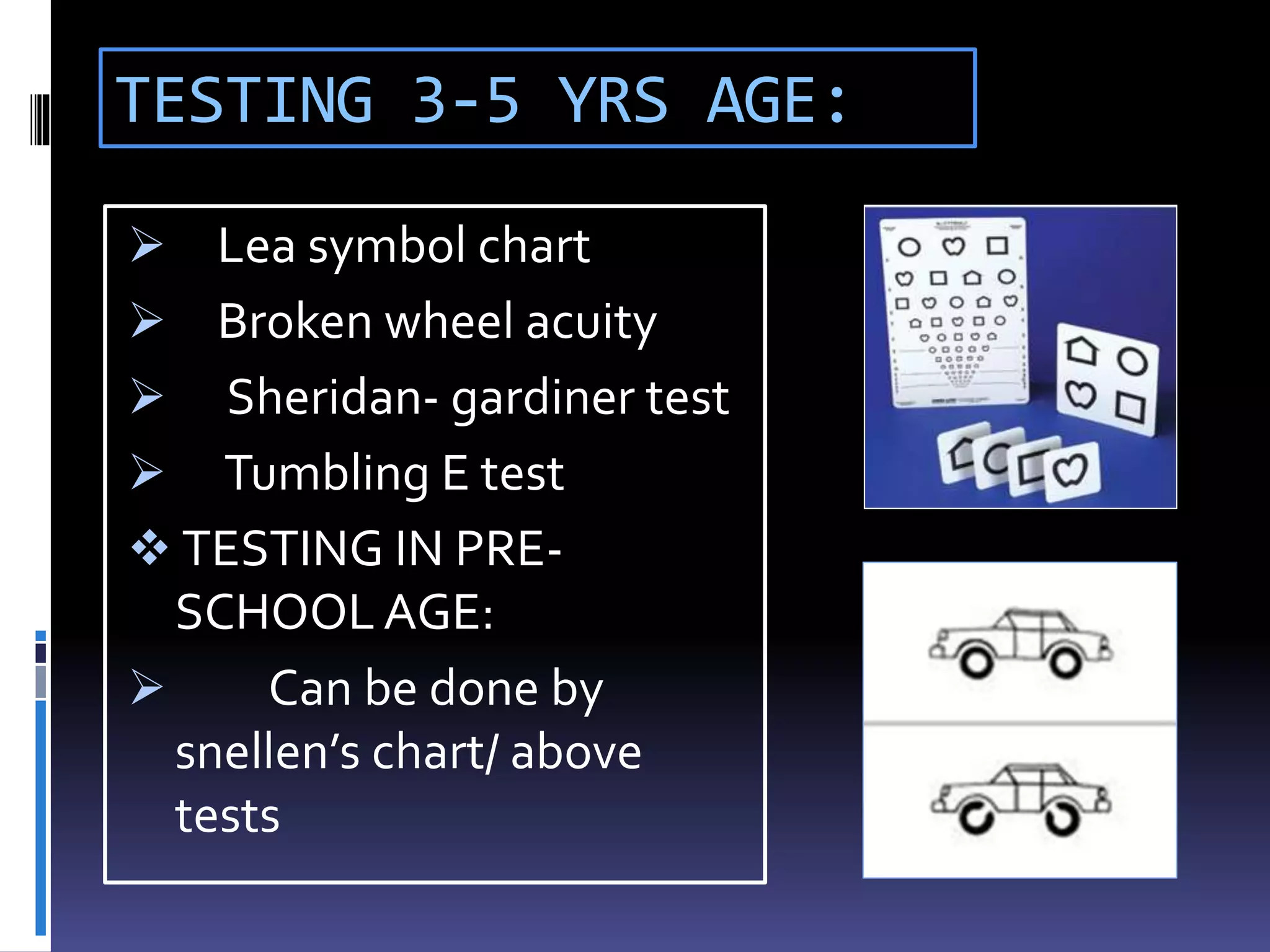
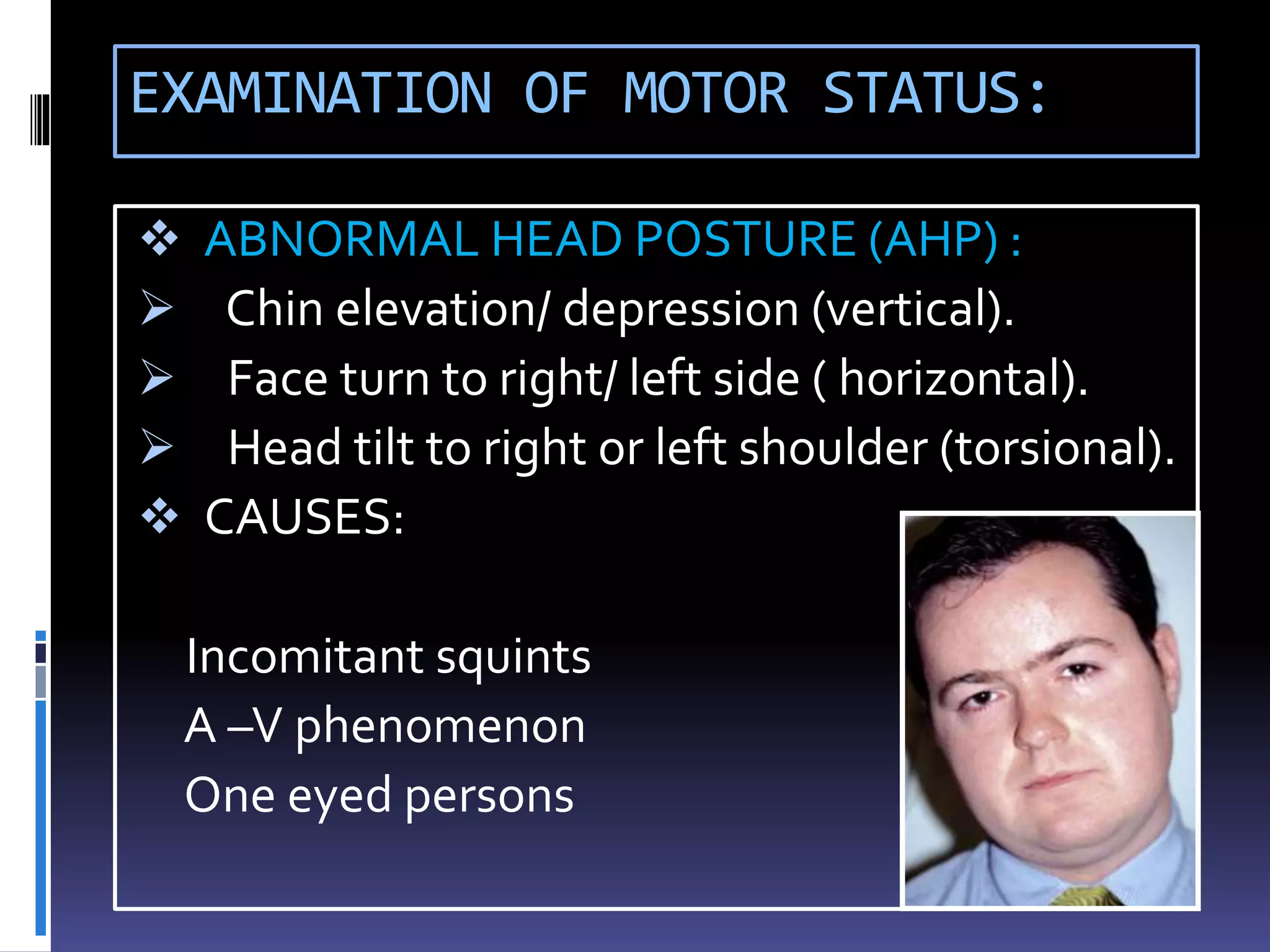
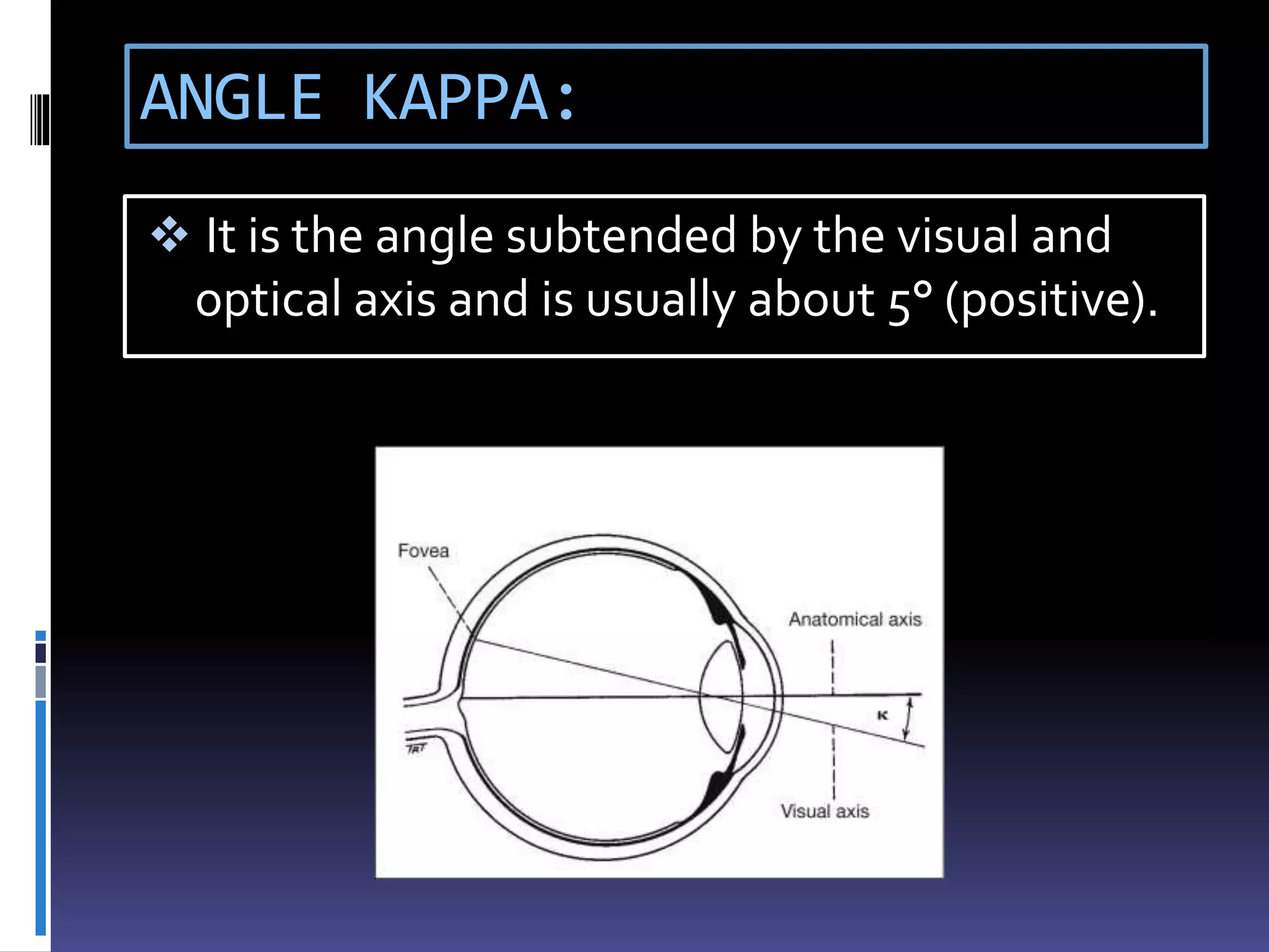
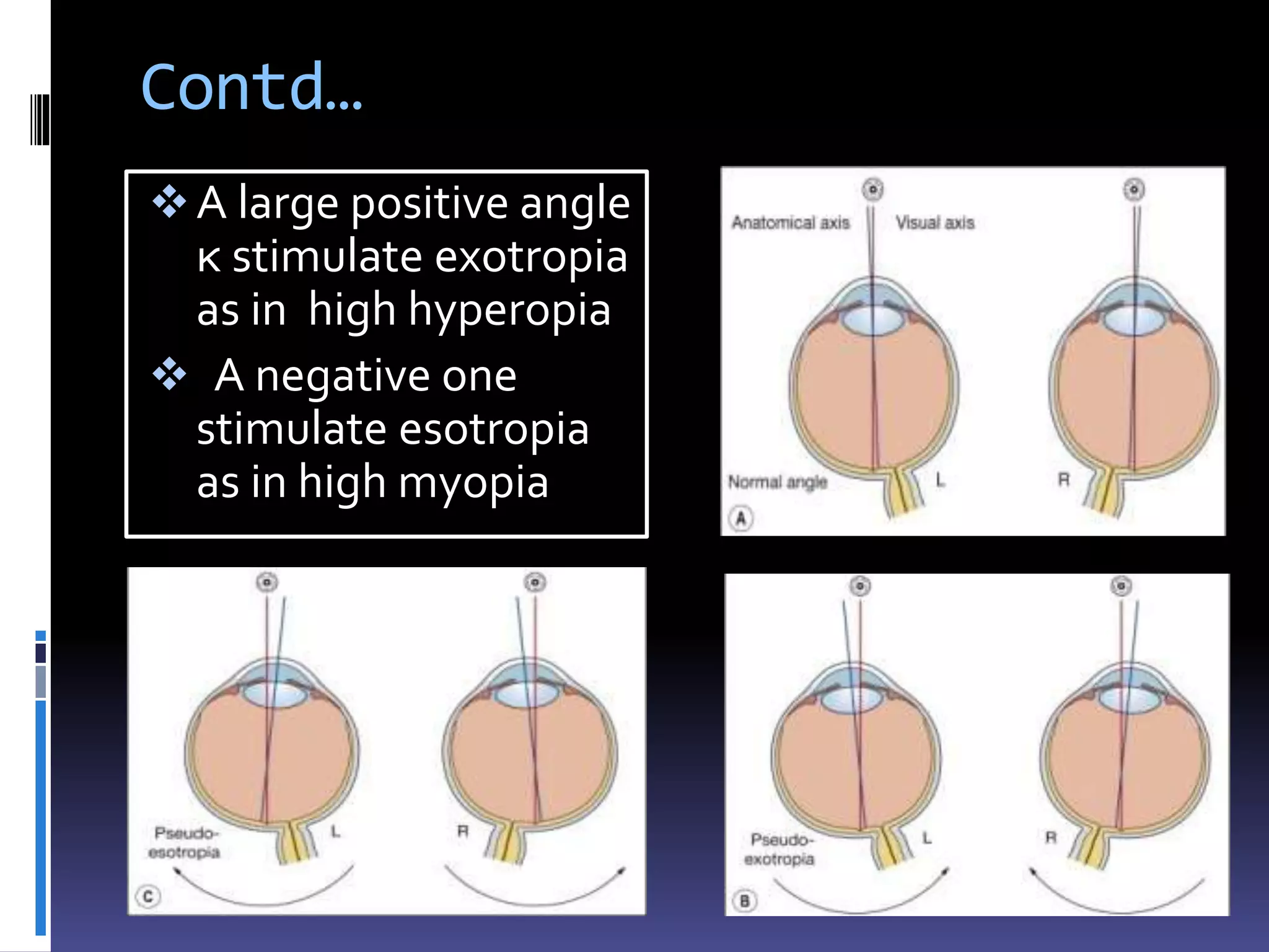
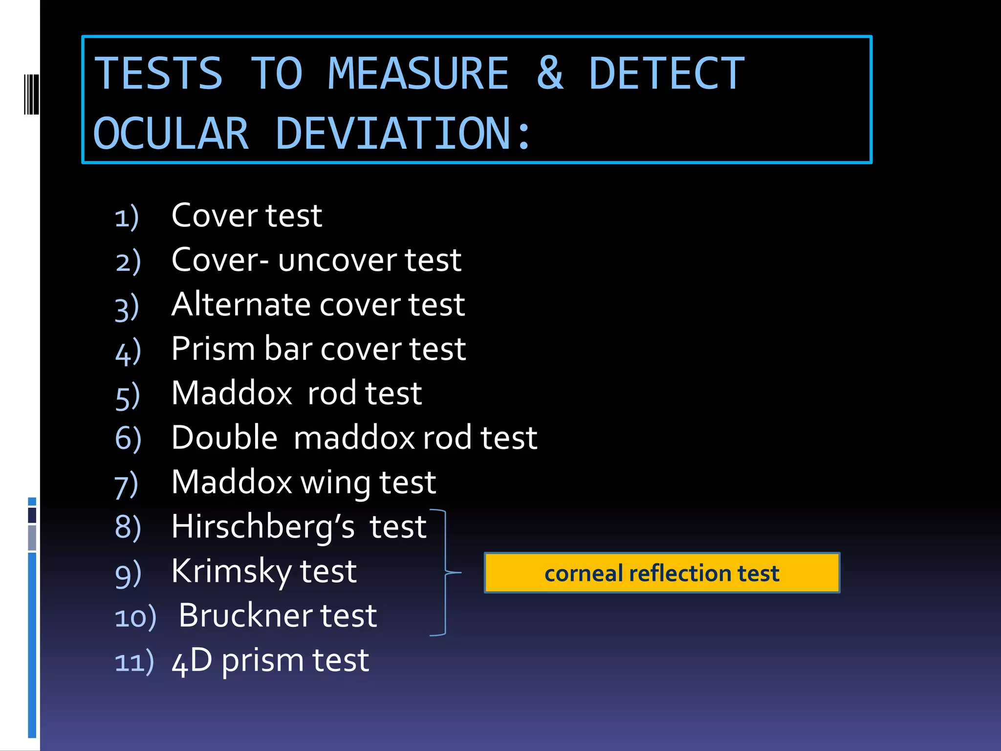
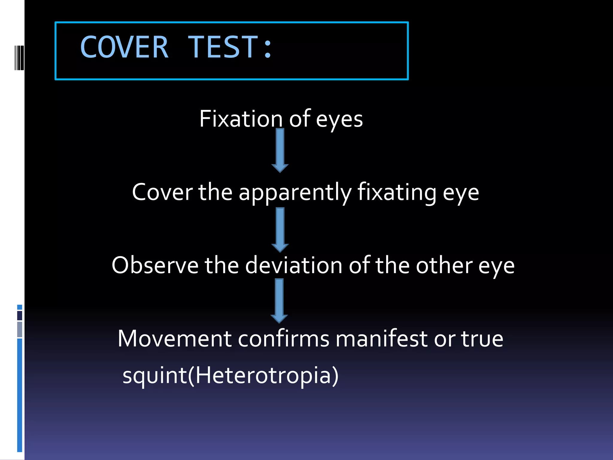
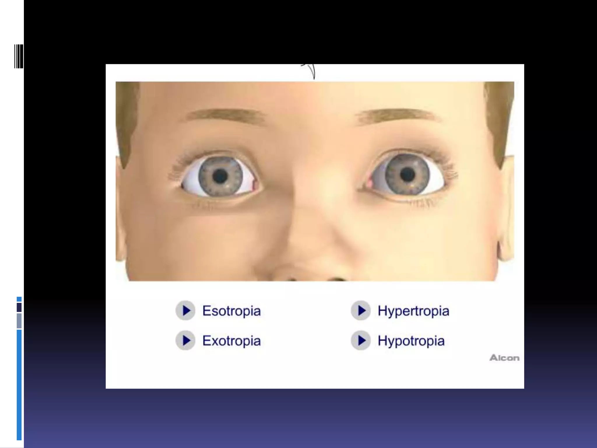
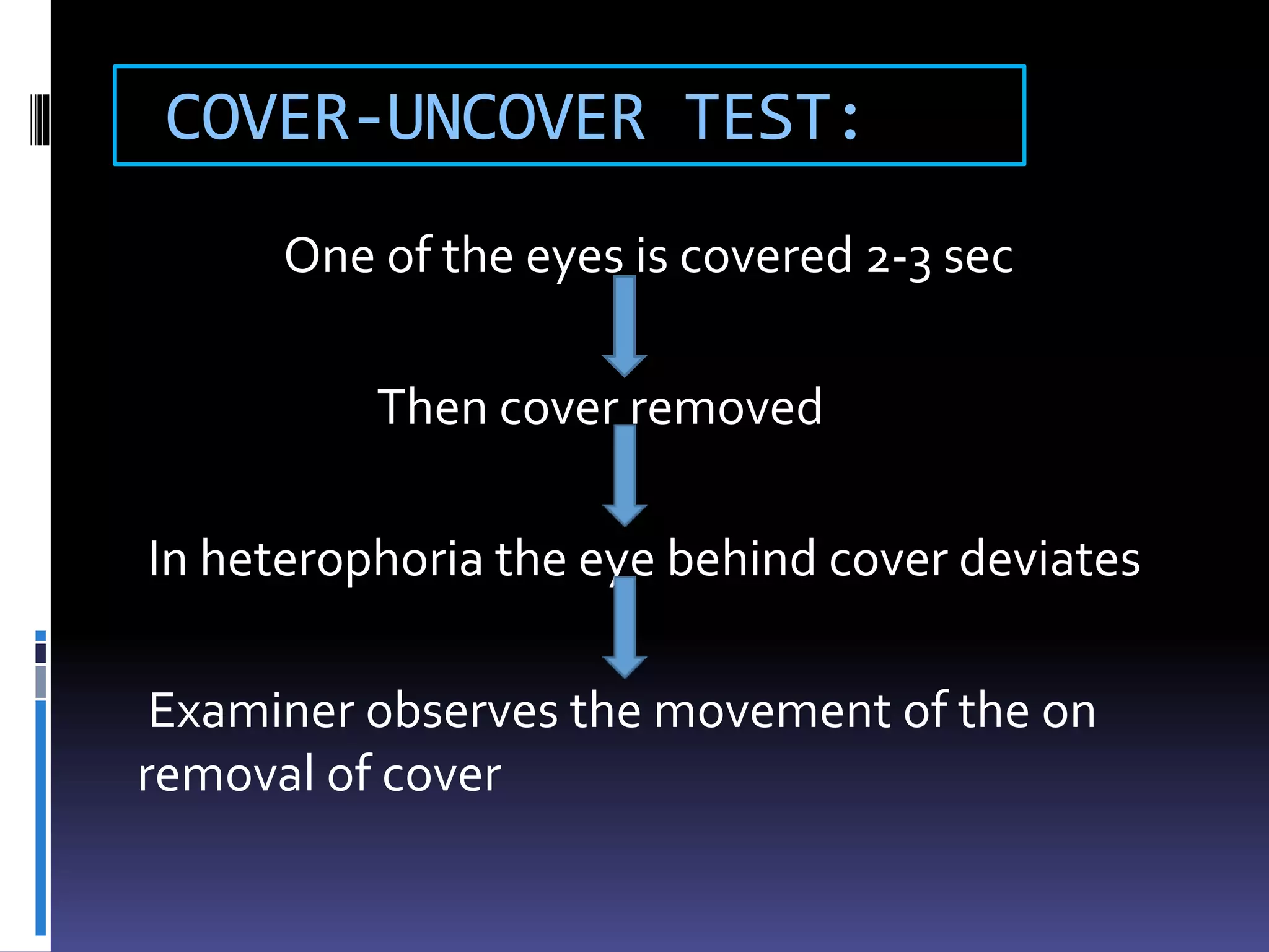
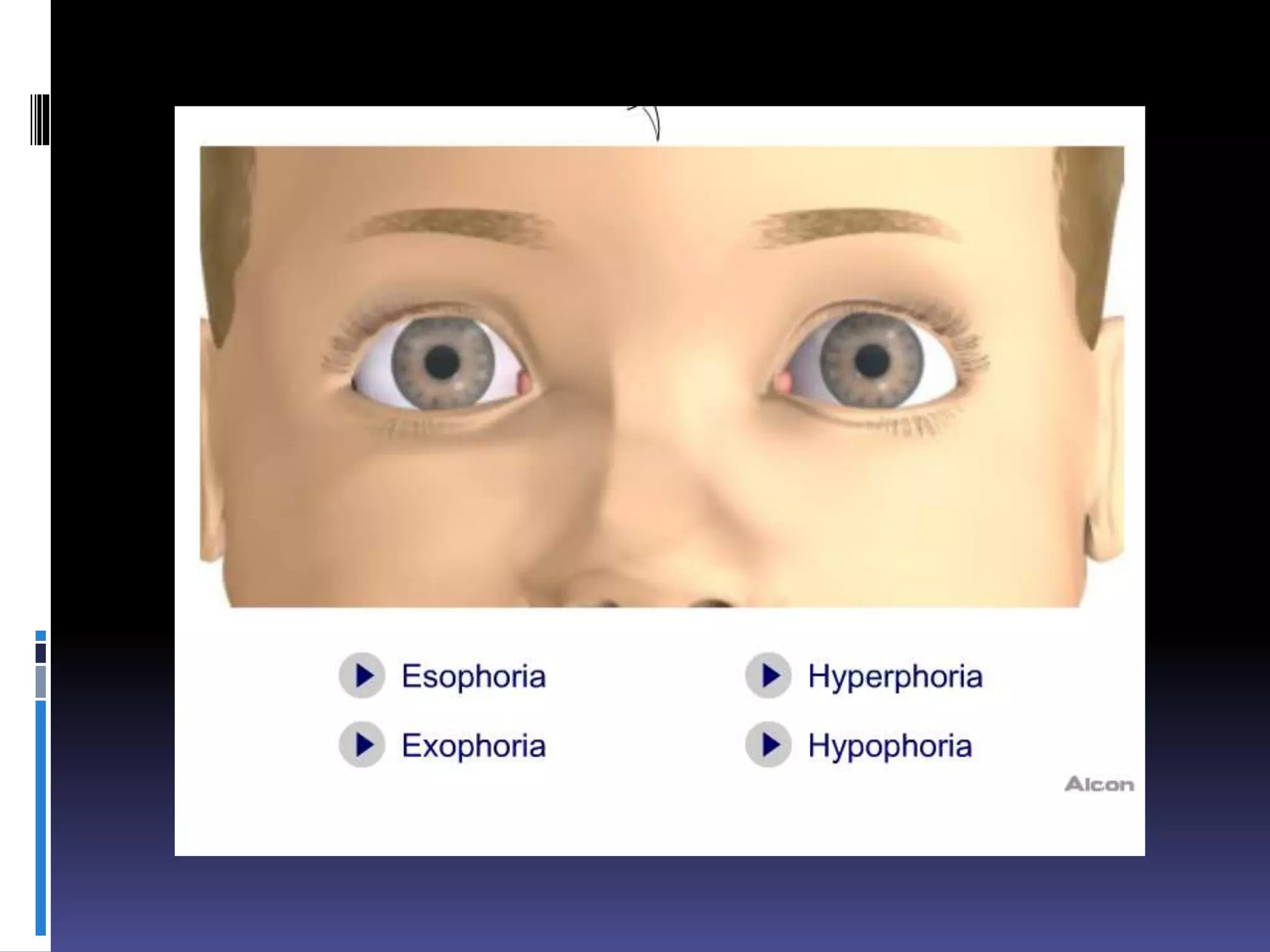
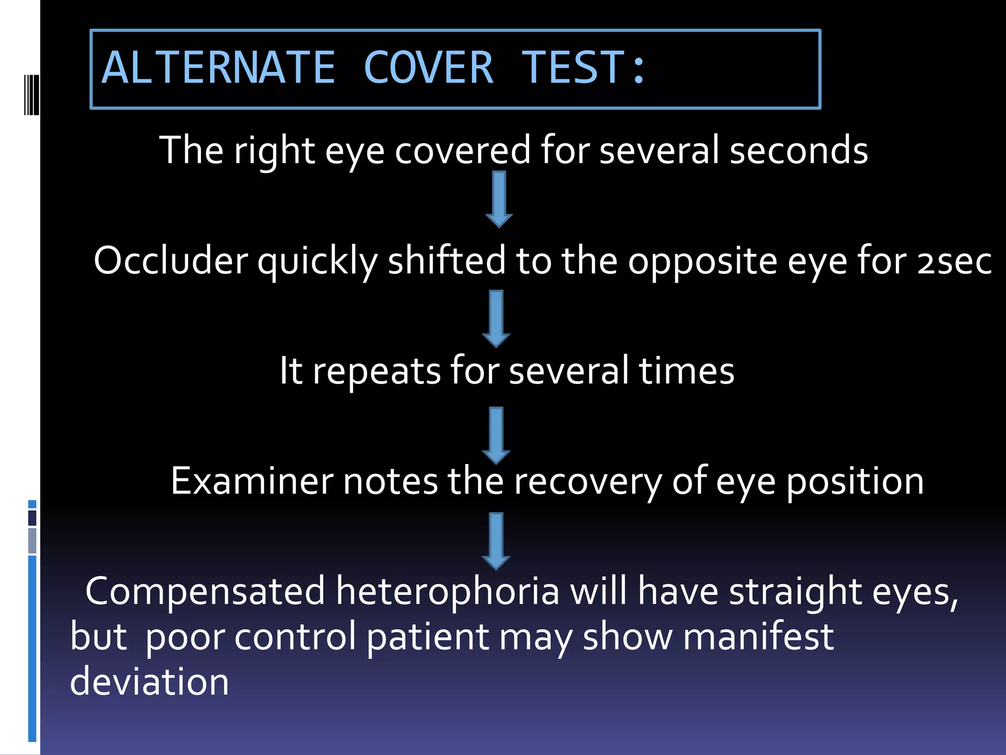
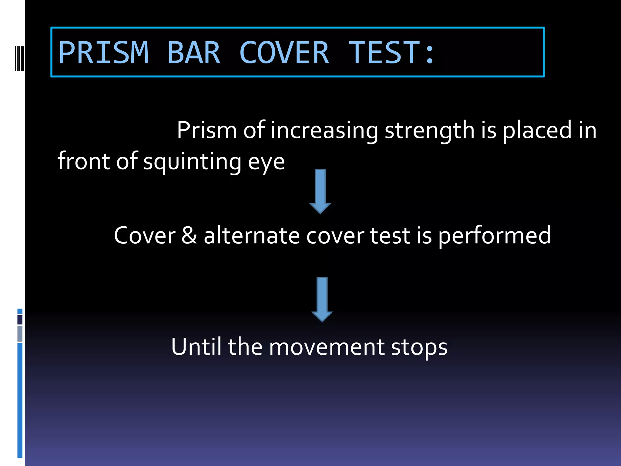
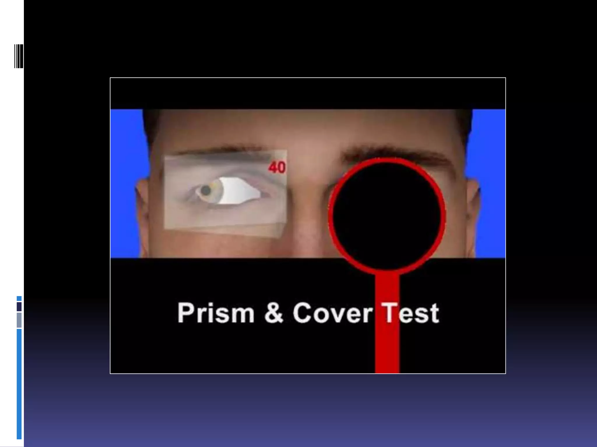
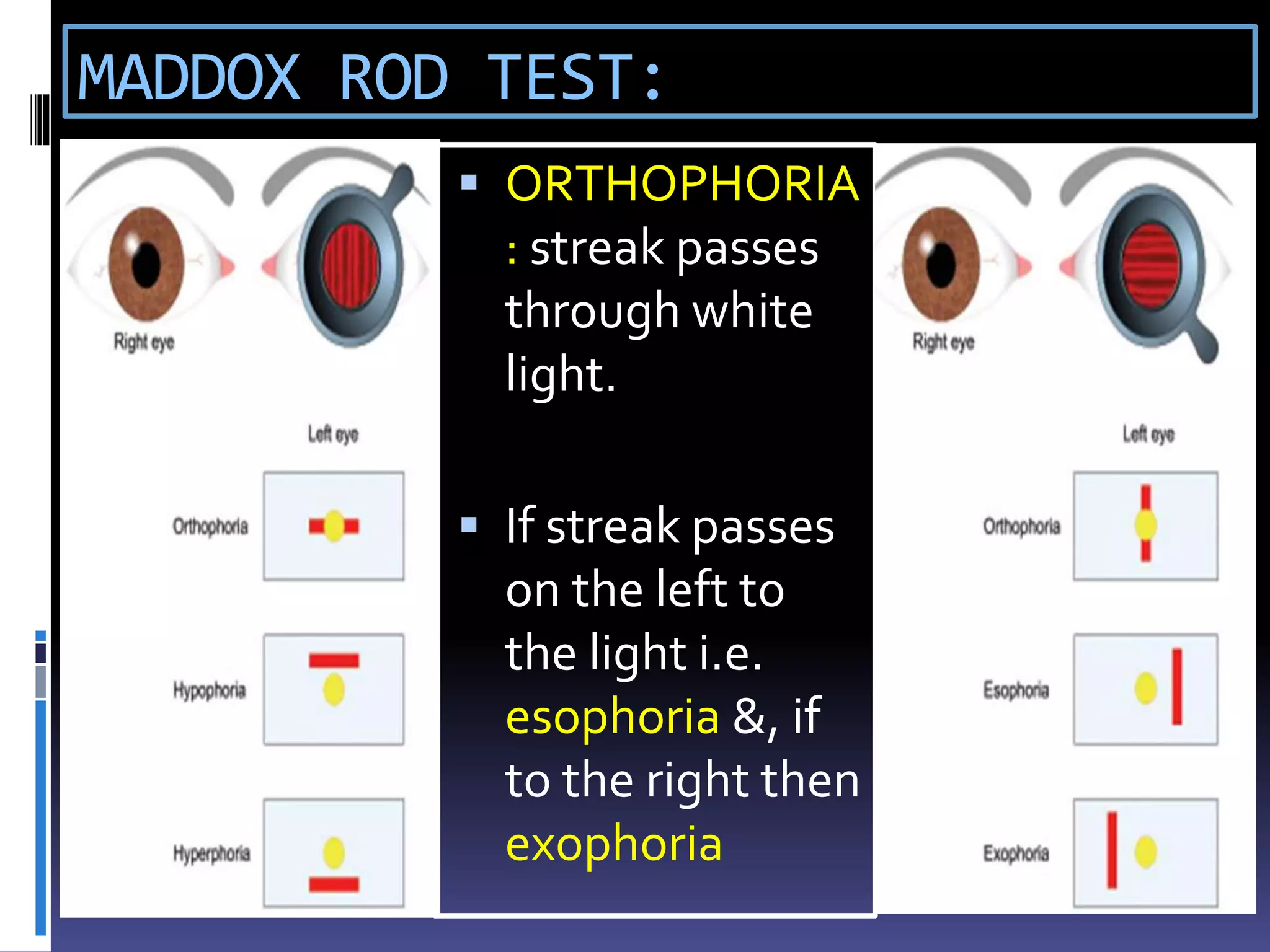
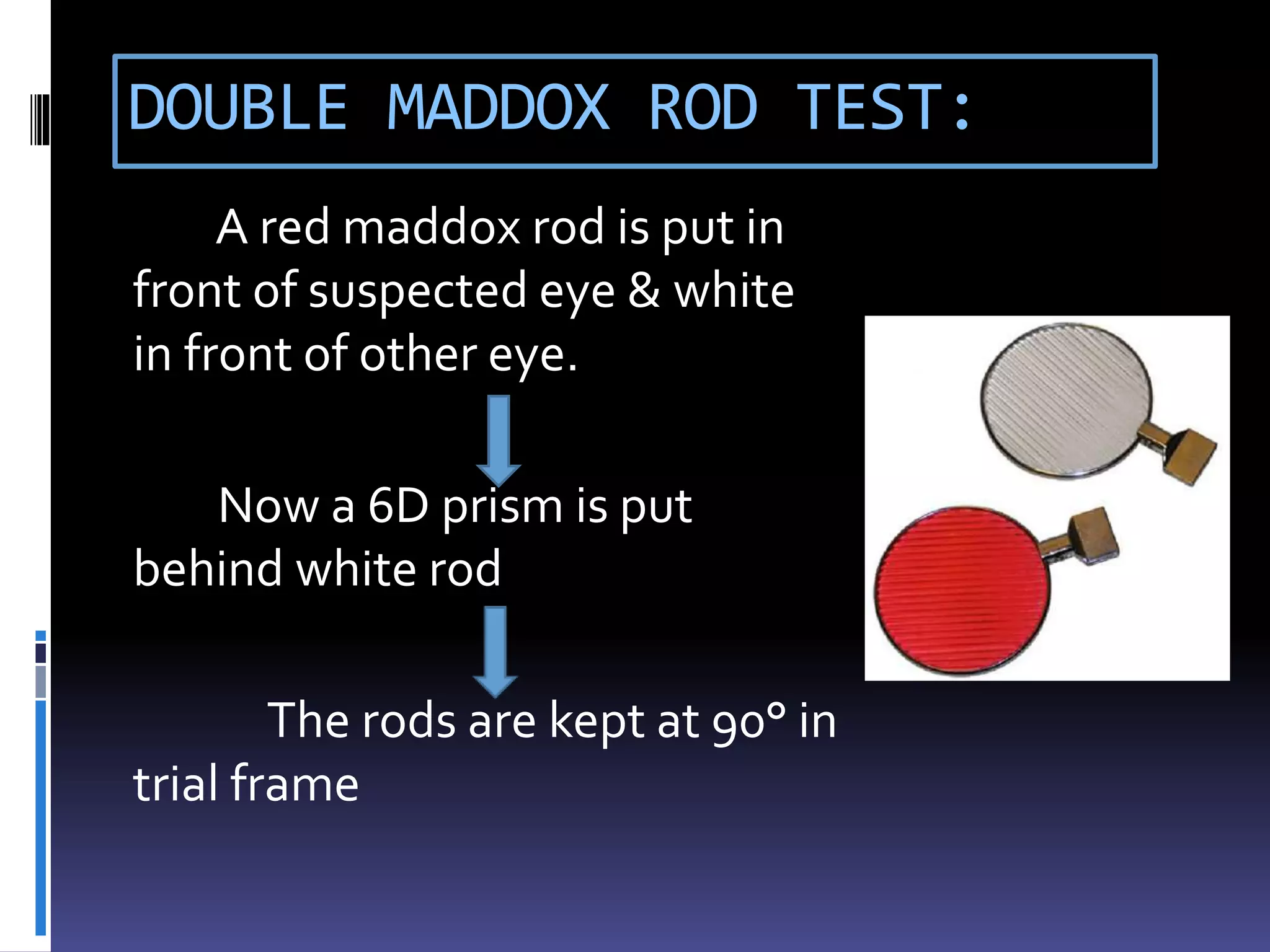
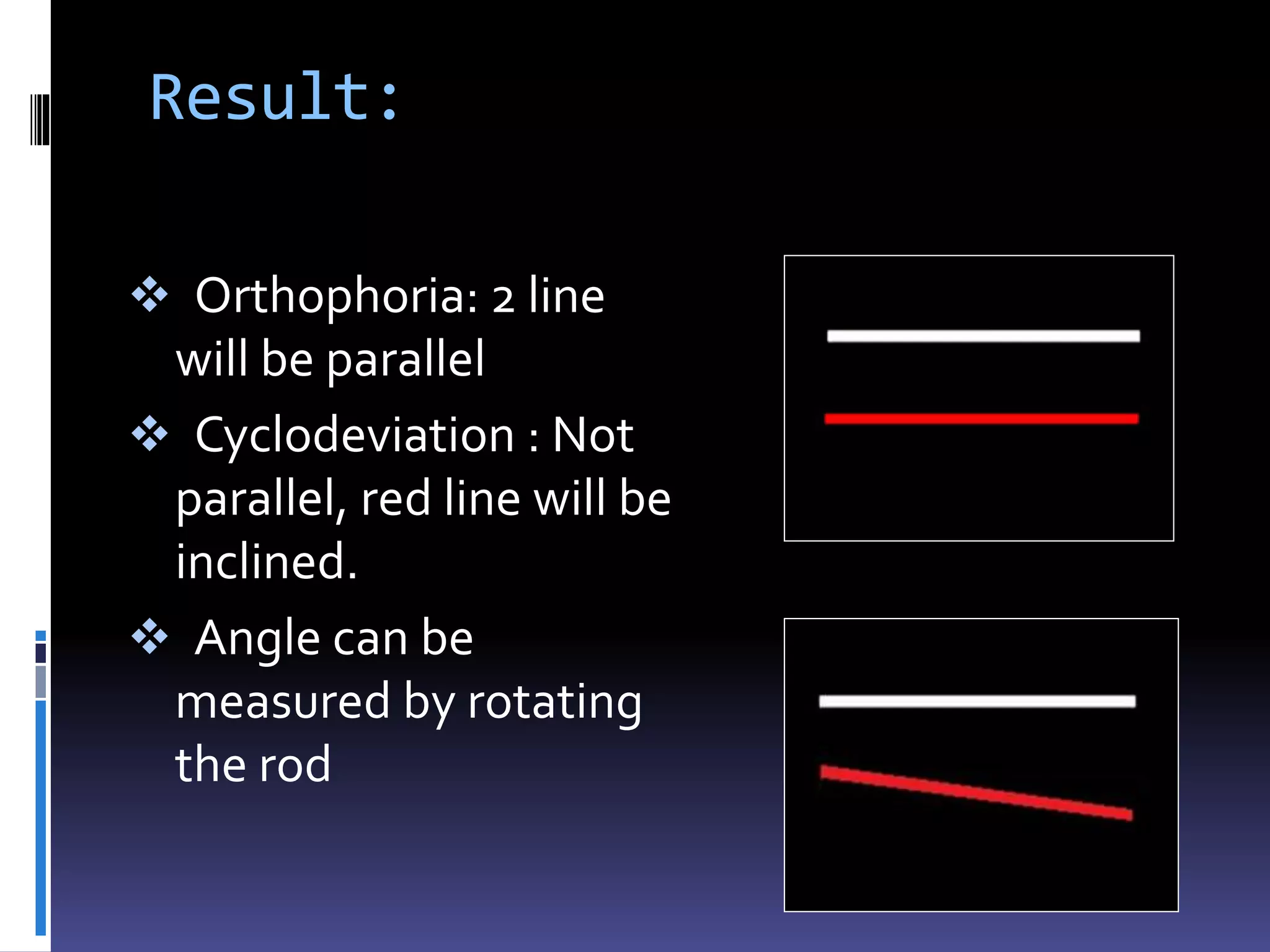
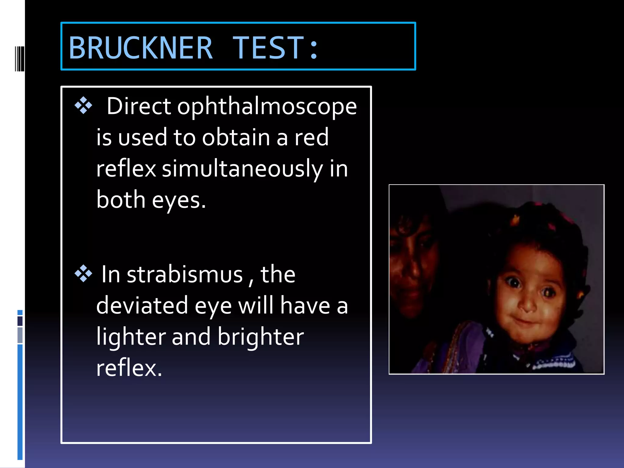
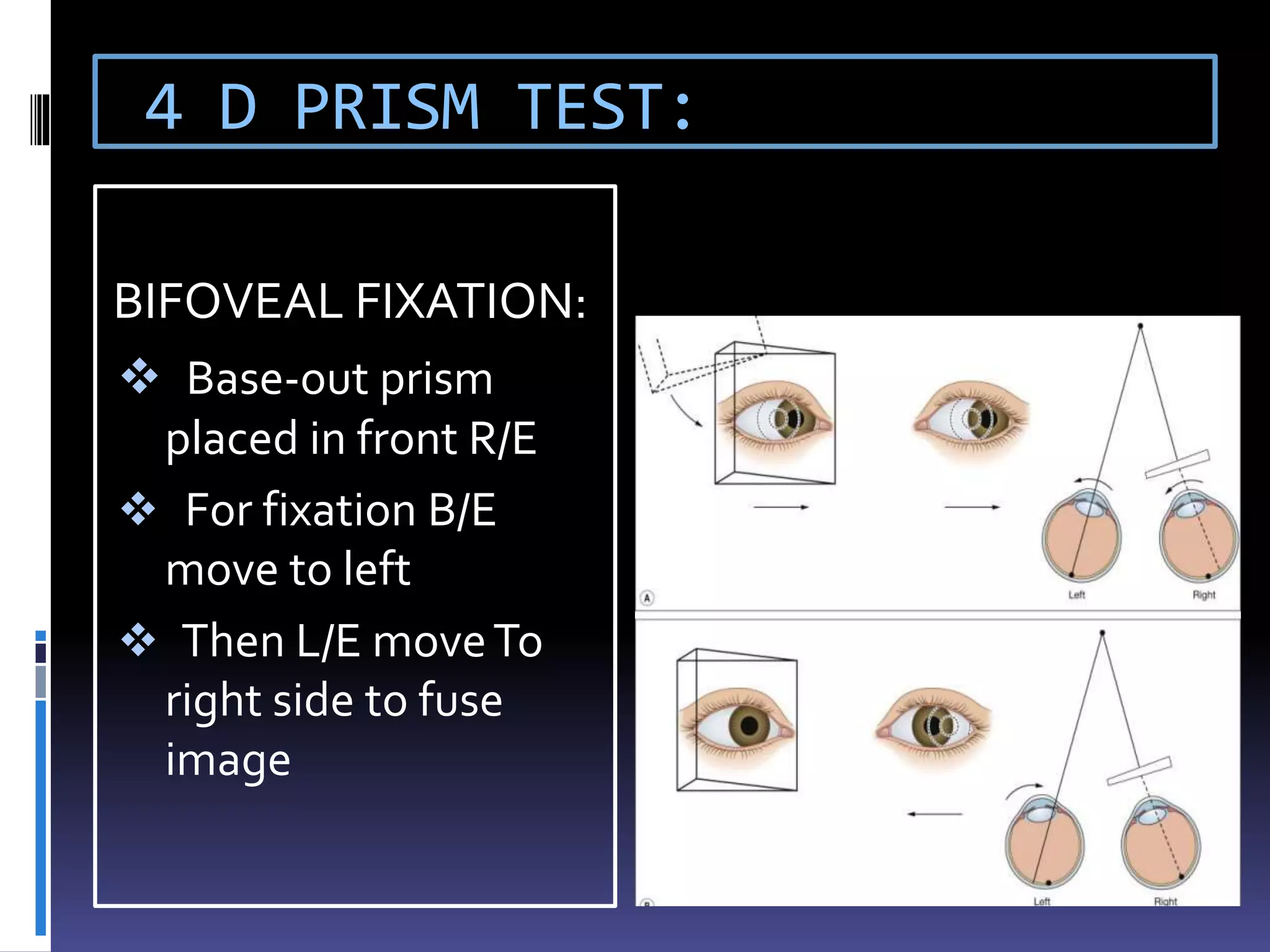
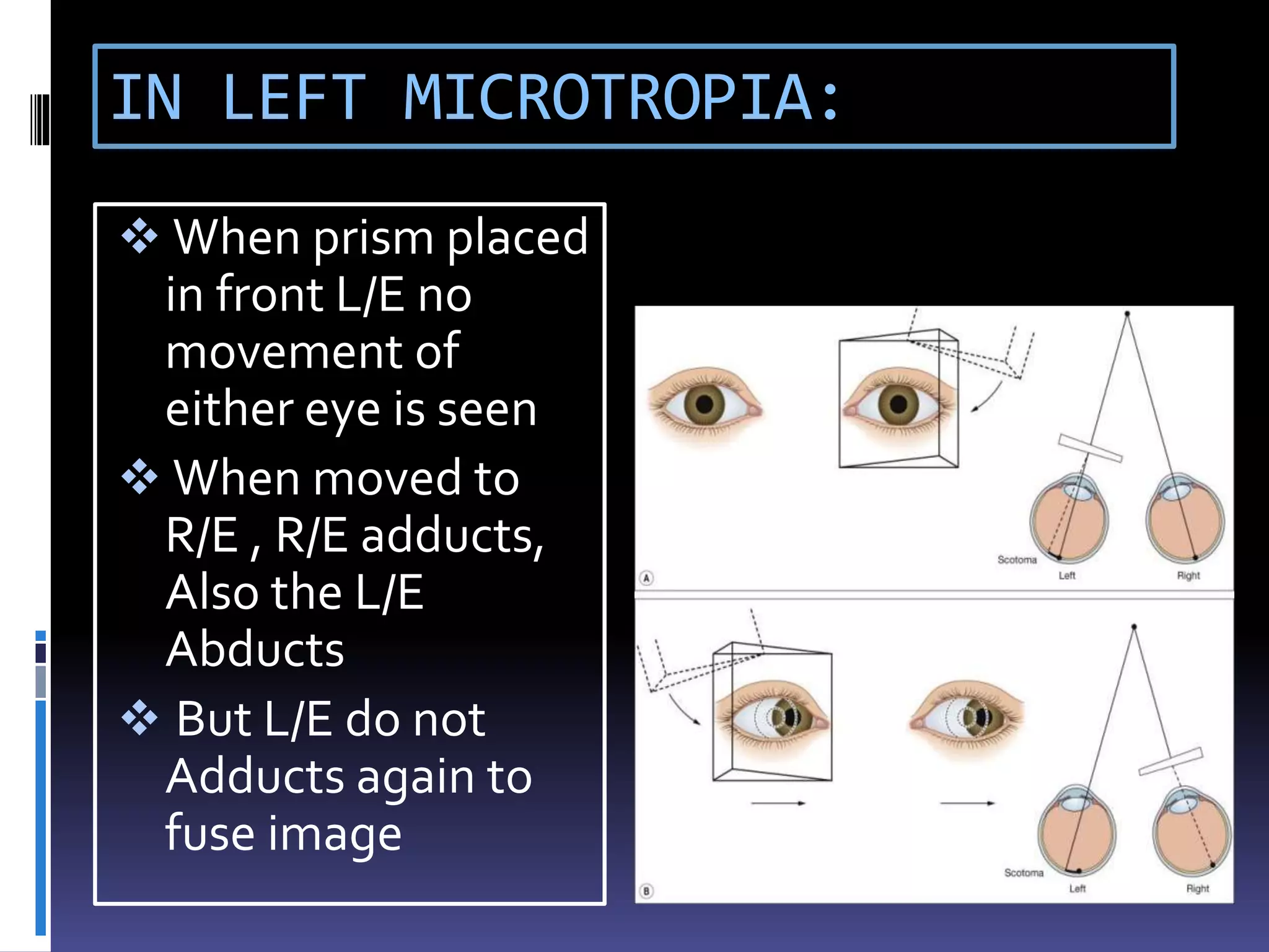
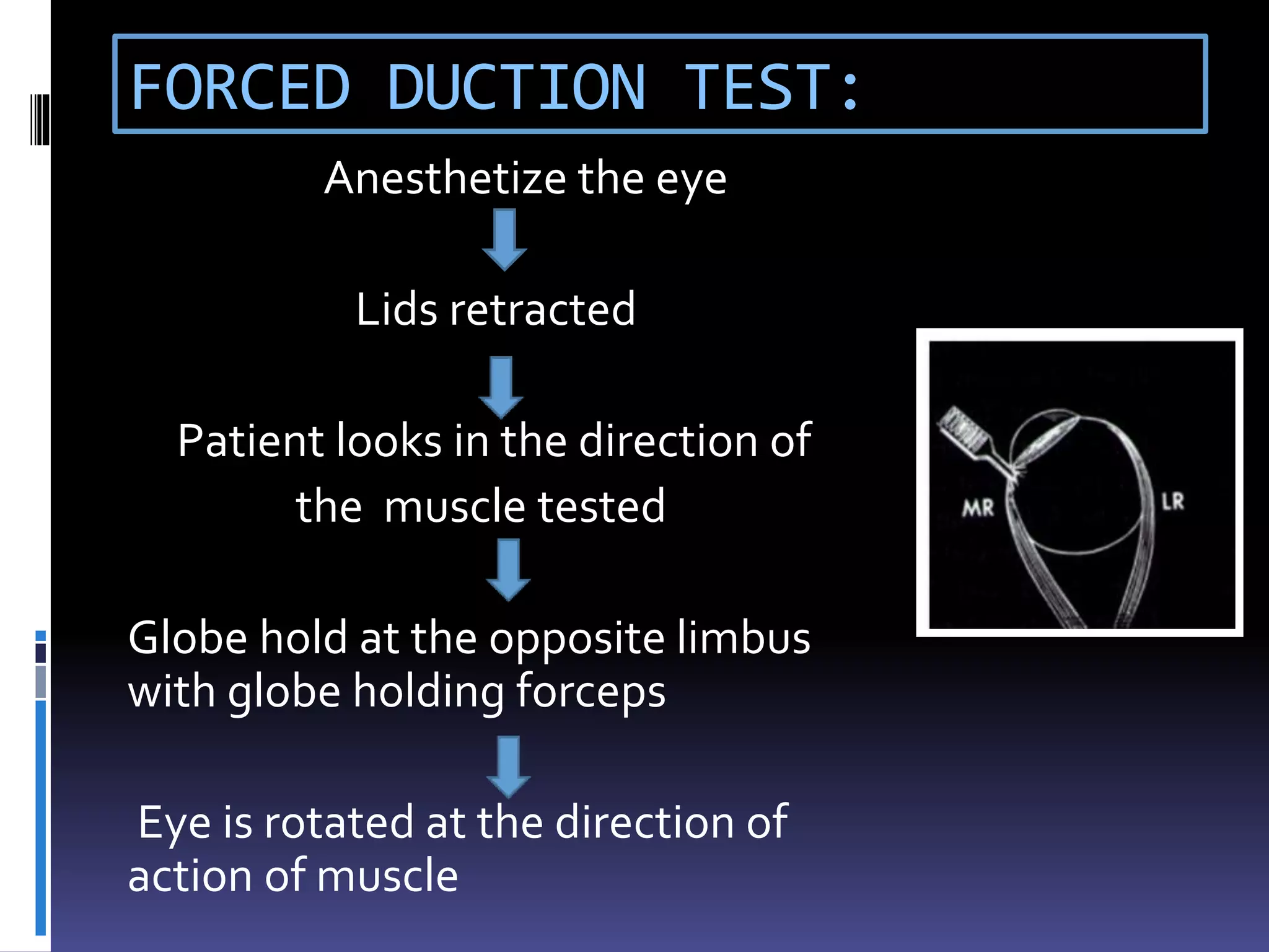
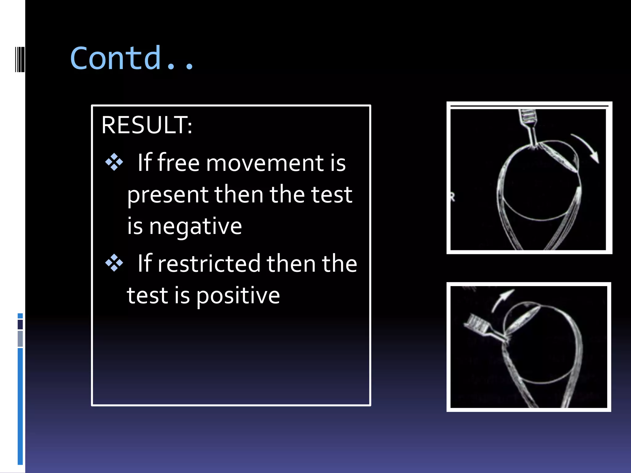
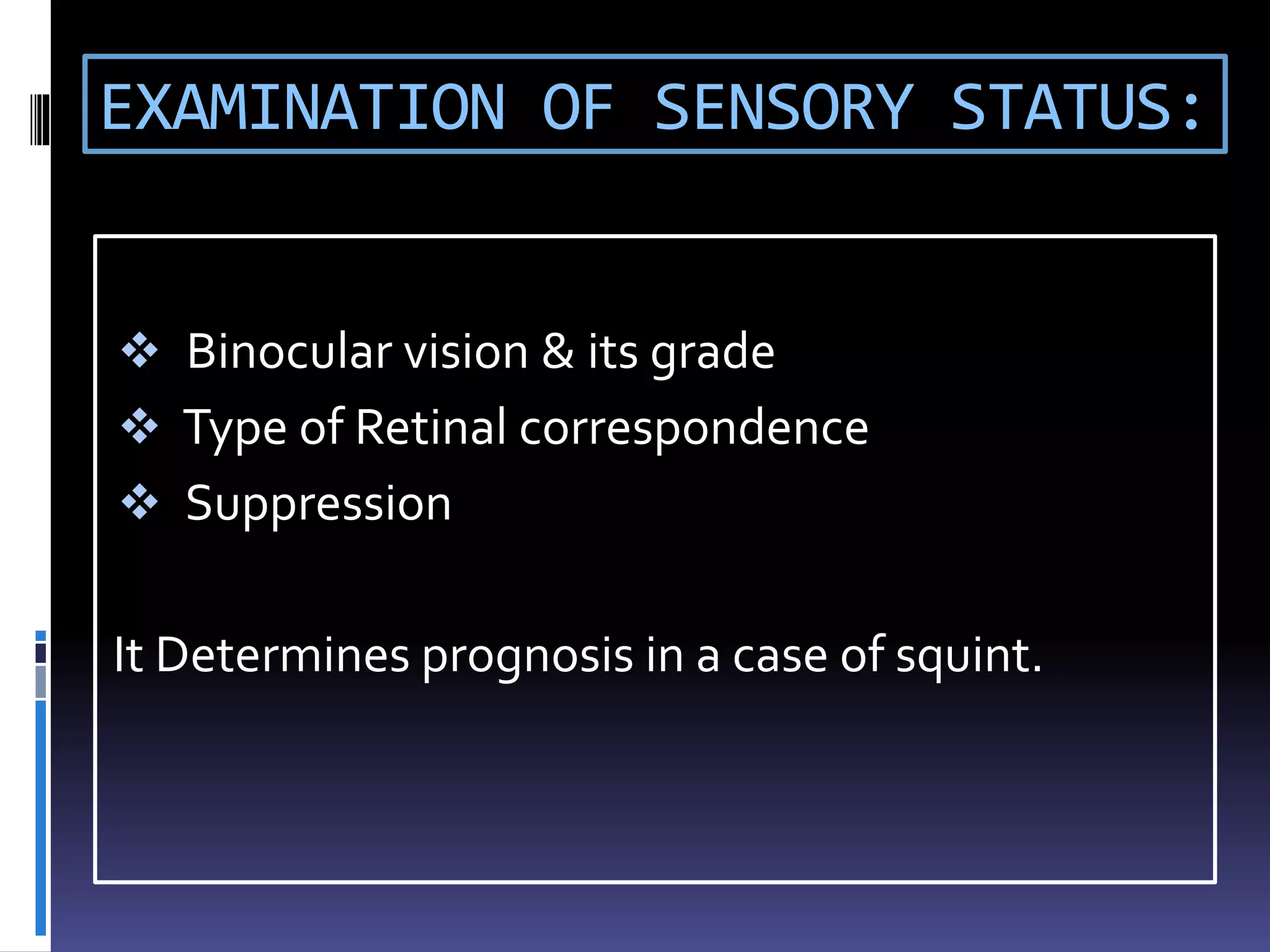
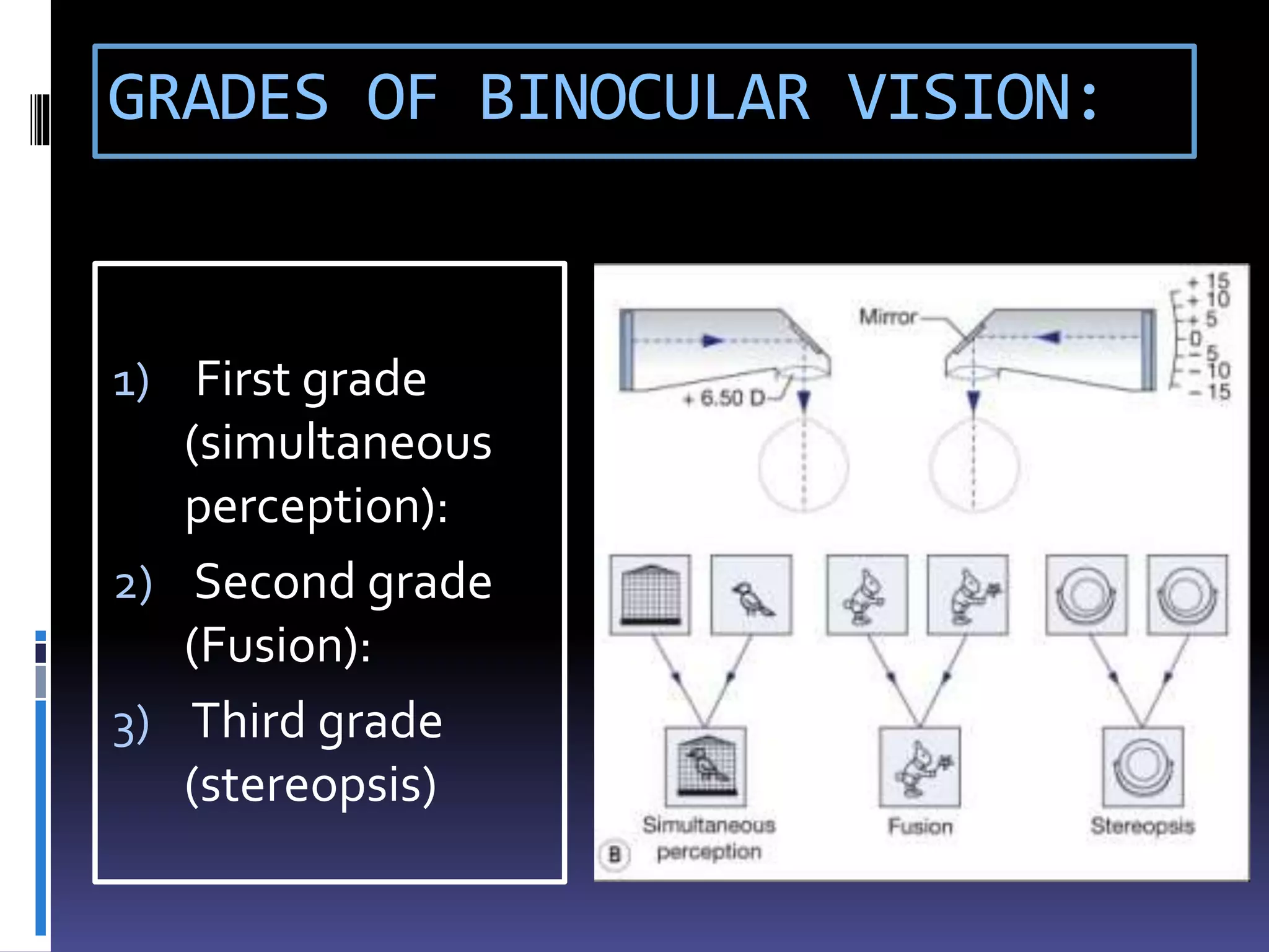
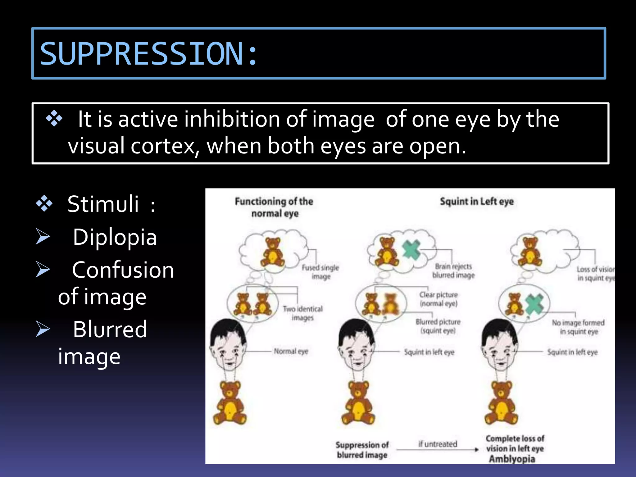
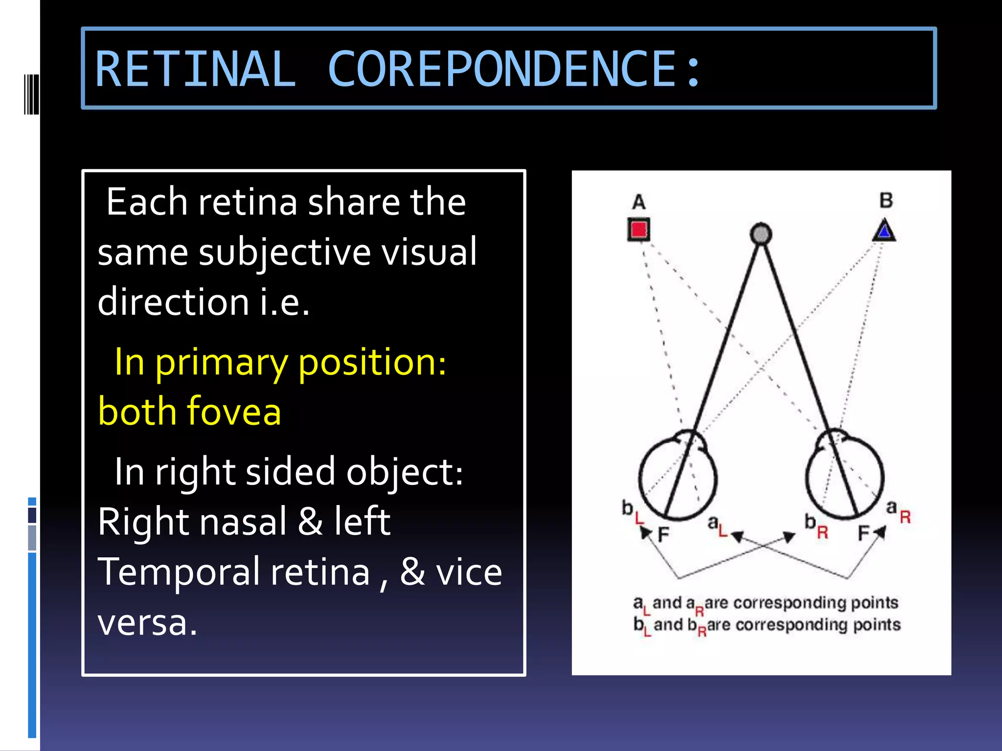
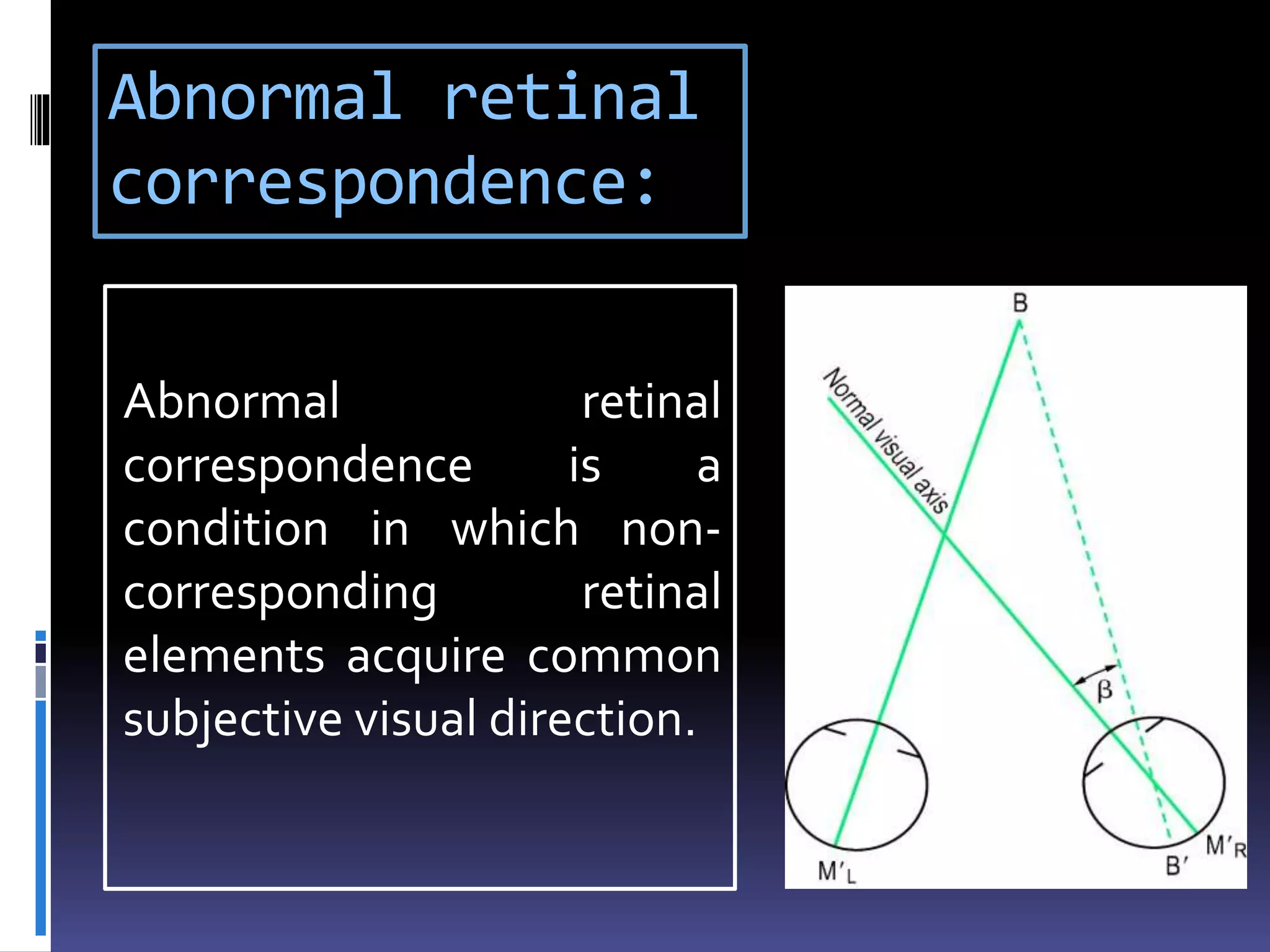
INTRODUCTION:
DEFINITION:
Strabismus is the
condition when
visual axis of two
eyes do not meet at
the point of fixation.
 COMITANT: Although misaligned they retain relation in
all direction of gaze
 INCOMITANT: Deviation is different in all position of gaze
 TROPIA: It is manifest ocular misalignment .
 PHORIA: It is latent ocular deviation.
 ALTERNATE : Fixation is retained by alternate eye
 UNILATERAL: Only one eye habitually fixes
 INTERMITTENT: when deviation remain only for some
time
Contd..
Tropia Or Phoria can
be,
 Esotropia /phoria
 Exotropia/phoria
 Hypertropia/phoria
 Hypotropia/phoria
 Cyclotropia/phoria
ASSESSMENT:
History
Visual Acuity
Examination:
1) Examination of motor status : a) Head posture
b) Ocular deviation
c) Ocular motility
d) Diplopia
2) Examination of sensory status & adaptation
 Refraction & fundoscopy
HISTORY
 The age of onset
 Gradual or sudden (old photographs may sometime
help)
 Symptoms, i.e. diplopia/ discomfort/ blurring/ eye
ache/ cosmetic
 Whether the symptoms occur during fatigue / illness/
stress/inattentive condition etc.
 Whether it occurs for near/ distant vision
 Whether it is unilateral/ alternate or constant/
intermittent.
 General health ( measles, whooping cough), birth
history( LBW) & family history( high refractive error,
strabismus)
 Previous treatment if any, & type & improvement after
it.
VISUAL ACUITY
TESTING IN < 1YR AGE:
 OKN test
 Catford drum test
 Preferential looking
test
 Teller acuity card test
 Visually evoked
potential
 Indirectly by, blinking &
fixation( central, steady
, maintained)
TESTING IN 1-3 YRS AGE:
 Cardiff acuity card test
 Marble game test
 STYCAR graded balls
vision test
 Coin test, Miniature toy
test, etc.
TESTING 3-5 YRS AGE:
 Lea symbol chart
 Broken wheel acuity
 Sheridan- gardiner test
 Tumbling E test
 TESTING IN PRE-
SCHOOLAGE:
 Can be done by
snellen’s chart/ above
tests
EXAMINATION OF MOTOR STATUS:
 ABNORMAL HEAD POSTURE (AHP) :
 Chin elevation/ depression (vertical).
 Face turn to right/ left side ( horizontal).
 Head tilt to right or left shoulder (torsional).
 CAUSES:
Incomitant squints
A –V phenomenon
One eyed persons
OCULAR DEVIATION:
 Is it a true squint/ pseudo
or apparent squint?
 Causes of pseudo
strabismus:
Telecanthus
Epicanthus
Hypertelorism
Ptosis
Lid retraction
Large angle κ
ANGLE KAPPA:
 It is the angle subtended by the visual and
optical axis and is usually about 5° (positive).
Contd…
A large positive angle
κ stimulate exotropia
as in high hyperopia
 A negative one
stimulate esotropia
as in high myopia
TESTS TO MEASURE & DETECT
OCULAR DEVIATION:
1) Cover test
2) Cover- uncover test
3) Alternate cover test
4) Prism bar cover test
5) Maddox rod test
6) Double maddox rod test
7) Maddox wing test
8) Hirschberg’s test
9) Krimsky test
10) Bruckner test
11) 4D prism test
corneal reflection test
COVER TEST:
Fixation of eyes
Cover the apparently fixating eye
Observe the deviation of the other eye
Movement confirms manifest or true
squint(Heterotropia)
COVER-UNCOVER TEST:
One of the eyes is covered 2-3 sec
Then cover removed
In heterophoria the eye behind cover deviates
Examiner observes the movement of the on
removal of cover
ALTERNATE COVER TEST:
The right eye covered for several seconds
Occluder quickly shifted to the opposite eye for 2sec
It repeats for several times
Examiner notes the recovery of eye position
Compensated heterophoria will have straight eyes,
but poor control patient may show manifest
deviation
PRISM BAR COVER TEST:
Prism of increasing strength is placed in
front of squinting eye
Cover & alternate cover test is performed
Until the movement stops
MADDOX ROD TEST:
 ORTHOPHORIA
: streak passes
through white
light.
 If streak passes
on the left to
the light i.e.
esophoria &, if
to the right then
exophoria
DOUBLE MADDOX ROD TEST:
A red maddox rod is put in
front of suspected eye & white
in front of other eye.
Now a 6D prism is put
behind white rod
The rods are kept at 90° in
trial frame
Result:
 Orthophoria: 2 line
will be parallel
 Cyclodeviation : Not
parallel, red line will be
inclined.
 Angle can be
measured by rotating
the rod
MADDOX DOUBLE PRISM:
 Patient will see 3 horizontal line parallel to
each other.
 In cyclodeviation Intermediate line will be
oblique.
MADDOX WING TEST:
R/E : white arrow
vertically And red
arrow horizontally
pointing left.
L/E :Horizontal row of
figures in white and
Vertical row in red
Contd…
 White arrow measures
horizontal tropia
 Red arrow measures
vertical tropia
 In cyclophoria red
scale is adjusted to
appear parallel to
horizontal scale.
HIRSCHBERG TEST:
 Corneal reflections
are usually symetrical
in absence of squint
 In esodeviation
reflex fall on
temporal cornea.
 Roughly 1mm shift
signifies 7° or 15
prism diopter.
KRIMSKY TEST:
A prism bar is placed in
font of the fixating eye
The power increased
until the reflections get
symmetrical.
BRUCKNER TEST:
 Direct ophthalmoscope
is used to obtain a red
reflex simultaneously in
both eyes.
 In strabismus , the
deviated eye will have a
lighter and brighter
reflex.
4 D PRISM TEST:
BIFOVEAL FIXATION:
 Base-out prism
placed in front R/E
 For fixation B/E
move to left
 Then L/E moveTo
right side to fuse
image
IN LEFT MICROTROPIA:
 When prism placed
in front L/E no
movement of
either eye is seen
 When moved to
R/E , R/E adducts,
Also the L/E
Abducts
 But L/E do not
Adducts again to
fuse image
OCULAR MOVEMENTS:
 Ductions: Monocular movement
 Versions: Binocular , simultaneous conjugate
movement.
 Vergences: Binocular, simultaneous,
disjugate movement .
ACTION OF EXTRA-OCULAR MUSCLES:
9 POSITION OF GAZE:
 Near point of
convergence:
Nearest point on which eye
can maintain binocular
fixation.
Measured by RAF rule
It should be Less than 10 cm.
 Near point of
accommodation:
Nearest point on which eyes
can maintain clear focus.
CONVERGENCE TEST:
Hold a pencil / a finger at distance of 30-40cm
Ask the patient to look
at its tip
Bring it nearer to
patient’s eyes.
kept at the eye level of patient till the eye develops
Diplopia or one eye Deviates
FUSIONAL AMPLITUDE:
It is the efficacy of vergence movements.
 It is done by increasing prism dioptre.
 Vertical fusional reserve: 1.5°-2.5°
 Horizontal negative fusional reserve
(abduction range): 3°-5°
 Horizontal positive fusional reserve
(adduction range) : 20°-40°
PRISM VERGENCE TEST:
Patient at 6m distance from light source
The highest Prism that permits single
vision gives
Verging Power
TESTS FOR DIPLOPIA:
 Diplopia charting
 Hess screen test
 Lees screen test
 PARK’S 3 step test
 Forced duction test
DIPLOPIA CHARTING:
 Maximum separation is in the quadrant in
which the muscle acts most
 The level of 2 image
 In which direction the image is deviated
(image displaced toward the direction of
action of the muscle)
LEFT LATERAL RECTUS PALSY:
HESS CHART:
 Patient is asked to
superimpose green light
on the red light.
 Procedure repeated with
red filter in front of left
eye.
 The points are marked by
examiner.
RESULT:
 In orthophoria: both
lights superimpose in 9
position of gaze.
Rt 6th nerve
palsy:
R/E 4th nerve palsy:
LESS SCREEN:
 Consists of two
opalescent glass
screens at right-
angles to each other,
bisected by a two-
sided plane mirror
which dissociates the
two eyes
 interpretation done
as like hess chart
PARK’S 3 step test:
 Use to identify cyclovertical muscle paralysis.
 Performed by measuring the vertical
alignment in
1)primary position,
2) In right and left gaze and
3) In head tilt to the right and to the left.
RIGHT HYPERTROPIA:
1) Circle the muscles which pull R/E down or L/E
up
2) Determine whether vertical deviation is more
on left/ right gaze
3) If worse when head tilted to right, tilt the
circles to the right
FORCED DUCTION TEST:
Anesthetize the eye
Lids retracted
Patient looks in the direction of
the muscle tested
Globe hold at the opposite limbus
with globe holding forceps
Eye is rotated at the direction of
action of muscle
Contd..
RESULT:
 If free movement is
present then the test
is negative
 If restricted then the
test is positive
EXAMINATION OF SENSORY STATUS:
 Binocular vision & its grade
 Type of Retinal correspondence
 Suppression
It Determines prognosis in a case of squint.
GRADES OF BINOCULAR VISION:
1) First grade
(simultaneous
perception):
2) Second grade
(Fusion):
3) Third grade
(stereopsis)
SUPPRESSION:
 It is active inhibition of image of one eye by the
visual cortex, when both eyes are open.
 Stimuli :
 Diplopia
 Confusion
of image
 Blurred
image
RETINAL COREPONDENCE:
Each retina share the
same subjective visual
direction i.e.
In primary position:
both fovea
In right sided object:
Right nasal & left
Temporal retina , & vice
versa.
Abnormal retinal
correspondence:
Abnormal retinal
correspondence is a
condition in which non-
corresponding retinal
elements acquire common
subjective visual direction.
TESTS FOR SENSORY STATUS:
1) Bagolini’s striated glass test
2) Worth four dot test
3) After-image test
4) Synoptophore
BAGOLINI STRIATED GLASS TEST:
Symmetrical cross
response
Asymmetrical cross
response
Single line present
 Cross response with
gap in one line
WORTH FOUR DOT TEST:
 All 4 lights seen –
NRC/ Harmonious
ARC
 Left suppression – 2
red light
 Right suppression – 3
green light
 Diplopia – 3 green &
2 red light
 Alternating
suppression –
Alternate green & red
light.
AFTER-IMAGE TEST(HERING-
BIELSCHOWSKY ):
A bright linear light is used
Patient views the target with one eye and the
light is flashed
For R/E flash held vertically & for L/E horizontally
Patient appreciate a plus sign.
Contd…
a) is consistent with normal retinal correspondence
(NRC), (b) shows uncrossed after-image(diplopia)
and anomalous retinal correspondence (ARC) and
(c) shows left supression
SYNOPTOPHORE:
 It is an instrument ,used for potential for
binocular function in presence of manifest
squint, & tests SP, FUSION, STEREOPSIS
TESTS FOR STEREOPSIS:
1) TNOTEST
2) FRISBYTEST
3) LANGTEST
4) TITMUSTEST
TNO RANDOM DOT TEST:
Consists of seven plates of
randomly distributed
paired red-green dots.
Test targets are only
visible to individuals who
have stereopsis .
FRISBY TEST:
 Consists of 3
transparent plates of,
each printed with 4
squares.
 1 of the squares
contains a hidden
circle, & the random
shapes are printed on
the reverse side.
 Patient needs to find
out the hidden circle.
LANG TEST:
 Here glass is not used.
Patient has to identify simple shape (star ) on
the card
TITMUS TEST:
Viewed through
polaroid spectacle
 Right plate contains
a picture of fly
 Left contains 9
squares, each
containing 4 circles,
of which 1 has
disparity
 test done at 40cm
distance
Contd…
FLY: Appears to stand
out from page
CIRCLES: 1 circle form
disparity & appear
forward.
THE ANIMALS : 3 rows
of animal ,one of
which appear forward.
Fundoscopy & refraction:
 Dilated fundoscopy is mandatory.
 To exclude any underlying ocular pathology
such as macular scarring, optic disc hypoplasia
or retinoblastoma.
 Proper Refractive correction should be given
by retinoscopy
CONCLUSION
It is a common childhood & adult problem.
It is extremely difficult to examine a child and
a tactful examination is to be done.
Quantification of the angle of deviation,
binocular vision, ocular movement, refraction is
important for diagnosis & proper treatment
purpose.
Assesssment of strabismus

Assesssment of strabismus

  • 2.
    INTRODUCTION: DEFINITION: Strabismus is the conditionwhen visual axis of two eyes do not meet at the point of fixation.
  • 3.
     COMITANT: Althoughmisaligned they retain relation in all direction of gaze  INCOMITANT: Deviation is different in all position of gaze  TROPIA: It is manifest ocular misalignment .  PHORIA: It is latent ocular deviation.  ALTERNATE : Fixation is retained by alternate eye  UNILATERAL: Only one eye habitually fixes  INTERMITTENT: when deviation remain only for some time
  • 4.
    Contd.. Tropia Or Phoriacan be,  Esotropia /phoria  Exotropia/phoria  Hypertropia/phoria  Hypotropia/phoria  Cyclotropia/phoria
  • 5.
    ASSESSMENT: History Visual Acuity Examination: 1) Examinationof motor status : a) Head posture b) Ocular deviation c) Ocular motility d) Diplopia 2) Examination of sensory status & adaptation  Refraction & fundoscopy
  • 6.
  • 7.
     The ageof onset  Gradual or sudden (old photographs may sometime help)  Symptoms, i.e. diplopia/ discomfort/ blurring/ eye ache/ cosmetic  Whether the symptoms occur during fatigue / illness/ stress/inattentive condition etc.  Whether it occurs for near/ distant vision  Whether it is unilateral/ alternate or constant/ intermittent.  General health ( measles, whooping cough), birth history( LBW) & family history( high refractive error, strabismus)  Previous treatment if any, & type & improvement after it.
  • 8.
  • 9.
    TESTING IN <1YR AGE:  OKN test  Catford drum test  Preferential looking test  Teller acuity card test  Visually evoked potential  Indirectly by, blinking & fixation( central, steady , maintained)
  • 10.
    TESTING IN 1-3YRS AGE:  Cardiff acuity card test  Marble game test  STYCAR graded balls vision test  Coin test, Miniature toy test, etc.
  • 11.
    TESTING 3-5 YRSAGE:  Lea symbol chart  Broken wheel acuity  Sheridan- gardiner test  Tumbling E test  TESTING IN PRE- SCHOOLAGE:  Can be done by snellen’s chart/ above tests
  • 12.
    EXAMINATION OF MOTORSTATUS:  ABNORMAL HEAD POSTURE (AHP) :  Chin elevation/ depression (vertical).  Face turn to right/ left side ( horizontal).  Head tilt to right or left shoulder (torsional).  CAUSES: Incomitant squints A –V phenomenon One eyed persons
  • 13.
    OCULAR DEVIATION:  Isit a true squint/ pseudo or apparent squint?  Causes of pseudo strabismus: Telecanthus Epicanthus Hypertelorism Ptosis Lid retraction Large angle κ
  • 14.
    ANGLE KAPPA:  Itis the angle subtended by the visual and optical axis and is usually about 5° (positive).
  • 15.
    Contd… A large positiveangle κ stimulate exotropia as in high hyperopia  A negative one stimulate esotropia as in high myopia
  • 16.
    TESTS TO MEASURE& DETECT OCULAR DEVIATION: 1) Cover test 2) Cover- uncover test 3) Alternate cover test 4) Prism bar cover test 5) Maddox rod test 6) Double maddox rod test 7) Maddox wing test 8) Hirschberg’s test 9) Krimsky test 10) Bruckner test 11) 4D prism test corneal reflection test
  • 17.
    COVER TEST: Fixation ofeyes Cover the apparently fixating eye Observe the deviation of the other eye Movement confirms manifest or true squint(Heterotropia)
  • 19.
    COVER-UNCOVER TEST: One ofthe eyes is covered 2-3 sec Then cover removed In heterophoria the eye behind cover deviates Examiner observes the movement of the on removal of cover
  • 21.
    ALTERNATE COVER TEST: Theright eye covered for several seconds Occluder quickly shifted to the opposite eye for 2sec It repeats for several times Examiner notes the recovery of eye position Compensated heterophoria will have straight eyes, but poor control patient may show manifest deviation
  • 22.
    PRISM BAR COVERTEST: Prism of increasing strength is placed in front of squinting eye Cover & alternate cover test is performed Until the movement stops
  • 24.
    MADDOX ROD TEST: ORTHOPHORIA : streak passes through white light.  If streak passes on the left to the light i.e. esophoria &, if to the right then exophoria
  • 25.
    DOUBLE MADDOX RODTEST: A red maddox rod is put in front of suspected eye & white in front of other eye. Now a 6D prism is put behind white rod The rods are kept at 90° in trial frame
  • 26.
    Result:  Orthophoria: 2line will be parallel  Cyclodeviation : Not parallel, red line will be inclined.  Angle can be measured by rotating the rod
  • 27.
    MADDOX DOUBLE PRISM: Patient will see 3 horizontal line parallel to each other.  In cyclodeviation Intermediate line will be oblique.
  • 28.
    MADDOX WING TEST: R/E: white arrow vertically And red arrow horizontally pointing left. L/E :Horizontal row of figures in white and Vertical row in red
  • 29.
    Contd…  White arrowmeasures horizontal tropia  Red arrow measures vertical tropia  In cyclophoria red scale is adjusted to appear parallel to horizontal scale.
  • 30.
    HIRSCHBERG TEST:  Cornealreflections are usually symetrical in absence of squint  In esodeviation reflex fall on temporal cornea.  Roughly 1mm shift signifies 7° or 15 prism diopter.
  • 31.
    KRIMSKY TEST: A prismbar is placed in font of the fixating eye The power increased until the reflections get symmetrical.
  • 32.
    BRUCKNER TEST:  Directophthalmoscope is used to obtain a red reflex simultaneously in both eyes.  In strabismus , the deviated eye will have a lighter and brighter reflex.
  • 33.
    4 D PRISMTEST: BIFOVEAL FIXATION:  Base-out prism placed in front R/E  For fixation B/E move to left  Then L/E moveTo right side to fuse image
  • 34.
    IN LEFT MICROTROPIA: When prism placed in front L/E no movement of either eye is seen  When moved to R/E , R/E adducts, Also the L/E Abducts  But L/E do not Adducts again to fuse image
  • 35.
    OCULAR MOVEMENTS:  Ductions:Monocular movement  Versions: Binocular , simultaneous conjugate movement.  Vergences: Binocular, simultaneous, disjugate movement .
  • 36.
  • 37.
  • 38.
     Near pointof convergence: Nearest point on which eye can maintain binocular fixation. Measured by RAF rule It should be Less than 10 cm.  Near point of accommodation: Nearest point on which eyes can maintain clear focus.
  • 39.
    CONVERGENCE TEST: Hold apencil / a finger at distance of 30-40cm Ask the patient to look at its tip Bring it nearer to patient’s eyes. kept at the eye level of patient till the eye develops Diplopia or one eye Deviates
  • 40.
    FUSIONAL AMPLITUDE: It isthe efficacy of vergence movements.  It is done by increasing prism dioptre.  Vertical fusional reserve: 1.5°-2.5°  Horizontal negative fusional reserve (abduction range): 3°-5°  Horizontal positive fusional reserve (adduction range) : 20°-40°
  • 41.
    PRISM VERGENCE TEST: Patientat 6m distance from light source The highest Prism that permits single vision gives Verging Power
  • 42.
    TESTS FOR DIPLOPIA: Diplopia charting  Hess screen test  Lees screen test  PARK’S 3 step test  Forced duction test
  • 43.
    DIPLOPIA CHARTING:  Maximumseparation is in the quadrant in which the muscle acts most  The level of 2 image  In which direction the image is deviated (image displaced toward the direction of action of the muscle)
  • 44.
  • 45.
    HESS CHART:  Patientis asked to superimpose green light on the red light.  Procedure repeated with red filter in front of left eye.  The points are marked by examiner. RESULT:  In orthophoria: both lights superimpose in 9 position of gaze.
  • 46.
  • 47.
  • 48.
    LESS SCREEN:  Consistsof two opalescent glass screens at right- angles to each other, bisected by a two- sided plane mirror which dissociates the two eyes  interpretation done as like hess chart
  • 49.
    PARK’S 3 steptest:  Use to identify cyclovertical muscle paralysis.  Performed by measuring the vertical alignment in 1)primary position, 2) In right and left gaze and 3) In head tilt to the right and to the left.
  • 50.
    RIGHT HYPERTROPIA: 1) Circlethe muscles which pull R/E down or L/E up
  • 51.
    2) Determine whethervertical deviation is more on left/ right gaze
  • 52.
    3) If worsewhen head tilted to right, tilt the circles to the right
  • 53.
    FORCED DUCTION TEST: Anesthetizethe eye Lids retracted Patient looks in the direction of the muscle tested Globe hold at the opposite limbus with globe holding forceps Eye is rotated at the direction of action of muscle
  • 54.
    Contd.. RESULT:  If freemovement is present then the test is negative  If restricted then the test is positive
  • 55.
    EXAMINATION OF SENSORYSTATUS:  Binocular vision & its grade  Type of Retinal correspondence  Suppression It Determines prognosis in a case of squint.
  • 56.
    GRADES OF BINOCULARVISION: 1) First grade (simultaneous perception): 2) Second grade (Fusion): 3) Third grade (stereopsis)
  • 57.
    SUPPRESSION:  It isactive inhibition of image of one eye by the visual cortex, when both eyes are open.  Stimuli :  Diplopia  Confusion of image  Blurred image
  • 58.
    RETINAL COREPONDENCE: Each retinashare the same subjective visual direction i.e. In primary position: both fovea In right sided object: Right nasal & left Temporal retina , & vice versa.
  • 59.
    Abnormal retinal correspondence: Abnormal retinal correspondenceis a condition in which non- corresponding retinal elements acquire common subjective visual direction.
  • 60.
    TESTS FOR SENSORYSTATUS: 1) Bagolini’s striated glass test 2) Worth four dot test 3) After-image test 4) Synoptophore
  • 61.
    BAGOLINI STRIATED GLASSTEST: Symmetrical cross response Asymmetrical cross response Single line present  Cross response with gap in one line
  • 62.
    WORTH FOUR DOTTEST:  All 4 lights seen – NRC/ Harmonious ARC  Left suppression – 2 red light  Right suppression – 3 green light  Diplopia – 3 green & 2 red light  Alternating suppression – Alternate green & red light.
  • 63.
    AFTER-IMAGE TEST(HERING- BIELSCHOWSKY ): Abright linear light is used Patient views the target with one eye and the light is flashed For R/E flash held vertically & for L/E horizontally Patient appreciate a plus sign.
  • 64.
    Contd… a) is consistentwith normal retinal correspondence (NRC), (b) shows uncrossed after-image(diplopia) and anomalous retinal correspondence (ARC) and (c) shows left supression
  • 65.
    SYNOPTOPHORE:  It isan instrument ,used for potential for binocular function in presence of manifest squint, & tests SP, FUSION, STEREOPSIS
  • 66.
    TESTS FOR STEREOPSIS: 1)TNOTEST 2) FRISBYTEST 3) LANGTEST 4) TITMUSTEST
  • 67.
    TNO RANDOM DOTTEST: Consists of seven plates of randomly distributed paired red-green dots. Test targets are only visible to individuals who have stereopsis .
  • 68.
    FRISBY TEST:  Consistsof 3 transparent plates of, each printed with 4 squares.  1 of the squares contains a hidden circle, & the random shapes are printed on the reverse side.  Patient needs to find out the hidden circle.
  • 69.
    LANG TEST:  Hereglass is not used. Patient has to identify simple shape (star ) on the card
  • 70.
    TITMUS TEST: Viewed through polaroidspectacle  Right plate contains a picture of fly  Left contains 9 squares, each containing 4 circles, of which 1 has disparity  test done at 40cm distance
  • 71.
    Contd… FLY: Appears tostand out from page CIRCLES: 1 circle form disparity & appear forward. THE ANIMALS : 3 rows of animal ,one of which appear forward.
  • 72.
    Fundoscopy & refraction: Dilated fundoscopy is mandatory.  To exclude any underlying ocular pathology such as macular scarring, optic disc hypoplasia or retinoblastoma.  Proper Refractive correction should be given by retinoscopy
  • 73.
    CONCLUSION It is acommon childhood & adult problem. It is extremely difficult to examine a child and a tactful examination is to be done. Quantification of the angle of deviation, binocular vision, ocular movement, refraction is important for diagnosis & proper treatment purpose.

Editor's Notes

  • #3 In normal condition as the visual axis meet at point of fixation, a clear image is formed, but in squint this doesn’t happen, so image get blurred or diplopia results.
  • #4 Means the deviation remain same, & no restriction of gazes, the muscles of both the eye can function normally.TROPIA: It is manifest ocular misalignment that can be constant or intermittent. PHORIA: It is latent ocular deviation that is controlled by fusional ability. Alternate: here both eyes have good vision, they fix alternately
  • #5 Where 12’o clock meridian is turned in orout
  • #6 To asses a case of squint we should start from a through history. Visual acuity testing & examination of motor & sensory status & their adaptations. & finally proper refraction with full cycloplegic & fundoscopy is mandatory .
  • #8 The age of onset, wether it is congenital or acquired, Whether gradual or sudden Symptoms, i.e. diplopia/ discomfort/ blurring ( mainly in pre-existent cases), diplopia is seen in incomitant one, wether blurring, eye strain in comitant cases. A phoria may became manifest in diseased condition bcoz of inability to use more fusion by the patient. This is important bcoz an accomodative etiology is responsible for such case. Variability (its imp, coz intermittent strabismus indicates some degree of binocularity, & if both eyes equally alternates tht means good vision in each eye) An intermittent long standing squint always has a better prognosis than a constant squintS General health, birth history & family history is important to know wether there is high refractive error or squint in family, any birth trauma, obstetric history is also important. Birth weight, other illness, maternal smoking history is alsoiportant Previous treatment if any, & type & improvement after it, any surgery, prism correction, occlusion therapy is important toassess the case.
  • #9 Assessment is a vital part , for prognosis alsoas patients may develop amblyopia during presentation .there r different tests by which VA can assess in children.
  • #10 OKN ia elicited by passing a succession of black & white white stripes through patients field of vision, the smallest strip patient elicit a nystagmus is the visualcaquity. In catford drum test use small dots instead of strips. In PLT is on the principle that child prefer to see more on a striated pattern rather than a homogenous one. VEP records the change in cortical cells by electrical stimulationof the retina Central means foveal fixation, steady means no nystagmus, maintained means viewing is converted from monoocular to binocular condition Forced choice preferential looking test
  • #11 The picture habe 2 border & made in such a way that can be seen in particular distance.In stycar different size of balls are rolled on agray background ,the smallest ball the child pics up is VA. Marble game is also on same principle.
  • #12 Patient have 4 cards & he has to identify which one is on the board. Child has to point out whish wheel is not complete. Tumbling E is up, down, rt ,lt sided E,which child has to identify. Sheridan contains H,T,O,V, & child has to match them
  • #13 Examination of motor status is done to know motor adaptation. Abnormal head posture is a motor adaptation to eliminate diplopia &centralize the visual field . Head posture is to be observed as the patient enters the clinic, without any instruction given to him. Patient chooses a head posture toward the side of the action of affected muscle, in which the ocular deviation is least..& image can be fused. Like in a case of rt hypertropia due to superior oblique palsy he will depress the chin, in lt LR palsy head will turned to the left side.
  • #14 Ocular deviation is important firstly to detect the squint & then to quantify the angle of deviation .Many a time the squint complaint by patient is not true, rather is is apparent. So before going for proper examination we should find out whether it is a apparent or true squint. There are some other pathological condition that may highlight or masquarade squint. Like a telecanthus or broad nasal bridge with or without epicanthic fold may mimic a esotropia or masquarade an exotropia.a hypertelerosim may momic exotrropia. A ptosis may masquarade hypo or highlight hypertropia. this patient is having a broad nasal bridge, telecanthus, & it seems he has esotropia. But see the corneal reflex is centered. So it is an apparent squint actually.
  • #15 Visual axis (line of vision) passes from the fovea, through the nodal point of the eye to the point of fixation Anatomical axis is a line passing from the posterior pole through the centre of the cornea. The angle is positive (normal) when the fovea is temporal to the centre of the posterior pole resulting in a nasal displacement of the corneal reflex, and negative when the converse applies.
  • #16 In high hyperopia the pupillary axis is more inside than the fovea, & in high myopia the pupillary axis is outside the visual axis
  • #18 We fix the eye of patient for both near & distant. Then the apparently fixed eye is covered & examiner observe the movement of the deviating eye.
  • #19 Here is a small video, wif we cover the fixed eye the other eye will deviate outward in exotropia.
  • #20 It detects latent squint/ heterophoria. Here we cover the deviating eye to break fusion & on uncovering the movement is observed. With a speilman occluder we can se movement when the eye is still covered.
  • #22 It reveals total deviation when fusion is suspended. Performed after cover- uncover test. patient with Compensated heterophoria will have straight eyes, but with poor control patient may show manifest deviation
  • #23 The previous tests were to qualify the deviation, but by this test we can quantify the deviation if further strong prism placed then opposite movement will be seen. Alternate cover test is performed first ( base opposite to the deviation/apex towards deviation)
  • #24 This is the PBCT. Infront of deviating eye we place increasing diopter of prism with the apex towards the deviation, until the movement of squinting eye stops.
  • #25 The Maddox rod consists of a series of fused cylindrical red glass rods which convert the appearance of a white spot of light into a red streak. In a dark room it is placed infront of right eye. In horizontal deviation we keep the maddox rod at 180 degree, & for vertical we keep at 90 degree Angel of deviation can also be measured by placing prism or by tangent scale Can’t differentiate between phoria & tropia
  • #26 It can measure cyclotropia or phoria. In orthophoria 2 streak will be parallel to each other. In cyclodeviation they will be oblique.then the red rod is rotated till the lines are paralel.this will give the magnitude & direction of cyclodeviation.
  • #27 It gives the magitude & direction of cyclodeviation. It do nor differentiate between tropia & phoria.
  • #28 2 clear 4D prism are mounted together, in a frame, held base to base. Double prism breaks a horizontal line & form 2 lines. So patient sees 2 lines through the prism, provided one eye is occluded. If the other eye kept open, he sees another line in between two. If no cyclophoria is present he will see 3 line parallel to each other. In cyclophoria the intermediate line will be oblique. It does not differentiate between tropia & phoria. & cant quantify the degee of deviation,but maddox double rod test can measure.
  • #29 It is a simple appliance to measure heterophoria for near fixation. It consists of a black plate, & on the other side face piece with nasal slot ,& two metal septa, one vertical & another oblique. It is used with its direction slightly downwards. The instrument is made in such a way that right eye can see the white arrow & red arrow & left eye can see the red arrow & horizontal arrow
  • #30  Patient is asked to look through the viewing hole & tell against which latter the red & white arrow are seen. Normally it should be at zero. In cyclophoria red scale is adjusted to appear parallel to horizontal scale. & measures in degree.
  • #31 The corneal first perkinjee image is used here to test occular deviation & estimation.
  • #32 When prism kept in front deviating eye, test is called prism reflection test
  • #33 Is performed by using direct ophthalmoscope to obtain a red reflex simultaneously in both eyes. If there is strabismus , the deviated eye will have a lighter and brighter reflex than the fixing eye. Brightness alters in: Media opacities, Refractive errors, Strabismus
  • #34 This is done in case of microtropia, where small esotropia is not usually detected by cover- uncover test. This test detects central suppression scotoma in microtropia. In bifoveal fixation when we place a 4D base out prism in front rt eye, the rt eye adduct & left abduct to take up fixation, then there is a corrective adduction of the left eye.
  • #35  but in microtropia when prism placed infront the eye with tropia, here the left eye, there will be no movement, if placed in front right eye there will be no corrective adduction movement .
  • #36 Ductions: Monocular movement around the axis of fick. Clinical assessment & recording of ocular movements
  • #38 For version movements 2 muscles are responsible, for dextroversion rt lateral rectus & left medial rectus, dor dextroelevation RSR & LIO
  • #39 The manifestation of squint only occurs if the latent tendency for squint does not overcome by the fusional vergence. So measurement of convergence, accommodation & fusional amplitude is important.
  • #41 Fusional amplitude: Done by increasing power of prism, base out for convergence, base in for divergence., vertical by base up/ down prism. When diplopia reported or one eye drifts the other way ,it indicates limit of vergence ability. Vertical vergence i.e. deorsumvergence & sursumvergence by base up & base down prism respectively.
  • #42 Increasing prisms are placed in front of eye. Until the patient reports diplopia.
  • #43 Patients of incomitant squint usually present with diplopia, that may be paralytic or restrictive origin. So diplopia is to be evaluated properly.we can do. … to know which muscle is involved, & the probable cause.
  • #44 A method in which subjective deviation is recorded by asking the subject to quantify the separation between double images which are dissociated by red green glass. A bar of light with a stenopaeic slit , is moved in the 9 position of gazes. This charting is important for diagnosis & further follow up We should note,
  • #45 1) Image of the left eye is more peripheral & more separated, 2) image is shifted to the left side, they r at same level, no cyclovertical muscle involvement so affected muscle is the lateral rectus of left eye
  • #46 Another way for documenting ocular deviation is hess charting. A gray screen marked with red spots. Pt is asked to superimpose with a green light, after wearing red- green glass. In comitant squint the field although displaced are equal & undistorted
  • #47 In the rt eye the chart is small, the LR is under acting, in the left eye the chart is large & the antagonist of the LR,means MR is acting more, so this is a rt lat rectus palsy.
  • #48 The right chart is smaller than the left Right chart shows under action of SO & over action of IO, the antagonist. Left chart shows over action of IR , the synergist, & under action of SR( inhibitionalpalsy)
  • #49  patient faces the non-illuminated screen with his chin stabilized on a chin rest attached to the mirror support and fixates the dots in the mirror. examiner indicates the dot required for the patient to plot.
  • #51 Narrows the suspecting muscle from 8 to 4. like in rt hypertropia may be eithe depressor of rt eye or elevator of left eye.
  • #52 Determine whether vertical deviation is more on left/ right gaze. If left gaze This implicates either intorter or extorter of left eye are possible which abduct eye.. So another 4 muscle of left side is circled.
  • #53 Head tilt to the right cause extorsion of left & intorsion of right. So if deviation increases on tilting to right, then circle the muscles of pt’s rt side
  • #54 This is a test to detect a restrictive element in incomitant strabismus. Patient is asked to look in the direction of action of the muscle tested( this relaxes the antagonist) Now by globe holding/ Pierse-Hoskin’s forceps , eye is rotated at the direction of action of muscle
  • #56 It is important to know there is any sensory adaptation present or not, it also has post-op importance. There is 2 mechanism of adaptation: 1) suppression & 2)abnormal retinal corespondence.
  • #57 These tests can be performed easily by synoptophore. For simultaneous perception, when 2 different image kept in different eye, pt will percept it as the bird is inside the cage. In fusion, a rabit without tail, & another without flower will be seen as single one with both tail & flower. For stereopsis the depth of the bucket will be seen.
  • #58 Normally patient have clear image in both the eyes,due to the function of both fovea. But in squint , the squinting eye will see a blur image , so eye try to avoid it, so the image is suppressed by brain & patient sees by one eye only, subsequently amblyopia develop. It should be know that in binocular condition patient will have supression. When fixating eye is occluded there will be clear view
  • #59 These points are called corresponding points & this is the basis of normal retinal correspondence Each retina share the same subjective visual direction i.e. In primary position: both fovea In right sided object: Right nasal & left Temporal retina , & vice versa.
  • #60 So under binocular condition fovea & extra foveal point share common subjective visual direction, but when normal eye is closed, the other eye deviates to take up fixation. the changes occur at cortical level, there is change in the synaptic connections from foveo-foveal to foveo-extrafoveal. Harmonious: in small angle squint, also called zero error, i.e.objective angle & angle of anomaly is equal. so pt get binocularity. In inharmonious form , objective angle exceeds angle of anomaly.
  • #61 These were sensory changes, measured by different tests
  • #62  these glasses convert a point light source to a streak, in one eye it is lept at 45, & in other 135 degree, a crosss is seen normally.Symmetrical cross response i.e. normal retinal corespondence in absence of squint / anomalous retinal correspondence in presence of manifest squint Microtropia: cross response with gap due to central scotoma. 2 lines but not intersecting is seen in diplopia
  • #63 It differentiate between BSV,ARC, SUPPRESSION. Patient wears a red- green glass Then views a box with 2 green, 1 red, 1 white light.
  • #64 A bright, linear light source with a central fixation target is used for this test.The patient views the target with one eye and the light is flashed, creating a lasting retinal after-image.This afterimage can be considered a physical localizer of the fovea, temporarily ‘etched’ into consciousness at the fovea until the image finally fades a few minutes later. The flash is held vertically and is flashed while the right eye fixates,then held horizontally and flashed a second time while the lefteye fixates.
  • #66 It is also used for measurement of deviation, IPD, Cnvergence, accomodation, & for orthoptic treatment purpose also.
  • #68 Dots are displaced horizontally in relation to their paired part. So, retinal disparity forms. viewed through a red-green spectacle. Dots are displaced horizontally in relation to their paired part. So, retinal disparity forms.
  • #69 Spectacle is not needed bcoz disparity is created by thickess. Consists of 3 transparent plates of varying thickness, each printed with 4 squares. Available of 6mm,3mm, 1.5 mm thickness respectively of 600 arc, 300 arc, 150 arc.. The squares are printed with random blue triangle.
  • #70  A prismatic film laminated over pictures ensures disparity. So can be used in children who are not willing to wear glass. One image is seen by right eye, another by left eye. & when fused in spite of disparity a 3D vision achieved. Different type of card contains different picture, there is 3 type of card.
  • #71  Contains 3D polaroid vectograph consisting of 2 plates
  • #72 & child may try to pickupthe tips of wings. If patient perceives circle to shifted, then they are not appreciating stereopsis