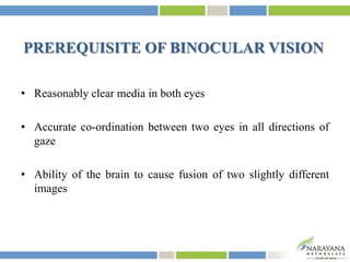
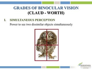
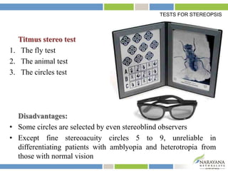
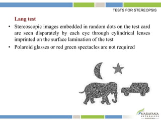
BINOCULAR VISION
SUJAY CHAUHAN
Narayana Nethralaya
Bengaluru
“We ‘see’ with our
brain,
not with our eyes;
but there is no
‘vision’
State of simultaneous vision with two seeing eyes (neither of
which needs necessarily be normal) when an individual fixes his
visual attention on an object of regard
Romano PE, Romano JA. Fusion: A new classification and methods for determining the
level of sensory binocular cooperation. Survey Ophthalmol. 1973;17:458.
BINOCULAR VISION
PREREQUISITE OF BINOCULAR VISION
• Reasonably clear media in both eyes
• Accurate co-ordination between two eyes in all directions of
gaze
• Ability of the brain to cause fusion of two slightly different
images
GRADES OF BINOCULAR VISION
(CLAUD - WORTH)
I. SIMULTANEOUS PERCEPTION
Power to see two dissimilar objects simultaneously
II. FUSION
Ability of the two eyes to produce a composite picture from
two similar pictures, each of which is incomplete in one small
detail.
GRADES OF BINOCULAR VISION
SENSORY FUSION
Unification of visual excitations from corresponding retinal images
into a single visual image
MOTOR FUSION
Ability to maintain single image with corrective movement of eyes
to bring the fovea round to the necessary position
GRADES OF BINOCULAR VISION
III. STEREOPSIS (Gk: Stepeos = solid, opsis = look)
Ability to obtain an impression of depth by the superimposition
of two pictures of the same object taken from slightly different
angles
GRADES OF BINOCULAR VISION
• Stereopsis develops when
horizontally disparate elements
are stimulated simultaneously
• Their fusion results in a single
visual impression perceived in
depth
(provided the fused image lies
within Panum’s area)
• Vertical disparity produces no
stereoscopic effect
GRADES OF BINOCULAR VISION
STEREOPSIS AND FUSION
• Sensory fusion is required for stereopsis, but not absolutely
required
• Stereopsis is also possible with diplopia
• Presence of sensory fusion does not guarantee presence of
stereopsis
STEREOSCOPIC ACUITY
Minimal disparity beyond which no stereoscopic effect is produced
GRADES OF BINOCULAR VISION
ADVANTAGES OF BINOCULAR VISION
• Stereopsis
• Increased field of vision
• Enhanced visual acuity, contrast sensitivity, visual motor skills
• Optical defects in one eye are made less obvious by normal
image of other eye
• Defective vision in any part of one visual field is masked, e.g.
blind spot
• Safety factor against partial or complete loss of vision
EVOLUTION OF BINOCULAR VISION
• Lower animals - eyes point in opposite directions - 360°field
of view
• Most predatory animals (including human being) - eyes face
in same direction - stereopsis
PSYCHOPHYSICS AND SENSORYASPECTS
OF BINOCULAR VISION
LOCATION
Position of an object in physical (objective) space
LOCALISATION
Position of the object in visual (subjective) space
RETINAL ELEMENTS / POINTS / AREAS
The retinocerebral apparatus engaged in elaborating a sensation in
response to excitation of a unit area of retinal surface
RELATIVE SUBJECTIVE VISUAL DIRECTIONS
• Retinal area stimulated by light - stimulus perceived in terms of
certain brightness, colour, form and localization in a certain
direction in visual space
• Each retinal element localizes the stimulus as a visual percept in
a specific visual direction - not absolute - relative to visual
direction of fovea
• Fovea - Principal visual direction (F)
• All other retinal elements - Secondary visual
directions (N & P)
Stable relationship - orderly visual field
RETINOMOTOR VALUES
Appearance of an object in periphery of visual field
Signal from retinal periphery to brain
(Visual direction of the object, relative to foveal visual direction)
Corresponding impulses to the extraocular muscles
Necessary ocular rotation (saccade)
• This retinomotor value of the retinal elements increases from
the center towards the periphery
• Retinomotor value of fovea = zero i.e. once an image is on
fovea, there is no incentive for ocular rotation
Clinical application - measurement of ocular deviations by
prism bar cover test (PBCT)
RETINOMOTOR VALUES
COMMON RELATIVE SUBJECTIVE VISUAL DIRECTIONS
Object fixated binocularly is
seen not in the direction of the
principal line of direction of
either eye but in a direction
coinciding with the median
plane of the head
This visual direction is common to both fovea
Every retinal element has a partner in the fellow retina
which share a common relative subjective visual direction
RETINAL CORRESPONDENCE
• Retinal elements of two eyes sharing a common subjective visual
direction - Corresponding retinal points
• All other retinal elements - Non-corresponding or disparate (with
respect to a given retinal element in fellow eye)
All common subjective visual directions
intersect at one point with the principal
visual direction
Subjective equivalent of two physical eyes
Third central imaginary eye
Binoculus
Cyclopean eye
ANOMALOUS RETINAL CORRESPONDENCE
• Active cortical adjustment in directional values of two eyes which
occurs in a child with early onset of squint
• Attempt to restore BSV as far as possible and avoid diplopia and
confusion
• Two foveae no longer have a common visual direction
• Fovea of one eye and a peripheral
retinal element of other eye acquire
a common visual direction
• Formation of pseudo macula
• Develops more commonly and is more severe when child is
small, with smaller degree of squint
• Longer the duration of squint more severe is the ARC
• Esotropia > Exotropia
• Less common in vertical deviations and in true alternating squints
with equal vision
2 types:
1. Harmonious: Angle of anomaly = Angle of squint i.e. subjective
angle of squint is zero
2. Unharmonious: Subjective angle < Objective angle
ANOMALOUS RETINAL CORRESPONDENCE
HOROPTER
• Aguilonius, 1613
• ‘Horizon of vision’
• Imaginary surface in space all points lying on which stimulate
corresponding retinal elements and seen as one
• Vieth-Muller circle (Theoretical / Mathematical / Geometrical
horopter) - Joining the object points and passing through fixation
point and pupils of two eyes
• Empirical horopter curve - flatter than Vieth-Muller circle
(Distribution of elements corresponding to each other is not same
in nasal and temporal parts of the two retinas)
HOROPTER
PANUM’S AREA OF SINGLE BINOCULAR VISION
• Region around the horopter in which single vision is present
• Panum’s theory of fusional areas - Retinal element in one eye
corresponds not only with a single point in the other but with an
elliptical area surrounding the exactly corresponding point
Object penetrating beyond
this band
Stimulation of retinal elements
outside the limits of Panum’s area
Physiological diplopia
PHYSIOLOGICAL DIPLOPIA
All points not lying on horopter curve - imaged disparately and
seen double
Test:
• A pencil at reading distance in front of head in mid-plane
• An object on the wall in line with pencil
• Fixating the distant object - pencil seen
double
• Shutting each eye alternately - C/L double
image of pencil disappears
• i.e., fixating a distant object, near object is
seen in crossed diplopia (near object in
temporal disparity with reference to fovea)
• Fixating the pencil - distant object doubles up
• Alternately closing each eye - I/L double image of distant object
vanishes
• i.e., fixating a near object, distant object
is seen in uncrossed diplopia (distant
object is imaged in nasal disparity
with reference to fovea)
PHYSIOLOGICAL DIPLOPIA
CLINICAL SIGNIFICANCE
• Becoming aware of physiological diplopia accidentally, double
vision would appear as an abnormal situation to a person
• Ruling out the presence of acute paresis of an extraocular muscle
and any other cause of diplopia, an ophthalmologist must conclude
that all the patient has experienced is physiological double vision
and explain it to the patient
• Presence of physiologic diplopia indicates that the patient is
capable of using both eyes in cooperation
• Orthoptic treatment of comitant strabismus
PHYSIOLOGICAL DIPLOPIA
FIXATION DISPARITY
• During binocular fixation, point of fixation is rarely ever imaged
exactly on corresponding points of two foveae
• Primary line of sight of one eye misses the fixation point very
slightly, being under-converged or over-converged
• Disparity is less than size of Panum's area - No diplopia
• At birth - eyes not associated with each other - act as two
independent sense organs
• Binocular vision - an acquired faculty
• Starts developing by 6 weeks of age - beginning of fixation reflex
• Refixation reflex - develops by 4 - 6 months of age
• By age of 6 years, fovea develops fully and child has almost 6/6
vision in each eye with BSV and stereopsis if eyes are straight
DEVELOPMENT OF BINOCULAR VISION
PREREQUISITE OF
DEVELOPMENT OF BINOCULAR VISION
• Good visual acuity in either eye
• Proper fixation at two fovea
• Normal retinal correspondence
• Visual fields of two eyes must overlap to a large extent
• Image formed on each retina must be approx. similar - size, shape,
colour and intensity
• Intact postural, fixation and kinetic reflexes
• Normal visual pathway
DEVELOPMENT OF BINOCULAR VISION
• By trial and error, child learns that, when image of an object is
brought onto the two fovea simultaneously, image is most
detailed
• Once this has become an established habit, the relative space
perceptions of the child begin to take form
• Crossfiring of various sensory phenomena, such as touch with
vision, eventually leads to an accurate determination of the
child's space
DEVELOPMENT OF BINOCULAR VISION
THEORY OF CORRESPONDENCE AND DISPARITY
• Most widely accepted theory of binocular vision
• A given retinal element in one retina shares a common subjective
visual direction with an element in other retina
• These corresponding elements, when stimulated simultaneously by
one object point, transmit single visual impressions that have no
depth quality
Retinal/binocular rivalry
When corresponding retinal elements are stimulated simultaneously
by two different objects, fusion becomes impossible and confusion
arises
OD OS
BINOCULAR IMPRESSION
(MOSAIC OF PIECES)
Diplopia
• When non-corresponding (disparate) retinal elements are stimulated
by one object point
• Image in the non-fixing eye falls on a location outside the macula
and is projected to a different
point in space
• Image in non-fixing eye is more
faint than that in fixing eye
However, if horizontal disparity remains within limits of Panum’s
area, a single visual impression is elicited - quality of relative
depth or stereopsis
Suppression
• Develops in immature visual cortex as a response to differing
inputs from each eye and is a barrier to development of fusion
• Eliminates visual confusion and diplopia by removing an
unwanted image
• May also eliminate fusion and stereopsis in mature visual system
in adult, e.g. in long-standing unilateral cataract
• Important aspect to consider in surgery - in presence of
supression, there is no diplopia; however, overcorrection of
strabismus may evoke diplopia
SUPPRESSION
2 types:
1. Facultative suppression
• Develops in the eye when it deviates
• The moment the eye takes up fixation - suppression disappears
• In alternating deviation
2. Obligatory suppression
• Suppression present even when deviating eye takes up fixation
• MC seen in monocular esotropia
• In initial stages, suppression is facultative; becomes obligatory
later
Horror fusionis
Intractable form of diplopia where there is both:
• Loss of ability to maintain fusion
• Absence of suppression
May occur after-
• Head injuries
• Surgery for monocular cataract in adults with previous
suppression
• After prolonged disruption of fusion
TESTS FOR STEREOPSIS
SYNOPTOPHORE OR STEREOSCOPE TESTS
• Presenting a set of 3 eccentric circles to each eye, outer circles
imaged on corresponding retinal elements are fused and serve as
a frame of reference for other 2 circles, which are also fused
• They appear in front or in back of the outer circle, depending on
the direction in which their centers have been shifted
• If displaced away from each other, they are imaged in nasal
disparity and seen in back of the outer circle; and vice versa
• Greater the displacement of inner circles, farther away from the
outer circle they are localized
VECTOGRAPH TEST
• Polaroid material - two targets are imprinted so that each target
is polarized at 90º with respect to other
• Vectograph dissociates the eyes optically
• Using properly oriented polaroid spectacles each target is seen
separately with the two eyes
TESTS FOR STEREOPSIS
Titmus stereo test
1. The fly test
2. The animal test
3. The circles test
Disadvantages:
• Some circles are selected by even stereoblind observers
• Except fine stereoacuity circles 5 to 9, unreliable in
differentiating patients with amblyopia and heterotropia from
those with normal vision
TESTS FOR STEREOPSIS
RANDOM DOT STEREOGRAM TESTS
Devoid of monocular clues - truer measurement of stereopsis than
Titmus test
Random dot E-test
3 cards:
1. Bas relief model
2. ‘E’ stereo figure with a random
dot background
3. Stereoblank with an identical random dot background
• Held 50 cm in front of the patient
• Quantitated by increasing distance
TESTS FOR STEREOPSIS
TNO random dot test
• 7 plates
2 types of figures:
i. Perceived when viewed binocularly with red-green spectacles
by normal subject having stereopsis
ii. Seen with and without spectacles even in absence of stereopsis
• First 3 plates - to establish presence of gross stereopsis quickly
• Remaining 4 plates - to quantitate level of stereopsis
TESTS FOR STEREOPSIS
Lang test
• Stereoscopic images embedded in random dots on the test card
are seen disparately by each eye through cylindrical lenses
imprinted on the surface lamination of the test
• Polaroid glasses or red green spectacles are not required
TESTS FOR STEREOPSIS
Frisby test
• 3 plastic cards each containing 4 squares of small random shapes
• One square in each plate contains a hidden circle which is seen
disparately
• Disparity is created by displacement of random shapes by
thickness of the plate
• Does not require use of glasses
TESTS FOR STEREOPSIS
SIMPLE MOTOR TASK TEST BASED ON STEREOPSIS
Lang’s two pencil test
• Stereopsis +nt - Able to do
• Stereopsis -nt or one eye closed - Unable to do
TESTS FOR STEREOPSIS
REFERENCES
• Noorden GK von, Campos EC. Binocular Vision and Ocular Motility:
Theory and Management of Strabismus. 6th edition. Mosby. 2002:7-37.
• Billson FA. Fundamentals of Clinical Ophthalmology: Strabismus. BMJ
Books. 2003:8-19.
• Khurana AK. Anatomy and Physiology of Eye. 2nd edition. CBS Pub.
2006:327-364.
• Mukherjee PK. Pediatric Ophthalmology. New Age Int Pub. 2005:637-672.
Binocular vision

Binocular vision

  • 1.
  • 2.
    “We ‘see’ withour brain, not with our eyes; but there is no ‘vision’
  • 3.
    State of simultaneousvision with two seeing eyes (neither of which needs necessarily be normal) when an individual fixes his visual attention on an object of regard Romano PE, Romano JA. Fusion: A new classification and methods for determining the level of sensory binocular cooperation. Survey Ophthalmol. 1973;17:458. BINOCULAR VISION
  • 4.
    PREREQUISITE OF BINOCULARVISION • Reasonably clear media in both eyes • Accurate co-ordination between two eyes in all directions of gaze • Ability of the brain to cause fusion of two slightly different images
  • 5.
    GRADES OF BINOCULARVISION (CLAUD - WORTH) I. SIMULTANEOUS PERCEPTION Power to see two dissimilar objects simultaneously
  • 6.
    II. FUSION Ability ofthe two eyes to produce a composite picture from two similar pictures, each of which is incomplete in one small detail. GRADES OF BINOCULAR VISION
  • 7.
    SENSORY FUSION Unification ofvisual excitations from corresponding retinal images into a single visual image MOTOR FUSION Ability to maintain single image with corrective movement of eyes to bring the fovea round to the necessary position GRADES OF BINOCULAR VISION
  • 8.
    III. STEREOPSIS (Gk:Stepeos = solid, opsis = look) Ability to obtain an impression of depth by the superimposition of two pictures of the same object taken from slightly different angles GRADES OF BINOCULAR VISION
  • 9.
    • Stereopsis developswhen horizontally disparate elements are stimulated simultaneously • Their fusion results in a single visual impression perceived in depth (provided the fused image lies within Panum’s area) • Vertical disparity produces no stereoscopic effect GRADES OF BINOCULAR VISION
  • 10.
    STEREOPSIS AND FUSION •Sensory fusion is required for stereopsis, but not absolutely required • Stereopsis is also possible with diplopia • Presence of sensory fusion does not guarantee presence of stereopsis STEREOSCOPIC ACUITY Minimal disparity beyond which no stereoscopic effect is produced GRADES OF BINOCULAR VISION
  • 11.
    ADVANTAGES OF BINOCULARVISION • Stereopsis • Increased field of vision • Enhanced visual acuity, contrast sensitivity, visual motor skills • Optical defects in one eye are made less obvious by normal image of other eye • Defective vision in any part of one visual field is masked, e.g. blind spot • Safety factor against partial or complete loss of vision
  • 12.
    EVOLUTION OF BINOCULARVISION • Lower animals - eyes point in opposite directions - 360°field of view • Most predatory animals (including human being) - eyes face in same direction - stereopsis
  • 14.
    PSYCHOPHYSICS AND SENSORYASPECTS OFBINOCULAR VISION LOCATION Position of an object in physical (objective) space LOCALISATION Position of the object in visual (subjective) space RETINAL ELEMENTS / POINTS / AREAS The retinocerebral apparatus engaged in elaborating a sensation in response to excitation of a unit area of retinal surface
  • 15.
    RELATIVE SUBJECTIVE VISUALDIRECTIONS • Retinal area stimulated by light - stimulus perceived in terms of certain brightness, colour, form and localization in a certain direction in visual space • Each retinal element localizes the stimulus as a visual percept in a specific visual direction - not absolute - relative to visual direction of fovea • Fovea - Principal visual direction (F) • All other retinal elements - Secondary visual directions (N & P) Stable relationship - orderly visual field
  • 16.
    RETINOMOTOR VALUES Appearance ofan object in periphery of visual field Signal from retinal periphery to brain (Visual direction of the object, relative to foveal visual direction) Corresponding impulses to the extraocular muscles Necessary ocular rotation (saccade)
  • 17.
    • This retinomotorvalue of the retinal elements increases from the center towards the periphery • Retinomotor value of fovea = zero i.e. once an image is on fovea, there is no incentive for ocular rotation Clinical application - measurement of ocular deviations by prism bar cover test (PBCT) RETINOMOTOR VALUES
  • 18.
    COMMON RELATIVE SUBJECTIVEVISUAL DIRECTIONS Object fixated binocularly is seen not in the direction of the principal line of direction of either eye but in a direction coinciding with the median plane of the head This visual direction is common to both fovea Every retinal element has a partner in the fellow retina which share a common relative subjective visual direction
  • 19.
    RETINAL CORRESPONDENCE • Retinalelements of two eyes sharing a common subjective visual direction - Corresponding retinal points • All other retinal elements - Non-corresponding or disparate (with respect to a given retinal element in fellow eye) All common subjective visual directions intersect at one point with the principal visual direction Subjective equivalent of two physical eyes Third central imaginary eye Binoculus Cyclopean eye
  • 20.
    ANOMALOUS RETINAL CORRESPONDENCE •Active cortical adjustment in directional values of two eyes which occurs in a child with early onset of squint • Attempt to restore BSV as far as possible and avoid diplopia and confusion • Two foveae no longer have a common visual direction • Fovea of one eye and a peripheral retinal element of other eye acquire a common visual direction • Formation of pseudo macula
  • 21.
    • Develops morecommonly and is more severe when child is small, with smaller degree of squint • Longer the duration of squint more severe is the ARC • Esotropia > Exotropia • Less common in vertical deviations and in true alternating squints with equal vision 2 types: 1. Harmonious: Angle of anomaly = Angle of squint i.e. subjective angle of squint is zero 2. Unharmonious: Subjective angle < Objective angle ANOMALOUS RETINAL CORRESPONDENCE
  • 22.
    HOROPTER • Aguilonius, 1613 •‘Horizon of vision’ • Imaginary surface in space all points lying on which stimulate corresponding retinal elements and seen as one
  • 23.
    • Vieth-Muller circle(Theoretical / Mathematical / Geometrical horopter) - Joining the object points and passing through fixation point and pupils of two eyes • Empirical horopter curve - flatter than Vieth-Muller circle (Distribution of elements corresponding to each other is not same in nasal and temporal parts of the two retinas) HOROPTER
  • 24.
    PANUM’S AREA OFSINGLE BINOCULAR VISION • Region around the horopter in which single vision is present • Panum’s theory of fusional areas - Retinal element in one eye corresponds not only with a single point in the other but with an elliptical area surrounding the exactly corresponding point Object penetrating beyond this band Stimulation of retinal elements outside the limits of Panum’s area Physiological diplopia
  • 25.
    PHYSIOLOGICAL DIPLOPIA All pointsnot lying on horopter curve - imaged disparately and seen double Test: • A pencil at reading distance in front of head in mid-plane • An object on the wall in line with pencil • Fixating the distant object - pencil seen double • Shutting each eye alternately - C/L double image of pencil disappears • i.e., fixating a distant object, near object is seen in crossed diplopia (near object in temporal disparity with reference to fovea)
  • 26.
    • Fixating thepencil - distant object doubles up • Alternately closing each eye - I/L double image of distant object vanishes • i.e., fixating a near object, distant object is seen in uncrossed diplopia (distant object is imaged in nasal disparity with reference to fovea) PHYSIOLOGICAL DIPLOPIA
  • 27.
    CLINICAL SIGNIFICANCE • Becomingaware of physiological diplopia accidentally, double vision would appear as an abnormal situation to a person • Ruling out the presence of acute paresis of an extraocular muscle and any other cause of diplopia, an ophthalmologist must conclude that all the patient has experienced is physiological double vision and explain it to the patient • Presence of physiologic diplopia indicates that the patient is capable of using both eyes in cooperation • Orthoptic treatment of comitant strabismus PHYSIOLOGICAL DIPLOPIA
  • 28.
    FIXATION DISPARITY • Duringbinocular fixation, point of fixation is rarely ever imaged exactly on corresponding points of two foveae • Primary line of sight of one eye misses the fixation point very slightly, being under-converged or over-converged • Disparity is less than size of Panum's area - No diplopia
  • 29.
    • At birth- eyes not associated with each other - act as two independent sense organs • Binocular vision - an acquired faculty • Starts developing by 6 weeks of age - beginning of fixation reflex • Refixation reflex - develops by 4 - 6 months of age • By age of 6 years, fovea develops fully and child has almost 6/6 vision in each eye with BSV and stereopsis if eyes are straight DEVELOPMENT OF BINOCULAR VISION
  • 30.
    PREREQUISITE OF DEVELOPMENT OFBINOCULAR VISION • Good visual acuity in either eye • Proper fixation at two fovea • Normal retinal correspondence • Visual fields of two eyes must overlap to a large extent • Image formed on each retina must be approx. similar - size, shape, colour and intensity • Intact postural, fixation and kinetic reflexes • Normal visual pathway DEVELOPMENT OF BINOCULAR VISION
  • 31.
    • By trialand error, child learns that, when image of an object is brought onto the two fovea simultaneously, image is most detailed • Once this has become an established habit, the relative space perceptions of the child begin to take form • Crossfiring of various sensory phenomena, such as touch with vision, eventually leads to an accurate determination of the child's space DEVELOPMENT OF BINOCULAR VISION
  • 32.
    THEORY OF CORRESPONDENCEAND DISPARITY • Most widely accepted theory of binocular vision • A given retinal element in one retina shares a common subjective visual direction with an element in other retina • These corresponding elements, when stimulated simultaneously by one object point, transmit single visual impressions that have no depth quality
  • 33.
    Retinal/binocular rivalry When correspondingretinal elements are stimulated simultaneously by two different objects, fusion becomes impossible and confusion arises OD OS BINOCULAR IMPRESSION (MOSAIC OF PIECES)
  • 34.
    Diplopia • When non-corresponding(disparate) retinal elements are stimulated by one object point • Image in the non-fixing eye falls on a location outside the macula and is projected to a different point in space • Image in non-fixing eye is more faint than that in fixing eye
  • 35.
    However, if horizontaldisparity remains within limits of Panum’s area, a single visual impression is elicited - quality of relative depth or stereopsis
  • 36.
    Suppression • Develops inimmature visual cortex as a response to differing inputs from each eye and is a barrier to development of fusion • Eliminates visual confusion and diplopia by removing an unwanted image • May also eliminate fusion and stereopsis in mature visual system in adult, e.g. in long-standing unilateral cataract • Important aspect to consider in surgery - in presence of supression, there is no diplopia; however, overcorrection of strabismus may evoke diplopia
  • 37.
    SUPPRESSION 2 types: 1. Facultativesuppression • Develops in the eye when it deviates • The moment the eye takes up fixation - suppression disappears • In alternating deviation 2. Obligatory suppression • Suppression present even when deviating eye takes up fixation • MC seen in monocular esotropia • In initial stages, suppression is facultative; becomes obligatory later
  • 38.
    Horror fusionis Intractable formof diplopia where there is both: • Loss of ability to maintain fusion • Absence of suppression May occur after- • Head injuries • Surgery for monocular cataract in adults with previous suppression • After prolonged disruption of fusion
  • 39.
    TESTS FOR STEREOPSIS SYNOPTOPHOREOR STEREOSCOPE TESTS • Presenting a set of 3 eccentric circles to each eye, outer circles imaged on corresponding retinal elements are fused and serve as a frame of reference for other 2 circles, which are also fused • They appear in front or in back of the outer circle, depending on the direction in which their centers have been shifted • If displaced away from each other, they are imaged in nasal disparity and seen in back of the outer circle; and vice versa • Greater the displacement of inner circles, farther away from the outer circle they are localized
  • 40.
    VECTOGRAPH TEST • Polaroidmaterial - two targets are imprinted so that each target is polarized at 90º with respect to other • Vectograph dissociates the eyes optically • Using properly oriented polaroid spectacles each target is seen separately with the two eyes TESTS FOR STEREOPSIS
  • 41.
    Titmus stereo test 1.The fly test 2. The animal test 3. The circles test Disadvantages: • Some circles are selected by even stereoblind observers • Except fine stereoacuity circles 5 to 9, unreliable in differentiating patients with amblyopia and heterotropia from those with normal vision TESTS FOR STEREOPSIS
  • 42.
    RANDOM DOT STEREOGRAMTESTS Devoid of monocular clues - truer measurement of stereopsis than Titmus test Random dot E-test 3 cards: 1. Bas relief model 2. ‘E’ stereo figure with a random dot background 3. Stereoblank with an identical random dot background • Held 50 cm in front of the patient • Quantitated by increasing distance TESTS FOR STEREOPSIS
  • 43.
    TNO random dottest • 7 plates 2 types of figures: i. Perceived when viewed binocularly with red-green spectacles by normal subject having stereopsis ii. Seen with and without spectacles even in absence of stereopsis • First 3 plates - to establish presence of gross stereopsis quickly • Remaining 4 plates - to quantitate level of stereopsis TESTS FOR STEREOPSIS
  • 44.
    Lang test • Stereoscopicimages embedded in random dots on the test card are seen disparately by each eye through cylindrical lenses imprinted on the surface lamination of the test • Polaroid glasses or red green spectacles are not required TESTS FOR STEREOPSIS
  • 45.
    Frisby test • 3plastic cards each containing 4 squares of small random shapes • One square in each plate contains a hidden circle which is seen disparately • Disparity is created by displacement of random shapes by thickness of the plate • Does not require use of glasses TESTS FOR STEREOPSIS
  • 46.
    SIMPLE MOTOR TASKTEST BASED ON STEREOPSIS Lang’s two pencil test • Stereopsis +nt - Able to do • Stereopsis -nt or one eye closed - Unable to do TESTS FOR STEREOPSIS
  • 47.
    REFERENCES • Noorden GKvon, Campos EC. Binocular Vision and Ocular Motility: Theory and Management of Strabismus. 6th edition. Mosby. 2002:7-37. • Billson FA. Fundamentals of Clinical Ophthalmology: Strabismus. BMJ Books. 2003:8-19. • Khurana AK. Anatomy and Physiology of Eye. 2nd edition. CBS Pub. 2006:327-364. • Mukherjee PK. Pediatric Ophthalmology. New Age Int Pub. 2005:637-672.