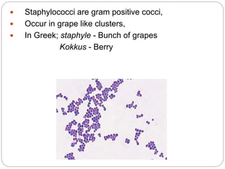
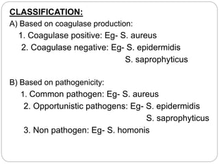
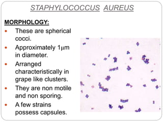
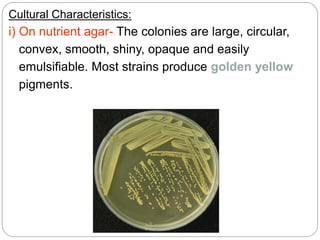
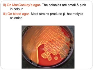
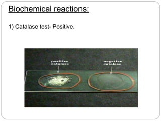
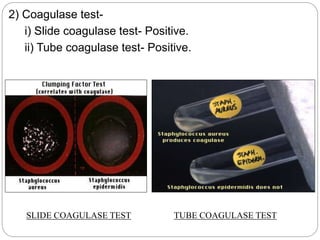
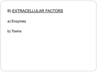
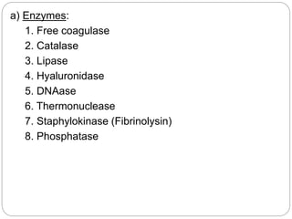
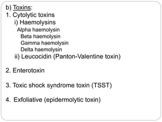
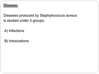
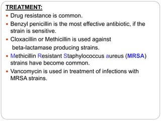
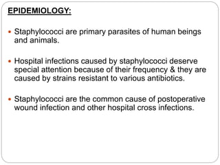
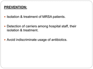
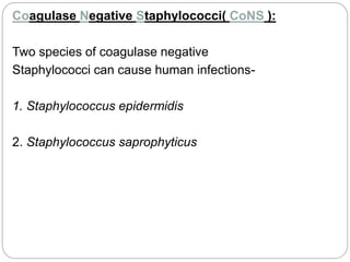
 Staphylococci are gram positive cocci,
 Occur in grape like clusters,
 In Greek; staphyle - Bunch of grapes
Kokkus - Berry
CLASSIFICATION:
A) Based on coagulase production:
1. Coagulase positive: Eg- S. aureus
2. Coagulase negative: Eg- S. epidermidis
S. saprophyticus
B) Based on pathogenicity:
1. Common pathogen: Eg- S. aureus
2. Opportunistic pathogens: Eg- S. epidermidis
S. saprophyticus
3. Non pathogen: Eg- S. homonis
STAPHYLOCOCCUS AUREUS
MORPHOLOGY:
 These are spherical
cocci.
 Approximately 1μm
in diameter.
 Arranged
characteristically in
grape like clusters.
 They are non motile
and non sporing.
 A few strains
possess capsules.
CULTURE:
Media used :-
i) Non selective media: Nutrient agar,
Blood agar,
MacConkey’s agar.
ii) Selective media: Salt-milk agar,
Ludlam’s medium
Cultural Characteristics:
i) On nutrient agar- The colonies are large, circular,
convex, smooth, shiny, opaque and easily
emulsifiable. Most strains produce golden yellow
pigments.
ii) On MacConkey’s agar- The colonies are small & pink
in colour.
iii) On blood agar- Most strains produce β- haemolytic
colonies.
Biochemical reactions:
1) Catalase test- Positive.
2) Coagulase test-
i) Slide coagulase test- Positive.
ii) Tube coagulase test- Positive.
SLIDE COAGULASE TEST TUBE COAGULASE TEST
3) Reduces nitrate to nitrite.
4) Ferments mannitol anaerobically with acid only.
5) Urea hydrolysis test- Positive.
6) Gelatin liquefaction test- Positive.
7) Produces Lipase.
8) Produces Phosphatase.
9) Produces Thermostable nuclease.
PATHOGENICITY:
Source of infection:
A) Exogenous: patients or carriers
B) Endogenous: From colonized site
Mode of transmission:
A) Contact: direct or indirect( through fomites)
B) Inhalation of air borne droplets
Virulence factors:
These include
A) Cell associated factors
B) Extracellular factors
A) CELL ASSOCIATED FACTORS:
a) Cell associated polymers
b) Cell surface proteins
a) CELL ASSOCIATED POLYMERS
1. Cell wall polysaccharide
2. Teichoic acid
3. Capsular polysaccharide
b) CELL SURFACE PROTEINS:
1. Protein A
2. Clumping factor (bound coagulase)
Structure of Staphylococcal cell wall
B) EXTRACELLULAR FACTORS
a) Enzymes
b) Toxins
a) Enzymes:
1. Free coagulase
2. Catalase
3. Lipase
4. Hyaluronidase
5. DNAase
6. Thermonuclease
7. Staphylokinase (Fibrinolysin)
8. Phosphatase
b) Toxins:
1. Cytolytic toxins
i) Haemolysins
Alpha haemolysin
Beta haemolysin
Gamma haemolysin
Delta haemolysin
ii) Leucocidin (Panton-Valentine toxin)
2. Enterotoxin
3. Toxic shock syndrome toxin (TSST)
4. . Exfoliative (epidermolytic toxin)
Disease:
Diseases produced by Staphylococcus aureus
is studied under 2 groups:
A) Infections
B) Intoxications
A) INFECTIONS:
Mechanism of pathogenesis:
Cocci gain access to damaged skin, mucosal or
tissue site
Colonize by adhering to cells or extracellular matrix
Evade the host defense mechanisms and multiply
Cause tissue damage
Common Staphylococcal infections are:
1) Skin and soft tissue: Folliculitis, furuncle (boil),
carbuncle, styes, abscess, wound infections, impetigo,
paronychia and less often cellulitis.
Folliculitis
Folliculitis
Furuncle (boil)
Carbuncle
Styes Abscess
Impetigo Paronychia
Cellulitis
Wound infection
2) Musculoskeletal: Osteomyelitis, arthritis, bursitis,
pyomyositis.
osteomyelitis
3) Respiratory: Tonsillitis, pharyngitis, sinusitis, otitis,
bronchopneumonia, lung abscess, empyema, rarely
pneumonia.
4) Central nervous system: Abscess, meningitis,
intracranial thrombophlebitis.
5) Endovascular: Bacteremia, septicemia, pyemia,
endocarditis.
Endocarditis
6) Urinary: Urinary tract infection.
B) INTOXICATIOINS:
The disease is caused by the bacterial exotoxins,
which are produced either in the infected host
or preformed in vitro.
There are 3 types-
1. Food poisoning
2. Toxic shock syndrome
3. Staphylococcal scalded skin syndrome
1) Food poisoning:
 Enterotoxin is responsible for manifestations of
staphylococcal food poisoning.
 Eight types of enterotoxin are currently known,
named A, B, C1-3, D, E, and H.
 It usually occurs when preformed toxin is ingested
with contaminated food.
 The toxin acts directly on the autonomic nervous
system to cause the illness, rather than gut
mucosa.
 The common food items responsible are - milk and milk
products, meat, fish and ice cream.
 Source of infection- food handler who is a carrier.
 Incubation period- 2 to 6 hours.
 Clinical symptoms- nausea, vomiting and diarrhoea.
 The illness is usually self limited, with recovery in a day
or so.
2) Staphylococcal Toxic shock syndrome (STSS):
 STSS is associated with infection of mucosal or
sequestered sites by TSST( formerly known as
enterotoxin type F) producing S.aureus.
 It is fatal multisystem disease presenting with fever,
hypotension, myalgia, vomiting, diarrhoea, mucosal
hyperemia and erythematous rash which desquamates
subsequently.
2 types of STSS known:
i) Menstrual associated STSS: Here colonization of
S.aureus occurs in the vagina of menstruating woman
who uses highly absorbent vaginal tampons.
ii) Non menstrual associated STSS: Here colonization of
S.aureus occurs in other sites like surgical wound.
3) Staphylococcal scalded skin syndrome
(SSSS):
 Exfoliative toxin produced by S.aureus is responsible for
this.
 It is a skin disease in which outer layer of epidermis gets
separated from the underlying tissues.
Types of SSSS:
Severe form Milder form
In new born - Ritter’s disease - Pemphigus
neonatorum
In older patients - Toxic epidermal - Bullous
necrolysis impetigo
Toxic epidermal necrolysis
Ritter’s disease
Bullous impetigo
Pemphigus neonatorum
LAB DIAGNOSIS:
Specimens collected: Depends on the type of infection.
 Suppurative lesion- Pus,
 Respiratory infection- Sputum,
 Bacteremia & septicemia- Blood,
 Food poisoning- Feces, vomit & the remains of
suspected food,
 For the detection of carriers- Nasal swab.
I) Direct microscopy:
 Direct microscopy with
Gram stained smear is
useful in case of pus,
where cocci in clusters
are seen.
 This is of no value for
specimen like sputum
where mixed flora are
normally present.
Methods of examination:
c) Gram staining: Smears
are examined from the
culture plate and reveals
Gram positive cocci(1μm
in diameter) arranged in
grape like clusters.
II) Culture:
a) Media used:
b) Cultural Characteristics:
d) Biochemical reactions:
III) Antibiotic sensitivity tests done as a guide to
treatment.
IV) Bacteriophage typing is done for epidemiological
purposes.
V) Serological tests are not useful.
TREATMENT:
 Drug resistance is common.
 Benzyl penicillin is the most effective antibiotic, if the
strain is sensitive.
 Cloxacillin or Methicillin is used against
beta-lactamase producing strains.
 Methicillin Resistant Staphylococcus aureus (MRSA)
strains have become common.
 Vancomycin is used in treatment of infections with
MRSA strains.
EPIDEMIOLOGY:
 Staphylococci are primary parasites of human beings
and animals.
 Hospital infections caused by staphylococci deserve
special attention because of their frequency & they are
caused by strains resistant to various antibiotics.
 Staphylococci are the common cause of postoperative
wound infection and other hospital cross infections.
PREVENTION:
 Isolation & treatment of MRSA patients.
 Detection of carriers among hospital staff, their
isolation & treatment.
 Avoid indiscriminate usage of antibiotics.
Coagulase Negative Staphylococci( CoNS ):
Two species of coagulase negative
Staphylococci can cause human infections-
1. Staphylococcus epidermidis
2. Staphylococcus saprophyticus
S. Epidermidis:
 It is a common cause of stitch abscesses.
 It has predilection for growth on implanted foreign
bodies such as artificial valves, shunts, intravascular
catheters and prosthetic appliances leading to
bacteraemia.
 In persons with structural abnormalities of urinary
tract, it can cause cystitis.
 Endocarditis may be caused, particularly in drug
addicts.
S.saprophyticus:
 It causes urinary tract infections, mostly in sexually active
young women.
 The infection is symptomatic and may involve the upper
urinary tract also.
 Men are infected much less often.
 It is one of the few frequently isolated CoNS that is
resistant to Novobiocin.
Characters S.aureus S.epidermididis S.saprophyticus
Coagulase + - -
Novobiocin
sensitivity
Sensitive Sensitive Resistant
Acid from
mannitol
fermentation
anaerobically
+ - -
Phosphatase + + -
Distinguishing features of the major species of
staphylococcus
Novobiocin sensitivity test
THANK YOU

Staphylococcus.ppt.........ali.rasool.badr

  • 2.
     Staphylococci aregram positive cocci,  Occur in grape like clusters,  In Greek; staphyle - Bunch of grapes Kokkus - Berry
  • 3.
    CLASSIFICATION: A) Based oncoagulase production: 1. Coagulase positive: Eg- S. aureus 2. Coagulase negative: Eg- S. epidermidis S. saprophyticus B) Based on pathogenicity: 1. Common pathogen: Eg- S. aureus 2. Opportunistic pathogens: Eg- S. epidermidis S. saprophyticus 3. Non pathogen: Eg- S. homonis
  • 4.
    STAPHYLOCOCCUS AUREUS MORPHOLOGY:  Theseare spherical cocci.  Approximately 1μm in diameter.  Arranged characteristically in grape like clusters.  They are non motile and non sporing.  A few strains possess capsules.
  • 5.
    CULTURE: Media used :- i)Non selective media: Nutrient agar, Blood agar, MacConkey’s agar. ii) Selective media: Salt-milk agar, Ludlam’s medium
  • 6.
    Cultural Characteristics: i) Onnutrient agar- The colonies are large, circular, convex, smooth, shiny, opaque and easily emulsifiable. Most strains produce golden yellow pigments.
  • 7.
    ii) On MacConkey’sagar- The colonies are small & pink in colour. iii) On blood agar- Most strains produce β- haemolytic colonies.
  • 8.
  • 9.
    2) Coagulase test- i)Slide coagulase test- Positive. ii) Tube coagulase test- Positive. SLIDE COAGULASE TEST TUBE COAGULASE TEST
  • 10.
    3) Reduces nitrateto nitrite. 4) Ferments mannitol anaerobically with acid only. 5) Urea hydrolysis test- Positive. 6) Gelatin liquefaction test- Positive. 7) Produces Lipase. 8) Produces Phosphatase. 9) Produces Thermostable nuclease.
  • 11.
    PATHOGENICITY: Source of infection: A)Exogenous: patients or carriers B) Endogenous: From colonized site Mode of transmission: A) Contact: direct or indirect( through fomites) B) Inhalation of air borne droplets
  • 12.
    Virulence factors: These include A)Cell associated factors B) Extracellular factors
  • 13.
    A) CELL ASSOCIATEDFACTORS: a) Cell associated polymers b) Cell surface proteins a) CELL ASSOCIATED POLYMERS 1. Cell wall polysaccharide 2. Teichoic acid 3. Capsular polysaccharide b) CELL SURFACE PROTEINS: 1. Protein A 2. Clumping factor (bound coagulase)
  • 14.
  • 15.
    B) EXTRACELLULAR FACTORS a)Enzymes b) Toxins
  • 16.
    a) Enzymes: 1. Freecoagulase 2. Catalase 3. Lipase 4. Hyaluronidase 5. DNAase 6. Thermonuclease 7. Staphylokinase (Fibrinolysin) 8. Phosphatase
  • 17.
    b) Toxins: 1. Cytolytictoxins i) Haemolysins Alpha haemolysin Beta haemolysin Gamma haemolysin Delta haemolysin ii) Leucocidin (Panton-Valentine toxin) 2. Enterotoxin 3. Toxic shock syndrome toxin (TSST) 4. . Exfoliative (epidermolytic toxin)
  • 18.
    Disease: Diseases produced byStaphylococcus aureus is studied under 2 groups: A) Infections B) Intoxications
  • 19.
    A) INFECTIONS: Mechanism ofpathogenesis: Cocci gain access to damaged skin, mucosal or tissue site Colonize by adhering to cells or extracellular matrix Evade the host defense mechanisms and multiply Cause tissue damage
  • 20.
    Common Staphylococcal infectionsare: 1) Skin and soft tissue: Folliculitis, furuncle (boil), carbuncle, styes, abscess, wound infections, impetigo, paronychia and less often cellulitis. Folliculitis Folliculitis
  • 21.
  • 22.
  • 23.
  • 24.
    2) Musculoskeletal: Osteomyelitis,arthritis, bursitis, pyomyositis. osteomyelitis 3) Respiratory: Tonsillitis, pharyngitis, sinusitis, otitis, bronchopneumonia, lung abscess, empyema, rarely pneumonia.
  • 25.
    4) Central nervoussystem: Abscess, meningitis, intracranial thrombophlebitis. 5) Endovascular: Bacteremia, septicemia, pyemia, endocarditis. Endocarditis 6) Urinary: Urinary tract infection.
  • 26.
    B) INTOXICATIOINS: The diseaseis caused by the bacterial exotoxins, which are produced either in the infected host or preformed in vitro. There are 3 types- 1. Food poisoning 2. Toxic shock syndrome 3. Staphylococcal scalded skin syndrome
  • 27.
    1) Food poisoning: Enterotoxin is responsible for manifestations of staphylococcal food poisoning.  Eight types of enterotoxin are currently known, named A, B, C1-3, D, E, and H.  It usually occurs when preformed toxin is ingested with contaminated food.  The toxin acts directly on the autonomic nervous system to cause the illness, rather than gut mucosa.
  • 28.
     The commonfood items responsible are - milk and milk products, meat, fish and ice cream.  Source of infection- food handler who is a carrier.  Incubation period- 2 to 6 hours.  Clinical symptoms- nausea, vomiting and diarrhoea.  The illness is usually self limited, with recovery in a day or so.
  • 29.
    2) Staphylococcal Toxicshock syndrome (STSS):  STSS is associated with infection of mucosal or sequestered sites by TSST( formerly known as enterotoxin type F) producing S.aureus.  It is fatal multisystem disease presenting with fever, hypotension, myalgia, vomiting, diarrhoea, mucosal hyperemia and erythematous rash which desquamates subsequently.
  • 31.
    2 types ofSTSS known: i) Menstrual associated STSS: Here colonization of S.aureus occurs in the vagina of menstruating woman who uses highly absorbent vaginal tampons. ii) Non menstrual associated STSS: Here colonization of S.aureus occurs in other sites like surgical wound.
  • 32.
    3) Staphylococcal scaldedskin syndrome (SSSS):  Exfoliative toxin produced by S.aureus is responsible for this.  It is a skin disease in which outer layer of epidermis gets separated from the underlying tissues.
  • 33.
    Types of SSSS: Severeform Milder form In new born - Ritter’s disease - Pemphigus neonatorum In older patients - Toxic epidermal - Bullous necrolysis impetigo
  • 34.
    Toxic epidermal necrolysis Ritter’sdisease Bullous impetigo Pemphigus neonatorum
  • 35.
    LAB DIAGNOSIS: Specimens collected:Depends on the type of infection.  Suppurative lesion- Pus,  Respiratory infection- Sputum,  Bacteremia & septicemia- Blood,  Food poisoning- Feces, vomit & the remains of suspected food,  For the detection of carriers- Nasal swab.
  • 36.
    I) Direct microscopy: Direct microscopy with Gram stained smear is useful in case of pus, where cocci in clusters are seen.  This is of no value for specimen like sputum where mixed flora are normally present. Methods of examination:
  • 37.
    c) Gram staining:Smears are examined from the culture plate and reveals Gram positive cocci(1μm in diameter) arranged in grape like clusters. II) Culture: a) Media used: b) Cultural Characteristics:
  • 38.
    d) Biochemical reactions: III)Antibiotic sensitivity tests done as a guide to treatment. IV) Bacteriophage typing is done for epidemiological purposes. V) Serological tests are not useful.
  • 39.
    TREATMENT:  Drug resistanceis common.  Benzyl penicillin is the most effective antibiotic, if the strain is sensitive.  Cloxacillin or Methicillin is used against beta-lactamase producing strains.  Methicillin Resistant Staphylococcus aureus (MRSA) strains have become common.  Vancomycin is used in treatment of infections with MRSA strains.
  • 40.
    EPIDEMIOLOGY:  Staphylococci areprimary parasites of human beings and animals.  Hospital infections caused by staphylococci deserve special attention because of their frequency & they are caused by strains resistant to various antibiotics.  Staphylococci are the common cause of postoperative wound infection and other hospital cross infections.
  • 41.
    PREVENTION:  Isolation &treatment of MRSA patients.  Detection of carriers among hospital staff, their isolation & treatment.  Avoid indiscriminate usage of antibiotics.
  • 42.
    Coagulase Negative Staphylococci(CoNS ): Two species of coagulase negative Staphylococci can cause human infections- 1. Staphylococcus epidermidis 2. Staphylococcus saprophyticus
  • 43.
    S. Epidermidis:  Itis a common cause of stitch abscesses.  It has predilection for growth on implanted foreign bodies such as artificial valves, shunts, intravascular catheters and prosthetic appliances leading to bacteraemia.  In persons with structural abnormalities of urinary tract, it can cause cystitis.  Endocarditis may be caused, particularly in drug addicts.
  • 44.
    S.saprophyticus:  It causesurinary tract infections, mostly in sexually active young women.  The infection is symptomatic and may involve the upper urinary tract also.  Men are infected much less often.  It is one of the few frequently isolated CoNS that is resistant to Novobiocin.
  • 45.
    Characters S.aureus S.epidermididisS.saprophyticus Coagulase + - - Novobiocin sensitivity Sensitive Sensitive Resistant Acid from mannitol fermentation anaerobically + - - Phosphatase + + - Distinguishing features of the major species of staphylococcus
  • 46.
  • 47.