This document discusses bacterial motility and the different types and mechanisms of bacterial movement. It covers:
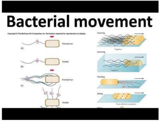
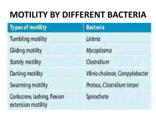
- Motility is the ability of cells to move on their own using energy. The main types are flagellar movement and gliding movement.
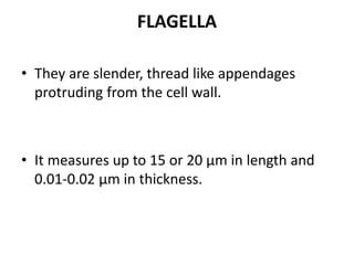
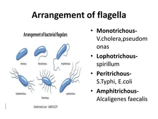
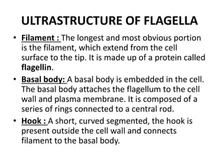
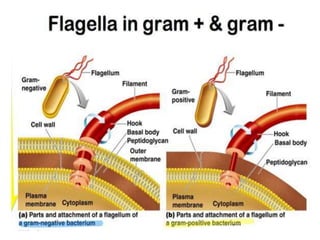
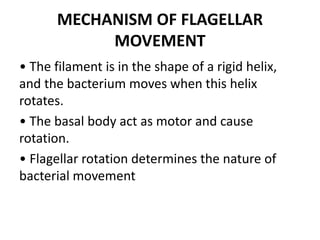
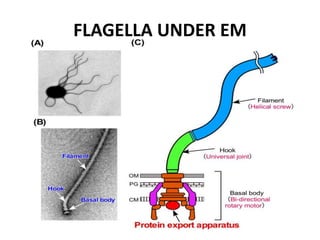
- Flagella are thin appendages that propel bacteria through rotation. Their structure includes a filament, basal body and hook. Arrangements can be monotrichous, lophotrichous or peritrichous.
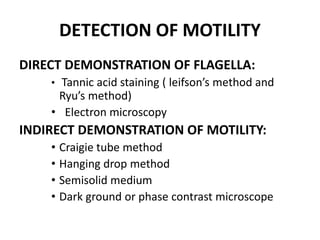
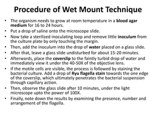
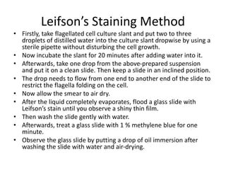
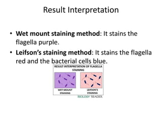
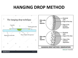
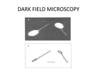
- Motility allows bacteria to find favorable environments and colonize host cells. Methods to detect motility include wet mount microscopy, staining techniques like Leifson's method, and using semi-solid media.


![Overview of Fungal Infections[1].ppt pptx](https://cdn.slidesharecdn.com/ss_thumbnails/overviewoffungalinfections1-250811070114-3c4fbad5-thumbnail.jpg?width=640&height=640&fit=bounds)


![Gas gangrene [Autosaved].pptxGas gangrene [Autosaved].pptx](https://cdn.slidesharecdn.com/ss_thumbnails/gasgangreneautosaved-250608063811-c85a18a4-thumbnail.jpg?width=640&height=640&fit=bounds)


