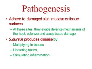
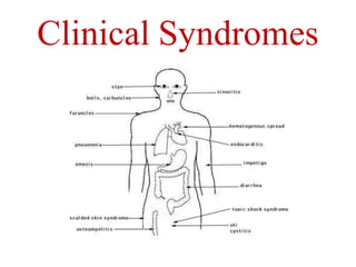
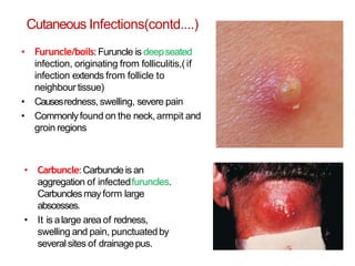
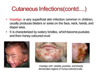
STAPHYLOCOCCI
INTRODUCTION
• Family: Micrococceae
(consists of Gram
positive cocci, arranged in
tetrads, clusters)
• Genus: Staphylococcus
• Term“staphylococcus”
derived from Greek :Staphyle=
bunch of grapes andKokkos = berry,
meaning bacteria occurring in
grapelike clusters or berry.
History
• Robert Koch(1878)- first to see
staphylococci in pusspecimen
• Louis Pasteur(1880)- first to
cultivate in liquidmedium
• SirAlexander Ongston (1881)-
named the bacteria as
“staphylococcus”
Classification
• Basedon pathogenecity:
– Pathogenic:- includes
only one i.e.,S.aureus
– Non-pathogenic:-
includes S.epidermidis,
S.saprophyticus, S.albus,
S. citrus, S.hominis,etc.
• Basedon coagulase
production:
– Coagulase positive: S.
aureus
– Coagulasenegative: S.
epidermidis, S.
saprophyticus
S. albus ,S. aureus ,S. citrus on NutrientAgar
•Basedon pigment production:
•S.aureus :-golden-yellow
pigmented colonies
•S.albus :- whitecolonies
•S.citrus :-lemonyellow
colonies
S.aureus
• Natural habitat:-Nostril andskin
Morphology:-
– Gram-positive, cocci, 0.5-1.5µm in diameter;
occur characteristically in group, also singlyand
in pairs
– Form irregular grapelike clusters (since divide in
3 planes)
– Non-motile, non- sporing and few strainsare
capsulated
Culture
• Aerobes andfacultative
anaerobes
• Opt. Temp.For growth= 37°C
• Opt. pHfor growth=7.5
• OnNutrient agar,
– golden yellow andopaque
colonies with smooth
glistening surface, 1-2 mm
in diameter (max. pigment
production@22 °C)
Culture (contd...)
• OnBlood agar,
– golden yellow colonies,
surrounded by aclear zoneof
hemolysis (beta-
hemolysis),esp. When
incubated in sheep or rabbit
blood agarin atmosphere of
20%CO2
• OnMacConkey agar,
– Smaller colonies than those
on NA(0.1-0.5 mm) and are
pink coloured due to lactose
fermentation
Culture (....contd)
• OnMannitol salt agar,
– S.aureus ferments
mannitol andappear
asyellow colonies
– MSAis auseful
selective medium for
recovering S.aureus
from faecalspecimens,
when investigating
food poisoning
Biochemical Properties
• Catalasepositive;
oxidasenegative
• Ferment glucose,
lactose, maltose,
sucroseand
mannitol, with
production of acid
but nogas
• Mannitol
fermentation carries
diagnosissignificance
Biochemical Properties(....contd)
• Indole test= negative
• MRtest= positive
• VPtest=positive
• Ureasetest= positive
• Hydrolyse gelatin
• Reducesnitrate to nitrite
• Phospahatase=positive
• DNA-asetest= positive
• Coagulasetest= positive
Virulence Factors
Cellwall associated
structures
• Peptidoglycan
• Capsule
• proteinA
• Clumpingfactor
(bound
coagulase)
Extracellular
toxins
• Haemolysin
• Leukocidin
• Enterotoxin
• TSST
• Exfoliatin toxin
Coagulase
• staphylokinase
• DNAase
• Phosphatase
• lipase
• Phospholipase
• hyaluronidase
• serokinase
• protease
Virulence Factors
Virulence Factors(contd....)
Cellwallasssociated
structures
Capsule
Adhere to hostcell
Resistphagocytosis
Peptidoglycan Inhibitsinflammatory
response
ProteinA
Binds to Fcmoiety of IgG,
exerting antiopsomin(and
therefore strongly
antiphagocyticeffect)
Clumping
factor(bound
coagulase)
Causeorganism to clumpin
presence of plasma
Virulence Factors(contd....)
Extracellular
toxins
Haemolysin
(α,β,γ,δ)
Haemolytic dermo-necrotic and
leucocidal
Leucocidin
(Panton-
Valentinefactor)
Kills WBCSby producing holes in theirCM
Enterotoxin Act on ANSto causeillness
TSST(toxin
shocksyndrome
toxin)
Produce fever,skin
rashes,diarrhoea,conjunctivitis,andeath
to shock
Exfoliatintoxin
Breaks intracellular bridges in the
stratum granulosum of epidermis and
causes its separation fromunderlying
tissue, resulting in a blistering and
exfoliating disease ofskin
Virulence Factors(....contd)
Extracellular
Enzymes
Free
coagulase
Clots plasma by acting alongwith
CRFpresent in plasma, bindingto
prothrombin and converting
fibrinogen tofibrin
Staphylokinase Degrades fibrinclots
Hyaluronidase
Hydrolyze the acidic
mucopolyysaccharides
present in matrix of
connective tisues
DnAase,Lipase,
Phospholipase,
protease
Degrades DNA, lipid,phospholipid,
and proteinrespectively
Pathogenesis
• Adhere to damaged skin, mucosaor tissue
surfaces
– At these sites, they evade defence mechanismsof
the host, colonize and causetissue damage
• S.aureus produces diseaseby
– Multiplying in tissues
– Liberating toxins,
– Stimulating inflammation
Clinical Syndromes
Clinical Syndromes
1. Cutaneous infections
– Folliculitis
– Boils/furuncles
– Carbuncle
– Impetigo
– Woundinfections
2. Deep infections
– Osteomyelitis
– Periostitis
– endocarditis
3. Exfoliative diseases
4. Toxinshock syndrome
5. Staphylococcal food intoxication
1) Cutaneous Infections
• Folliculitis:It is inflammation of the
hair follicles.
• Asmall red bump or pimple developsat
infection sites of hairfollicle.
•Sty:Asty is folliculitis affecting one or more
hair follicles on the edge of the upper or lower
eyelid.
Cutaneous Infections(contd....)
• Furuncle/boils: Furuncle is deepseated
infection, originating from folliculitis,(if
infection extends from follicle to
neighbourtissue)
• Causesredness, swelling, severe pain
• Commonlyfound on the neck, armpit and
groin regions
• Carbuncle:Carbuncle is an
aggregation of infectedfuruncles.
Carbunclesmayform large
abscesses.
• It is alarge area of redness,
swelling and pain, punctuatedby
several sites of drainagepus.
Cutaneous Infections(contd....)
• Impetigo: a very superficial skin infection common in children,
usually produces blisters or sores on the face, neck, hands, and
diaper area.
• It is characterized by watery bristles, whichbecome pustules
and then honey colouredcrust
impetigo with vesicles, pustules, andsharply
demarcated regions of honey-coloredcrusts.
2) DeepInfections
• Osteomyelitis:
inflammation of bone
• Bacteria canget to thebone
– Via bloodstream
– Following an injury
Clinical features: pain, swelling,
deformity, defective healing,
in some casepus flow,
Diagnosis: X-ray,MRI, bone
aspirates
Deep Infections(contd....)
• Periostitis:inflammation
of periosteum
• Clinical features: fever,
localised pain,
leucocytosis
• Diagnosis: needle
aspiration of subperiosteal
fluid
Deep Infections(....contd)
• Endocarditis: It isan
inflammation of the
inner layer of the
heart, the
endocardium
• Endocarditis occurs
when bacteria enter
bloodstream, travel
to heart, and lodgeon
abnormal heart
valvesor damaged
heart tissue.
3)Exfoliative Disease
• (Exfoliate= scaling off tissues in layers)
• Also known as‘Staphylococcalskinscalded
syndrome’
• previously called dermatitis exfoliativa,
pemphigus neonatorum, Lyell’s diseaseand
Ritter’s disease
• Epidermal toxin produced by S.aureus atskin
and is carried by bloodstream to epidermis ,
where it causesasplit in acellular layer i.e.,
this toxin separatesouter layer of epidermis
from underlyingtissue
4) ToxicShockSyndrome
• Causedwhen Toxinshocksyndrome toxin (TSST)liberated by S.aureus
enters bloodstream
• It is amultisystem illness, characterizedby:
Vomiting Diarrhoea
Skinrashes Kidneyfailure
High Fever Headache
Conjunctival
reddening
Hypotension
5) Staphylococcal FoodPoisoning
• Causedwhen consuming food in whichS.aureus
hasmultiplied and formed endotoxin
• Symptoms:
– Nausea
– Vomiting
– Severeabdominal cramp
– Diarrhoea
– Sweating
– Headache,etc.
Mode OfTransmission
Personwith lesions Airborne droplets
Asymptomatic carrier Cross-infection
Mode of
transmission
Prevention
Washyour
hands
Keepwounds
covered
Reducetampon
risks
Avoid sharing
personal careitems
Cooking andstoring
food properly
Treatment and Drugs
Antibiotic
therapy
Wound
drainage
Deviceremoval Removal of
deadtissue
Laboratory Diagnosis
A. Haematological Investigation:
1. TLC(Total leukocyte count):
Normal:
In caseof infection:
4000-10000 cells/mm³
>10000 cells/mm³
2. DLC(Differential leukocyte count):
Normal neutrophil : 80%
In caseof infection: >80%
Laboratory Diagnosis(contd....)
B. Bacteriological
Investigation:
• Specimens:
– Pus:from woundor
abscessor burns]
– NasalSwab:from
suspectedcarrier
– Food: to diagnose
staphylococcalintoxication
– Blood: to diagnose
endocarditis and
bacteremia
– Sputum: to diagnoselower
respiratory tract infection
Laboratory Diagnosis
(contd....)
• Culture andisolation:
– Specimensare cultured on BA plate andare
incubated @37 °Cfor 24 hours
– After incubation, BAplate is observed for
significant bacterial growth (> 2mm in
diameter)
– Then, Gram-staining is performed of the
isolated organisms
– Then, subcultured on NAplate for further
biochemical tests
• Tubecoagulasetest:
– i. Mix 0.5ml of human plasma with 0.1ml of
an overnight broth culture ofS.aureus
– ii. Incubate the mix in awater bath@37°C
for 3-6hours
– Result: plasma clots and doesn’t flow if the
tube isinverted
MRSA
• Most strains of S.aureus, even those acquired incommunity,
are penicillin resistant
– Resistanceis attributable to beta-lactamase production due togenes
located on extrachromosomalplasmids.
• Someare resistant to the newer beta-lactamaseresistant
semisynthetic penicillins, such asmethicillin, oxacillin,
nafcillin.
– Resistanceis due to presence of unusualpenicillin-binding
protein(PBP)in the cellwall of resistantstrains
• Infection with MRSAis likely to be more severe and require
longer hospitalization, with incumbent increased costs than
infection with amethicillin susceptiblestrain.
CONS
• CoagulaseNegative Staphylococci(CONS) that
are commonly implicated aspathogens include
• Staphylococcus epidermidis: causesinfection of
native heart valves and intravascular
prosthesis.
• Staphylococcus saprophyticus: causesurinary
tract infections, mainly in sexually activewomen.
• CONS that are lesscommonly implicated as
pathogens include: S.hominis, S.haemolyticus,
S.cohnii, s.lugdunensis, S.saccharolyticus,
S.schleiferi, S.simulans and S.warneri
THANK U

STAPHYLOCOCCUS

  • 1.
  • 2.
    INTRODUCTION • Family: Micrococceae (consistsof Gram positive cocci, arranged in tetrads, clusters) • Genus: Staphylococcus • Term“staphylococcus” derived from Greek :Staphyle= bunch of grapes andKokkos = berry, meaning bacteria occurring in grapelike clusters or berry.
  • 3.
    History • Robert Koch(1878)-first to see staphylococci in pusspecimen • Louis Pasteur(1880)- first to cultivate in liquidmedium • SirAlexander Ongston (1881)- named the bacteria as “staphylococcus”
  • 4.
    Classification • Basedon pathogenecity: –Pathogenic:- includes only one i.e.,S.aureus – Non-pathogenic:- includes S.epidermidis, S.saprophyticus, S.albus, S. citrus, S.hominis,etc. • Basedon coagulase production: – Coagulase positive: S. aureus – Coagulasenegative: S. epidermidis, S. saprophyticus S. albus ,S. aureus ,S. citrus on NutrientAgar •Basedon pigment production: •S.aureus :-golden-yellow pigmented colonies •S.albus :- whitecolonies •S.citrus :-lemonyellow colonies
  • 5.
    S.aureus • Natural habitat:-Nostrilandskin Morphology:- – Gram-positive, cocci, 0.5-1.5µm in diameter; occur characteristically in group, also singlyand in pairs – Form irregular grapelike clusters (since divide in 3 planes) – Non-motile, non- sporing and few strainsare capsulated
  • 6.
    Culture • Aerobes andfacultative anaerobes •Opt. Temp.For growth= 37°C • Opt. pHfor growth=7.5 • OnNutrient agar, – golden yellow andopaque colonies with smooth glistening surface, 1-2 mm in diameter (max. pigment production@22 °C)
  • 7.
    Culture (contd...) • OnBloodagar, – golden yellow colonies, surrounded by aclear zoneof hemolysis (beta- hemolysis),esp. When incubated in sheep or rabbit blood agarin atmosphere of 20%CO2 • OnMacConkey agar, – Smaller colonies than those on NA(0.1-0.5 mm) and are pink coloured due to lactose fermentation
  • 8.
    Culture (....contd) • OnMannitolsalt agar, – S.aureus ferments mannitol andappear asyellow colonies – MSAis auseful selective medium for recovering S.aureus from faecalspecimens, when investigating food poisoning
  • 9.
    Biochemical Properties • Catalasepositive; oxidasenegative •Ferment glucose, lactose, maltose, sucroseand mannitol, with production of acid but nogas • Mannitol fermentation carries diagnosissignificance
  • 10.
    Biochemical Properties(....contd) • Indoletest= negative • MRtest= positive • VPtest=positive • Ureasetest= positive • Hydrolyse gelatin • Reducesnitrate to nitrite • Phospahatase=positive • DNA-asetest= positive • Coagulasetest= positive
  • 11.
    Virulence Factors Cellwall associated structures •Peptidoglycan • Capsule • proteinA • Clumpingfactor (bound coagulase) Extracellular toxins • Haemolysin • Leukocidin • Enterotoxin • TSST • Exfoliatin toxin Coagulase • staphylokinase • DNAase • Phosphatase • lipase • Phospholipase • hyaluronidase • serokinase • protease
  • 12.
  • 13.
    Virulence Factors(contd....) Cellwallasssociated structures Capsule Adhere tohostcell Resistphagocytosis Peptidoglycan Inhibitsinflammatory response ProteinA Binds to Fcmoiety of IgG, exerting antiopsomin(and therefore strongly antiphagocyticeffect) Clumping factor(bound coagulase) Causeorganism to clumpin presence of plasma
  • 14.
    Virulence Factors(contd....) Extracellular toxins Haemolysin (α,β,γ,δ) Haemolytic dermo-necroticand leucocidal Leucocidin (Panton- Valentinefactor) Kills WBCSby producing holes in theirCM Enterotoxin Act on ANSto causeillness TSST(toxin shocksyndrome toxin) Produce fever,skin rashes,diarrhoea,conjunctivitis,andeath to shock Exfoliatintoxin Breaks intracellular bridges in the stratum granulosum of epidermis and causes its separation fromunderlying tissue, resulting in a blistering and exfoliating disease ofskin
  • 15.
    Virulence Factors(....contd) Extracellular Enzymes Free coagulase Clots plasmaby acting alongwith CRFpresent in plasma, bindingto prothrombin and converting fibrinogen tofibrin Staphylokinase Degrades fibrinclots Hyaluronidase Hydrolyze the acidic mucopolyysaccharides present in matrix of connective tisues DnAase,Lipase, Phospholipase, protease Degrades DNA, lipid,phospholipid, and proteinrespectively
  • 16.
    Pathogenesis • Adhere todamaged skin, mucosaor tissue surfaces – At these sites, they evade defence mechanismsof the host, colonize and causetissue damage • S.aureus produces diseaseby – Multiplying in tissues – Liberating toxins, – Stimulating inflammation
  • 17.
  • 18.
    Clinical Syndromes 1. Cutaneousinfections – Folliculitis – Boils/furuncles – Carbuncle – Impetigo – Woundinfections 2. Deep infections – Osteomyelitis – Periostitis – endocarditis 3. Exfoliative diseases 4. Toxinshock syndrome 5. Staphylococcal food intoxication
  • 19.
    1) Cutaneous Infections •Folliculitis:It is inflammation of the hair follicles. • Asmall red bump or pimple developsat infection sites of hairfollicle. •Sty:Asty is folliculitis affecting one or more hair follicles on the edge of the upper or lower eyelid.
  • 20.
    Cutaneous Infections(contd....) • Furuncle/boils:Furuncle is deepseated infection, originating from folliculitis,(if infection extends from follicle to neighbourtissue) • Causesredness, swelling, severe pain • Commonlyfound on the neck, armpit and groin regions • Carbuncle:Carbuncle is an aggregation of infectedfuruncles. Carbunclesmayform large abscesses. • It is alarge area of redness, swelling and pain, punctuatedby several sites of drainagepus.
  • 21.
    Cutaneous Infections(contd....) • Impetigo:a very superficial skin infection common in children, usually produces blisters or sores on the face, neck, hands, and diaper area. • It is characterized by watery bristles, whichbecome pustules and then honey colouredcrust impetigo with vesicles, pustules, andsharply demarcated regions of honey-coloredcrusts.
  • 22.
    2) DeepInfections • Osteomyelitis: inflammationof bone • Bacteria canget to thebone – Via bloodstream – Following an injury Clinical features: pain, swelling, deformity, defective healing, in some casepus flow, Diagnosis: X-ray,MRI, bone aspirates
  • 23.
    Deep Infections(contd....) • Periostitis:inflammation ofperiosteum • Clinical features: fever, localised pain, leucocytosis • Diagnosis: needle aspiration of subperiosteal fluid
  • 24.
    Deep Infections(....contd) • Endocarditis:It isan inflammation of the inner layer of the heart, the endocardium • Endocarditis occurs when bacteria enter bloodstream, travel to heart, and lodgeon abnormal heart valvesor damaged heart tissue.
  • 25.
    3)Exfoliative Disease • (Exfoliate=scaling off tissues in layers) • Also known as‘Staphylococcalskinscalded syndrome’ • previously called dermatitis exfoliativa, pemphigus neonatorum, Lyell’s diseaseand Ritter’s disease • Epidermal toxin produced by S.aureus atskin and is carried by bloodstream to epidermis , where it causesasplit in acellular layer i.e., this toxin separatesouter layer of epidermis from underlyingtissue
  • 26.
    4) ToxicShockSyndrome • CausedwhenToxinshocksyndrome toxin (TSST)liberated by S.aureus enters bloodstream • It is amultisystem illness, characterizedby: Vomiting Diarrhoea Skinrashes Kidneyfailure High Fever Headache Conjunctival reddening Hypotension
  • 27.
    5) Staphylococcal FoodPoisoning •Causedwhen consuming food in whichS.aureus hasmultiplied and formed endotoxin • Symptoms: – Nausea – Vomiting – Severeabdominal cramp – Diarrhoea – Sweating – Headache,etc.
  • 28.
    Mode OfTransmission Personwith lesionsAirborne droplets Asymptomatic carrier Cross-infection Mode of transmission
  • 29.
  • 30.
  • 31.
    Laboratory Diagnosis A. HaematologicalInvestigation: 1. TLC(Total leukocyte count): Normal: In caseof infection: 4000-10000 cells/mm³ >10000 cells/mm³ 2. DLC(Differential leukocyte count): Normal neutrophil : 80% In caseof infection: >80%
  • 32.
    Laboratory Diagnosis(contd....) B. Bacteriological Investigation: •Specimens: – Pus:from woundor abscessor burns] – NasalSwab:from suspectedcarrier – Food: to diagnose staphylococcalintoxication – Blood: to diagnose endocarditis and bacteremia – Sputum: to diagnoselower respiratory tract infection
  • 33.
    Laboratory Diagnosis (contd....) • Cultureandisolation: – Specimensare cultured on BA plate andare incubated @37 °Cfor 24 hours – After incubation, BAplate is observed for significant bacterial growth (> 2mm in diameter) – Then, Gram-staining is performed of the isolated organisms – Then, subcultured on NAplate for further biochemical tests • Tubecoagulasetest: – i. Mix 0.5ml of human plasma with 0.1ml of an overnight broth culture ofS.aureus – ii. Incubate the mix in awater bath@37°C for 3-6hours – Result: plasma clots and doesn’t flow if the tube isinverted
  • 34.
    MRSA • Most strainsof S.aureus, even those acquired incommunity, are penicillin resistant – Resistanceis attributable to beta-lactamase production due togenes located on extrachromosomalplasmids. • Someare resistant to the newer beta-lactamaseresistant semisynthetic penicillins, such asmethicillin, oxacillin, nafcillin. – Resistanceis due to presence of unusualpenicillin-binding protein(PBP)in the cellwall of resistantstrains • Infection with MRSAis likely to be more severe and require longer hospitalization, with incumbent increased costs than infection with amethicillin susceptiblestrain.
  • 35.
    CONS • CoagulaseNegative Staphylococci(CONS)that are commonly implicated aspathogens include • Staphylococcus epidermidis: causesinfection of native heart valves and intravascular prosthesis. • Staphylococcus saprophyticus: causesurinary tract infections, mainly in sexually activewomen. • CONS that are lesscommonly implicated as pathogens include: S.hominis, S.haemolyticus, S.cohnii, s.lugdunensis, S.saccharolyticus, S.schleiferi, S.simulans and S.warneri
  • 36.